


Chapter 28: Sensory Input and Motor Output
28.1 The Senses
Living organisms and stimuli:
All living organisms respond to stimuli
Stimuli are environmental signals that provide information about the external or internal environment
Plant and animal responses:
Plants respond to external stimuli, such as light, by changing their growth pattern
Animal responses often result in motion
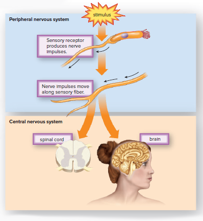
Complex animals rely on sensory receptors to provide information to the central nervous system
The central nervous system integrates sensory input before directing a motor response
Sensory receptors:
Sense organs are specialized to receive one kind of stimulus
Sensory receptors transform the stimulus into nerve impulses that reach a particular section of the cerebral cortex
Integration, the summing up of signals, occurs before sensory receptors initiate nerve signals
Sensory adaptation is a type of integration that results in a decrease in response to a stimulus
Brain and perception:
The brain is responsible for sensation and perception
Each part of the brain interprets impulses in only one way
If photoreceptors of the eye are stimulated by pressure and not light, the brain causes us to see "stars" or other visual patterns.
Chemical Senses
Sensory receptors have fundamental functions that help animals stay safe, find food, and find mates.
Chemoreceptors are responsible for detecting chemicals in the environment, which is believed to be our most primitive sense.
Chemoreceptors are present almost universally in animals.
Planarians have chemoreceptors all over their bodies, but they are found in higher concentrations on the auricles at the sides of the head.
Male moths have receptors for a sex attractant on their antennae.
The receptors on the antennae of the male silkworm moth are very sensitive, with only 40 out of 40,000 receptor proteins needing to be activated for the male to respond to a chemical released by the female.
Houseflies have chemoreceptors largely on their feet, which they use to taste instead of their mouth.
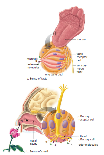
In mammals, the receptors for taste are located in the mouth, and the receptors for smell are in the nose.
Taste and Smell
Sensory receptors have fundamental functions that help animals stay safe, find food, and find mates.
Chemoreceptors are present almost universally in animals and give us the ability to detect chemicals in the environment.
Male moths have receptors for a sex attractant on their antennae, while houseflies have chemoreceptors largely on their feet.
In mammals, the receptors for taste are located in the mouth, and the receptors for smell are in the nose.
Taste buds, located primarily on the tongue, contain taste receptor cells, and the nose contains olfactory receptor cells.
There are at least five primary types of tastes: bitter, sour, salty, sweet, and umami.
Foods rich in certain amino acids produce the taste of umami.
The brain interprets the nerve impulses generated by receptor proteins as taste and smell.
Our senses of taste and smell have evolved to meet our physiological needs.
Smell is even more important to our survival than taste.
The olfactory bulbs have direct connections with the limbic system and its centers for emotions and memory.
Hearing and Balance
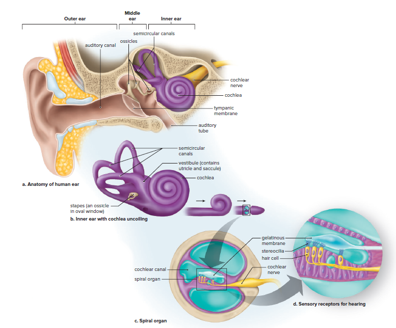
The human ear has two sensory functions: hearing and balance (equilibrium).
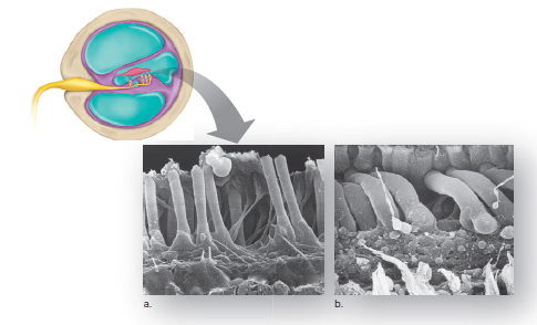
Sensory receptors for both functions consist of hair cells with long microvilli called stereocilia.
These microvilli are sensitive to mechanical stimulation and belong to a class of receptors called mechanoreceptors.
The sensory receptors for balance and hearing are similar and are present in the same organ.
This suggests an evolutionary relationship between them.
The sense organs of the mammalian ear may have evolved from a type of sense organ in fish.
Hearing
Most invertebrates cannot hear.
Some arthropods, including insects, have simple sound receptors.
In insects, the ear consists of a pair of air pockets enclosed by a membrane called the tympanic membrane.
The human ear has a tympanic membrane between the outer ear and middle ear.
The outer ear collects sound waves that cause the tympanic membrane to vibrate.
Three tiny bones in the middle ear amplify the sound about 20 times.
The last bone strikes the membrane of the oval window, causing it to vibrate and pass pressure to the fluid in the cochlea.
The sensory receptors for hearing are hair cells located in the cochlear canal of the cochlea.
The hair cells of the spiral organ synapse with the cochlear nerve, and nerve impulses travel to the brain stem and auditory areas of the cerebral cortex.
Each part of the spiral organ is sensitive to different wave frequencies, which correspond to the pitch of a sound.
Volume is a function of the amplitude of sound waves.
Hearing loss can be caused by infections, bone growth, or damage to the spiral organ.
Deafness due to middle ear damage is called conduction deafness, while deafness due to spiral organ damage is called nerve deafness.
Noise pollution can damage hearing, and noise-reduction earmuffs and earplugs are available.
Some medicines may damage the ability to hear.
Balance
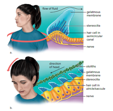
Humans have two senses of balance: rotational and gravitational.
Rotational equilibrium involves the semicircular canals, which respond to head movement in different planes of space.
Hair cells with stereocilia in a gelatinous membrane are embedded in the base of each canal.
Fluid within a semicircular canal flows over and displaces the gelatinous membrane, causing the stereocilia of the hair cells to bend and change the pattern of impulses carried to the CNS.
Gravitational equilibrium refers to the position of the head in relation to gravity.
It depends on the utricle and saccule, two membranous sacs located in the inner ear.
Both sacs contain hair cells with stereocilia in a gelatinous membrane.
Calcium carbonate granules called otoliths rest on this membrane.
When the head moves, the otoliths are displaced, and the membrane moves, bending the stereocilia of the hair cells and altering the frequency of nerve impulses to the CNS.
These data, usually supplemented by vision, tell the brain the direction of the movement of the head.
Similar Receptors in Other Animals
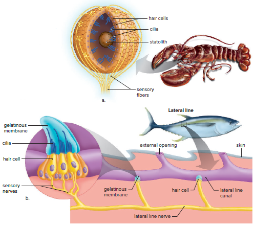
Gravitational equilibrium organs, called statocysts, are found in several types of invertebrates, including cnidarians, molluscs, and crustaceans.
Statocysts give information only about the position of the head, not the sensation of movement.
When the head stops moving, a small particle called a statolith stimulates the cilia of the closest hair cells, generating impulses that indicate the position of the head.
The lateral line system of fishes uses sense organs similar to those in the human inner ear.
In bony fishes, the system consists of sense organs located within a canal that has openings to the outside.
The sense organ is a collection of hair cells with cilia embedded in a gelatinous membrane.
Water currents and pressure waves from nearby objects cause the membrane and the cilia of the hair cells to bend.
The hair cells initiate nerve impulses that go to the brain.
Fishes use these data not for hearing or balance but to locate other fish, including predators, prey, and mates.
Vision
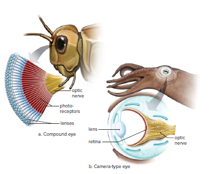
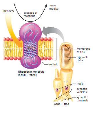
Photoreceptors are sensory receptors that are sensitive to light.
Planarians have eyespots that allow them to determine only the direction of light.
Other photoreceptors form actual images that provide detailed information about an object, including how far away it is.
Arthropods have compound eyes composed of many independent visual units, each with a lens to focus light rays on photoreceptors.
Vertebrates and certain mollusks have a camera-type eye with a single lens that focuses light on photoreceptors, which number in the millions and are closely packed together within a retina.
Insects have color vision, but they make use of a slightly shorter range of the electromagnetic spectrum than humans do.
Some fishes and most reptiles are believed to have color vision, but among mammals, only humans and other primates have expansive color vision.
Expansive color vision was adaptive for a diurnal lifestyle, which accounts for its retention in only a few mammals.
The Human Eye
Each eye views the same object from a slightly different angle.
This allows for binocular vision and the ability to perceive 3D images and depth.
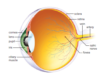
The human eye has numerous parts involved in preparing the stimulus for sensory receptors.
Light rays are brought to focus on the photoreceptors within the retina.
The cornea and lens are involved in focusing light rays on the photoreceptors.
The iris regulates the amount of light that enters the eye through the pupil.
The retina generates nerve impulses that are sent to the visual part of the cerebral cortex.
The brain forms an image of the object from this information.
The lens is controlled by the ciliary muscles and changes shape to focus on near or distant objects.
With normal aging, the lens loses its ability to accommodate for near objects, requiring reading glasses.
Aging and UV exposure can cause cataracts, which make the lens opaque and require surgery as the only viable treatment.
Photoreceptors of the Eye
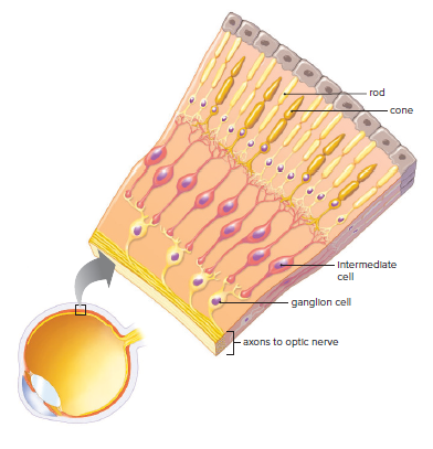
Rods and cones are photoreceptors in the human eye.
Rods are more numerous and sensitive to light than cones.
Cones are located primarily in the fovea and are responsible for color vision.
The retina has three layers of cells: photoreceptors, intermediate cells, and ganglion cells.
Information from rods and cones is processed by the intermediate cells and ganglion cells.
The optic nerve carries signals from the ganglion cells to the brain.
The blind spot is a small area on the retina where there are no photoreceptors.
Seeing in the Dark
Nocturnal animals use either vision or sonar to navigate in the dark.
Bats use echolocation to avoid obstacles and find prey.
Bats emit ultrasonic sounds in chirps to locate their prey.
Bats determine the distance to their prey by timing the echo's return.
A bat-inspired sonar walking stick is being developed for visually impaired people.
The walking stick emits ultrasonic chirps and detects echoes from nearby objects.
Buttons on the cane's handle vibrate to warn the user of obstacles.
A fast, strong signal indicates a close obstacle.
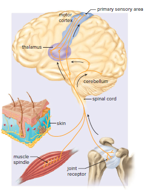
Cutaneous Receptors and Proprioceptors
Cutaneous receptors and proprioceptors provide sensory input from the skin, muscles, and joints to the primary sensory area of the cerebral cortex.
The cerebral cortex has areas that represent each part of the body.
Figure 28.11 shows the representation of each body part in the cerebral cortex.
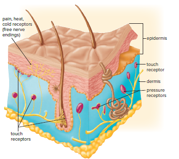
Cutaneous Receptors
Skin is the outermost covering of our body.
The skin contains numerous sensory receptors called cutaneous receptors.
Cutaneous receptors help us respond to changes in our environment, be aware of dangers, and communicate with others.
Sensory receptors in the skin are for touch, pressure, pain, and temperature.
The skin has two layers: epidermis and dermis.
Epidermis is packed with cells that become keratinized as they rise to the surface.
Free nerve endings responsive to cold or warmth are present in the epidermis.
Pain receptors (nociceptors) sensitive to extremes in temperature or pressure and to chemicals released by damaged tissue are present in the epidermis.
Stimulation of internal receptors is felt as pain in the skin called referred pain.
The Dermis of the skin contains sensory receptors for pressure and touch.
Pacinian corpuscles are onion-shaped pressure receptors that lie deep inside the dermis.
Several other cutaneous receptors detect touch.
Touch receptors are concentrated in parts of the body essential for sexual stimulation: the fingertips, palms, lips, tongue, nipples, penis, and clitoris.
Proprioceptors
Proprioceptors aid in maintaining equilibrium and posture despite gravity's force on the skeleton and muscles.
Muscle spindles are made up of sensory nerve endings wrapped around a few muscle cells in a connective tissue sheath.
Golgi tendons and other sensory receptors are situated in the joints.
The speed of nerve impulses from proprioceptors is directly related to the stretching of the organs they occupy.
A motor response causes the contraction of muscle fibers adjacent to the proprioception.
28.2 The Motor Systems
The muscular and skeletal systems are involved in the nervous system's motor response to stimuli.
The functions of the muscular and skeletal systems overlap; hence they are referred to as the musculoskeletal system.
The musculoskeletal system in humans performs the following functions:
Supports the body and enables the movement of body parts.
Protects internal organs such as the heart, lungs, brain, and spinal cord.
Aids the functioning of other systems, such as breathing, digestion, and blood circulation.
Skeletal muscle contraction assists in the movement of blood in veins and lymphatic vessels.
Skeletal muscles help maintain a constant body temperature.
Bones store fat and calcium, with calcium ions playing a major role in muscle contraction and nerve conduction.
Types of Skeletons
Vertebrates have an endoskeleton while arthropods have an exoskeleton.
Both skeletons are jointed and help animals live successfully on land.
The endoskeleton of humans is made of bone, which is living material and can grow.
The endoskeleton protects internal organs and grows as the animal grows.
Soft tissues protect the endoskeleton and are easier to repair than the skeleton itself.
The exoskeleton of arthropods is made of chitin, which is strong and flexible.
The exoskeleton protects against wear and tear, enemies, and prevents drying out.
The exoskeleton does not grow with the animal, and arthropods molt to rid themselves of an exoskeleton that has become too small.
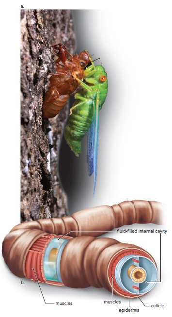
Animals without a hard skeleton have a fluid-filled internal cavity that acts as a hydrostatic skeleton.
A hydrostatic skeleton offers support and resistance to muscle contractions, allowing the animal to move and change shape.
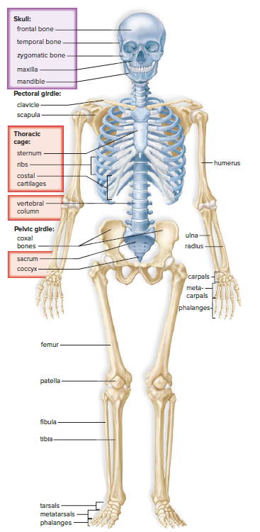
The Human Skeleton
The human skeleton is composed of 206 bones, which are divided into two main parts: the axial skeleton and the appendicular skeleton.
Axial Skeleton:
Bones in the midline of the body.
Consists of the skull (cranium and facial bones), vertebral column (spine), and rib cage (ribs and sternum).
Protects vital organs such as the brain, spinal cord, heart, and lungs.
Provides flexibility and support to the body.
Intervertebral discs separate the vertebrae and can cause pain if damaged.
The rib cage moves when we breathe.
Appendicular Skeleton:
Contains bones of two girdles and their attached limbs.
The pectoral girdle (shoulder) and upper limbs are specialized for flexibility.
The pelvic girdle (hip) and lower limbs are specialized for strength and protect internal organs.
The clavicle and scapula make up the shoulder girdle.
The Humerus of the arm articulates only with the scapula and is stabilized by tendons and ligaments that form a rotator cuff.
Radius and ulna contribute to the easy twisting motion of the forearm.
The pelvic girdle contains two massive coxal bones that articulate with the femurs (thighbones.)
Kneecap protects the knee, the tibia is the shinbone, and the fibula is the more slender bone in the leg.
Each foot contains bones of the ankle, instep, and five toes.
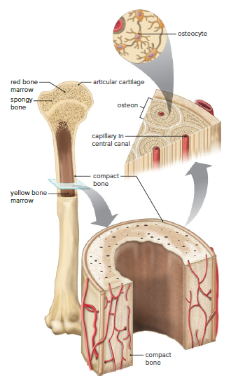
Structure of a Bone
Long bones have a cavity that is bounded by compact bone.
Compact bone contains osteons (Haversian systems), where osteocytes lie in tiny chambers arranged in concentric circles around central canals.
Osteoblasts deposit bone, and osteoclasts secrete enzymes that digest the matrix of bone and release calcium into the bloodstream.
Osteoporosis occurs when osteoclasts are working harder than osteoblasts.
Spongy bone is found at each end of a long bone and has numerous bony bars and plates separated by irregular spaces.
Red bone marrow is found in the spaces of spongy bone and produces red blood cells.
Yellow bone marrow is found in the cavity of a long bone and stores fat.
A thin shell of compact bone and a layer of hyaline cartilage are found beyond the spongy bone, which is important to healthy joints.
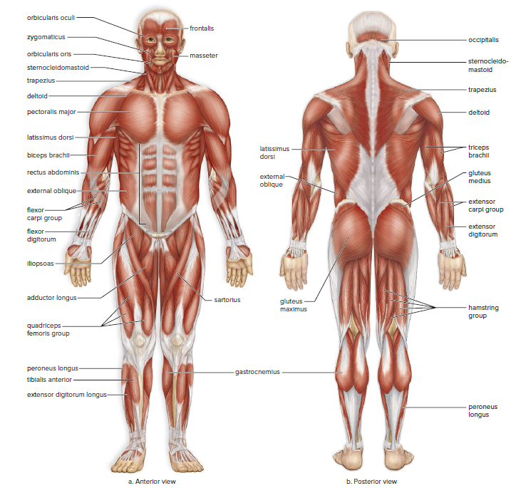
Skeletal Muscle Structure and Physiology
Three types of muscle: smooth, cardiac, and skeletal.
Smooth muscles:
Contain sheets of long, spindle-shaped cells.
Each cell has a single nucleus.
Cardiac muscles:
Striated and typically possess a single nucleus.
Branched chains of cells that interconnect, forming a lattice network.
Skeletal muscles:
Muscle fibers that are also striated.
Quite elongated and run the length of a skeletal muscle.
Arise during development when several cells fuse, resulting in one long, multinucleated cell.
Skeletal muscles are the major type of muscle in the body and are under the voluntary control of the nervous system.
Figure 28.16 illustrates some of the major skeletal muscles in the body.
Skeletal Muscle Contraction
Skeletal muscle fibers shorten when they contract.
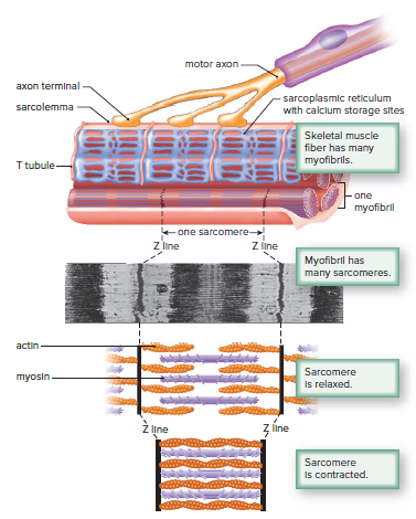
Nerve impulses travel down a motor axon and release acetylcholine (ACh) into a synaptic cleft.
ACh diffuses across the cleft and binds to receptors in the plasma membrane of a muscle fiber (sarcolemma).
The sarcolemma generates impulses that travel along its T tubules to the sarcoplasmic reticulum.
Calcium is released from calcium storage sites, causing muscle fibers to contract.
Muscle fibers contain many parallel myofibrils, which are striated due to the placement of protein filaments within sarcomeres.
Sarcomeres contain thick filaments made up of myosin and thin filaments made up of actin.
During muscle contraction, actin filaments slide past myosin filaments, shortening the sarcomere.
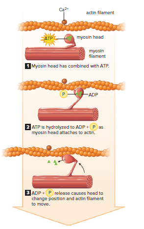
The sliding filament model of muscle contraction explains the movement of actin filaments in relation to myosin filaments.
Myosin heads attach to actin filaments when calcium is present, and the release of ADP and phosphorus P causes myosin to shift its position and pull the actin filament to the center of the sarcomere.
The cycle of attachment and detachment of myosin heads continues until the actin filaments move nearer and nearer the center of the sarcomere.
The contraction continues until nerve impulses cease and calcium ions are returned to their storage sites.
The sarcoplasmic reticulum contains active transport proteins that pump calcium ions back into the calcium storage sites, causing the muscle to relax.
Without ATP, the myosin heads cannot detach from actin, and calcium cannot be pumped back into the sarcoplasmic reticulum, resulting in rigor mortis.
Skeletal Muscles Move Bones at Joints
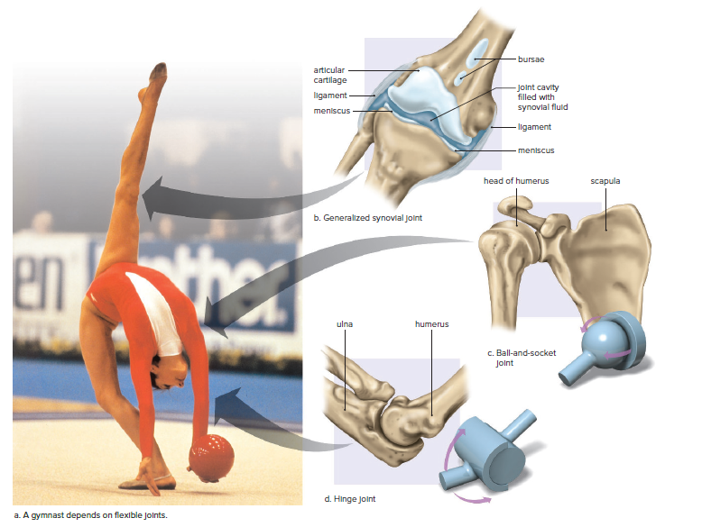
Joints are classified as immovable, slightly movable, and freely movable (synovial joints).
In synovial joints, ligaments bind the two bones together, providing strength and support and forming a capsule containing lubricating synovial fluid.
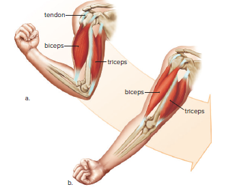
Muscles are attached to bones by tendons that span movable joints.
Muscles work in antagonistic pairs, meaning if one muscle flexes the joint and raises the limb, the other extends the joint and straightens the limb.
Synovial joints may include additional structures such as menisci and bursae.
Ball-and-socket joints at the hips and shoulders allow movement in all directions, even rotational movement.
Elbow and knee joints are synovial joints called hinge joints because they largely permit movement in one direction only.
Joint disorders include sprains, bursitis, and torn meniscus.
Arthroscopic surgery is used to remove cartilage fragments or to repair ligaments or cartilage in a less traumatic way than surgically opening the knee with long incisions.
Rheumatoid arthritis is less common than osteoarthritis, which is caused by overworking a joint.
Constant compression and abrasion damage articular cartilage, causing it to soften, crack, and wear away.
As the disease progresses, exposed bone thickens, and forms spurs that enlarge and restrict joint movement.
Weight loss can ease arthritis by reducing the load on joints.
A sensible exercise program helps build up muscles that stabilize joints.
Low-impact activities like biking and swimming are best for people with arthritis.
Damaged joints can be replaced with a prosthesis.
Glucosamine and chondroitin supplements can promote cartilage formation and repair.
Exercise improves muscular strength, endurance, and flexibility.
Exercise improves cardiorespiratory endurance and may lower blood cholesterol levels.
Exercise promotes the activity of osteoblasts, which helps prevent osteoporosis.
Exercise helps prevent weight gain and relieves depression while enhancing mood.