Tags & Description
Brain
The organ in your head
Made up of nerves that process information and control behaviour.
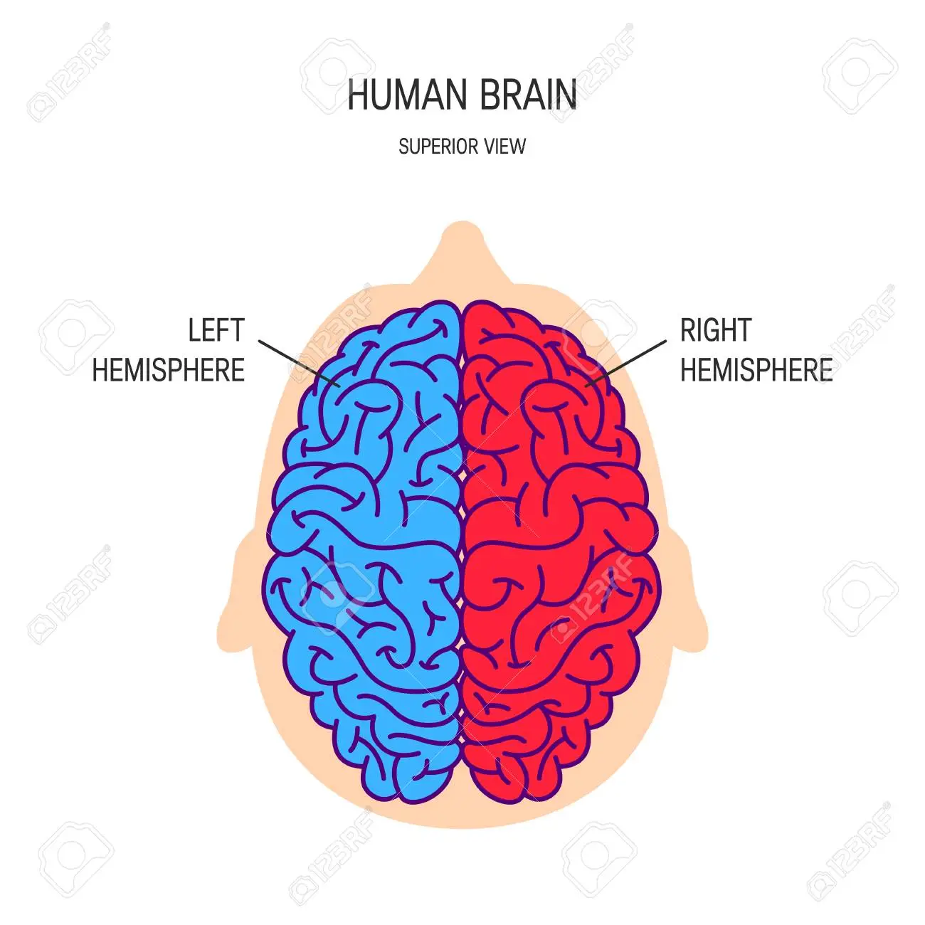
Hemisphere
Half of the brain
Right hemisphere on the right side
Left hemisphere on the left
Cerebrum
Largest part of the brain where higher processing happens
Upper part of the brain
Has an outer cortex
Cortex
Outer layer of the brain (a ‘shell‘)
Has lots of folds to increase its surface area
Technical terms for:
The ‘bumps‘ on the surface
Creases on the surface
Gyri (‘a gyrus’)
Sulci (‘a sulcus’)
What does the large surface area allow the human brain to have?
More nerve cells
This allows it to control more functions
Why are the surfaces of animal brains smoother than a human brain?
Animals have fewer behavioural functions than humans
So the need less surface area in the brain
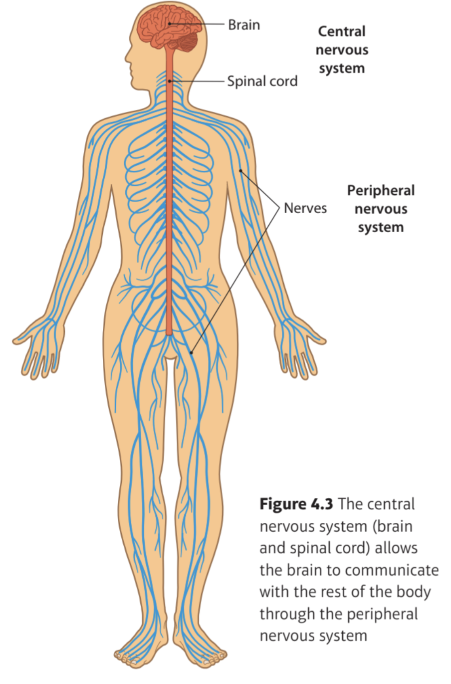
Spinal cord
A pathway of nerves inside the spine
It connects the brain to the rest of the body through the peripheral nervous system
Brainstem
The part of the brain that connects the spinal cord to the upper brain
Controls reflexes
Reflexes
Actions that are automatic
They don’t require conscious thought
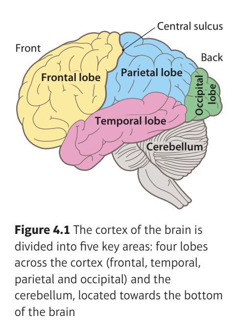
Frontal lobe
The area at the front of the brain responsible for decision-making and impulse control
Helps control problem-solving skills
Helps us concentrate and pay attention to different activities
Motor cortex
Towards the back of the frontal lobe
A large area just in front of the central sulcus
Plays a key role in the voluntary movements
Central sulcus
The crease that separates the frontal lobe from the parietal lobe
Temporal lobe
The area on the side of the brain that controls hearing and memory
Helps hear/understand sounds
Helps understand speech and create speech
Contains the auditory cortex (controls hearing)
Parietal lobe
The area at the top of the brain important for perception and sensations of touch
Contains the somatosensory cortex- sense of touch
Occipital lobe
The area at the back of the brain that controls vision
Contains the visual cortex
Helps us process visual information from our eyes
Helps make sense of this information so we understand what we are seeing
Cerebellum
An area of the brain near the brainstem that controls motor movements
Vital role in motor skills- movement, coordination and balance
Takes information from the senses, spinal cord and other parts of the brain and then combines them to coordinate behaviour
The message is sent via the spinal cord
How does the cerebellum coordinate behaviour if we are running and see an object in our way?
The cerebellum combines this information and sends a message back to the body
It tells it to move to avoid the object.
This message is sent via the spinal cord
It tells us to change direction while helping us keep our balance
So we do not fall as we dodge the object in our way
Structure of the brain
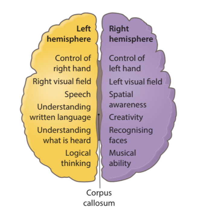
Lateralisation of function
The different jobs performed by each half of the brain
Each hemisphere will have different specialist roles
How is the brain asymmetrical?
The 2 hemispheres of the brain are not exactly the same, in terms of structure and function
Each hemisphere controls different functions and plays a larger or smaller role in a particular behaviour
What side of the brain controls each side of the body?
Each side of the brain controls the functions on the opposite side of the body
Right side of the brain controls functions on the left side of the body
Corpus callosum
A thick bundle of nerve fibres that connect the right and left hemisphere of the brain so that they can communicate with each other
So that the whole brain can work as one complete organ
The 2 hemispheres retain their own roles while working together to control behaviour in the whole body
Broca’s area
A part of the left hemisphere of the brain that controls speech production
What does the left hemisphere control?
Logical thinking
Broca’s area controls the production of speech
Broca’s area is linked to the control of nerve cells in our face that help us speak
Processing of language-based information
Ability to write and understand language
What might happen if someone’s Broca’s area was damaged?
They will find it difficult to talk
Stuttering
Difficult to identify objects verbally
Spatial awareness
The ability to negotiate space and navigate our way around our environment
What does the right hemisphere control?
Creativity
Spatial awareness
Our ability to recognise and perceive faces
Processing of music
Making sense of visual information that we see
Role of the corpus callosum
Allows messages to be passed from the left hemisphere to the right hemisphere and vice versa
Makes it easier for the brain to pass messages between the different areas of the brain
Makes connections between different types of information
How does the corpus callosum play a role when you hear something spoken in your left ear?
The information passes to the right hemisphere of the brain
The information is then passed to the left hemisphere to be decoded so that the brain understands what was said
The information is then be passed to the right hand for the person to write down what they have heard
Sex differences in brain lateralisation
Males and females have brains that work differently
Females → better at language skills (left-brain tasks)
Males → better at spatial skills (right-brain tasks)
Females may have a thicker corpus callosum
What does it mean if females have a thicker corpus callosum?
They use both sides of their brain for tasks
Males show dominance for 1 hemisphere for the same tasks
There is more activity in one hemisphere than the other, rather than an equal spread of activity
Strengths of lateralisation as an explanation of sex differences between males and females
Studies provided evidence to show that male and female brains work differently because of how the roles of different areas of the cortex are organised
A study by Harasty et al. (1997) showed that parts of the brain that process and produce language are slightly bigger in females compared to males
Explain why there is a view that females are better at tasks that use language skills.
Study by Rilea et al. (2005) found that males were better at spatial tasks, especially those that use a lot of activity in the right hemisphere.
Evidence that supports these differences were concluded from scientific methods such as brain scans and lab experiments.
So research was controlled and helps prevent the interference of extraneous variables.
This strengthens the explanation as the research evidence is scientific and so the explanation, developed from this evidence, is the same
Weaknesses of lateralisation as an explanation of sex differences between males and females
Research and evidence showing the 2 hemispheres of males and females work differently has weaknesses.
In the Rilea et al. (2005) study males did not always do better than females on the spatial tasks.
There were spatial tasks used in the study that did not use a lot of ‘right- brain’ activity.
So differences in how males and females use the right hemisphere for spatial tasks can’t account for all differences in performance.
Sommer et al. (2004) study showed that there was no strong evidence that females used both hemispheres for language tasks
So this isn’t a good explanation for girls being better at language skills than boy
What is the central nervous system (CNS) made up of?
The brain
Spinal cords
What does the central nervous system (CNS) do?
Helps the brain and body communicate with one another by relaying messages from the brain to the rest of the body to instruct it what to do
The sensory nerves in the body send messages to the brain via the spinal cord
The brain processes the information and then sends messages to the body down the spinal cord to make the body do something
Sensory nerves
Located in the skin, muscles or organs
Take information into the nervous
Peripheral nervous system (PNS)
Nerves that branch out from the brain and spinal cord
Activated by the spinal cord
Makes the body do the actions the brain is telling it to do
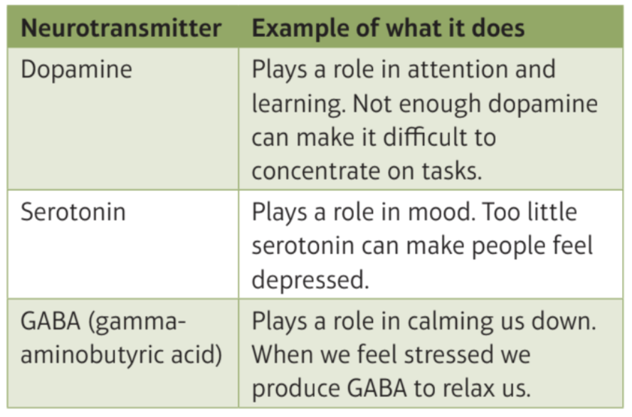
Neurotransmitters
Chemicals released from neurons to help them to pass messages from one cell to another across a synapse
Neuron
A nerve cell that transmits information
Neurotransmitters and their roles
Dopamine
Serotonin
GABA
When are neurotransmitters released and what happens to it?
When a nerve impulse reaches the end of a nerve fibre
The neurotransmitter is then picked up by another neuron to receive the message and continue the nerve impulse
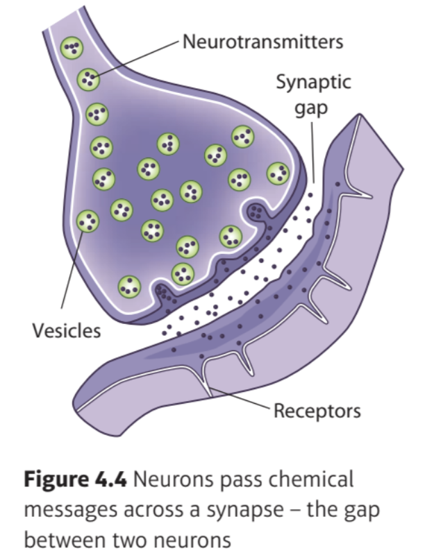
Synaptic transmission
The process by which neurotransmitters are released by a neuron, move across the synaptic gap and are then taken up by another neuron
Synapse
A gap between 2 neurons that allows messages, in the form of neurotransmitters, to pass from one cell to another
Axon
The long structure that connects the cell body of a neuron to the terminal button at the end of the cell
Terminal button
The end of a neuron
Filled with tiny sacs called vesicles
Contain neurotransmitters
Vesicles
Small sacs containing neurotransmitter (chemical) molecules
Receptors
Special sites on neurons that are designed to absorb neurotransmitter molecules
Synaptic functioning
Messages are passed throughout the nervous system, from one neuron to the next, by synaptic transmission
An electrical impulse is triggered inside the cell body of a neuron
The neuron then passes a small impulse along the axon towards the end of the nerve fibre
When the nerve impulse reaches the terminal button, the vesicles release the neurotransmitter molecules into the synapse
These molecules are ‘grabbed’ by the receptors on the next neuron to pass the message impulse on
Neurological damage
Damage to the body’s central and peripheral nervous system
What would happen if the brain is damaged?
Messages normally passed around in the nervous system might be interrupted
Agnosia
Inability to interpret sensations and so unable to make sense of the information/recognise something
Issue in the way the brain processes sensory information
Visual agnosia
Inability to recognise things that can be seen
Disorder of perception
Result of damage to the parietal lobe
Can see the object in front of them perfectly but their brain can’t make sense of this information.
Difficulties of a person with visual agnosia
Recognizing objects due to impairments in basic perceptual processing or higher-level recognition processes.
Understanding another person’s identity
Perceiving shapes or forms
Symptoms of visual agnosia
(different symptoms depending on the type of visual information the brain cannot understand)
Patients may not be able recognise:
The colour of an object
Objects and name them
Places they are familiar with.
Prosopagnosia
‘face-blindness’
Inability to recognise faces even though they can be seen
Eyes send information to the brain about the face, but the brain can’t recognise who the face belongs to
What can prosopagnosia be caused by?
Damage to the fusiform face area (FFA)
Near the back of the temporal lobe
Next to the occipital lobe
Symptoms of prosopagnosia
Patients find it difficult to identify people by their faces
Some patients:
See all faces as ‘the same’ and can’t tell faces apart
Can’t recognise faces of people that they know really well
Have trouble matching up pictures of faces that they don’t know
Where is the Pre-frontal cortex located?
Area of the brain’s cortex at the very front of the frontal lobe
Immediately behind the forehead
What does the Pre-frontal cortex control?
Helps us to control our impulses, decision-making, and rational thinking
Stops you from doing something like hitting someone when you are angry
Helps us to keep our emotions balanced so that we do not get too emotional
No matter what emotion we are feeling
What happens if the pre-frontal cortex is damaged?
People can become impulsive and aggressive.
Difficult for people to control their emotions
So their personality changes a lot
People are more likely to commit crimes that they would not have done before
What did Adrian Raine et al. (1997) study?
The brains of murderers
He compared these to a similar group of people who had not committed murder
What did Adrian Raine et al. (1997) find and what was it used to show?
There were differences in the pre-frontal cortex of the two groups.
Murderers had less activity in the pre-frontal cortex
Making them more impulsive and aggressive
Used as an explanation for why some people are more prone to violent and impulsive behaviour than others
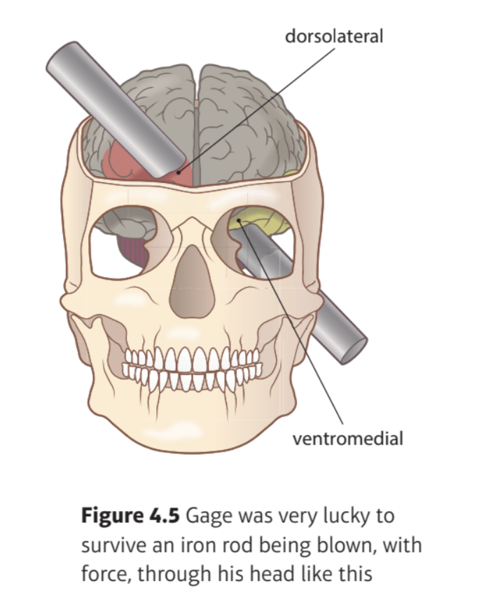
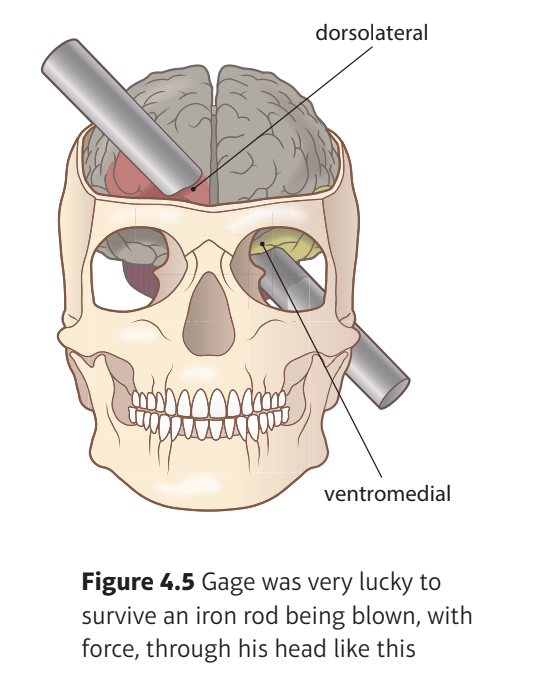
Background to: Damasio et al. (1994) The Return of Phineas Gage: Clues About the Brain from the Skull of a Famous Patient
In 1848, he suffered damage to his face + frontal lobe of his brain after an iron rod was fired through his head
His personality permanently changed
Calm + responsible → Irresponsible + rude
Psychologists used the evidence from his doctor, John Harlow, to understand the role of the frontal lobe
He died 12 years after his original injury due to a severe epilepsy as a result of the accident
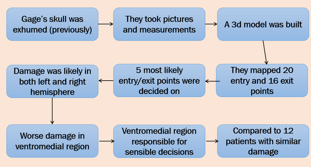
The body was exhumed so that he could study his skull
Aims to: Damasio et al. (1994) The Return of Phineas Gage: Clues About the Brain from the Skull of a Famous Patient
Damasio et al. wanted to:
Build a model of Gage’s skull to map out how the iron rod passed through his head (3D computer representation)
Identify which parts of his brain were most likely to have been badly damaged in the accident.
Discover if areas other than the frontal lobe had also been damaged
Procedure: Damasio et al. (1994) The Return of Phineas Gage: Clues About the Brain from the Skull of a Famous Patient
First, they took pictures and measurements of Gage’s skull
They built a virtual 3D replica model of a skull that matched the measurements of Gage’s skull.
Actual measurements of the rod were taken
3 cm diameter + 109 cm long.
This was compared to the damaged parts of the skull to work out the path the iron bar took
They did so by matching up the possible entry and exit points for the iron rod on their model.
20 entry + 16 exit points tested
They found the 5 most likely paths
They used the virtual replica of his brain to map out which areas would have been damaged in each case
White matter
Brain and spinal cord tissue
Consists mainly of nerve fibres (axons)
Where neurons pass messages along axon fibres
Results: Damasio et al. (1994) The Return of Phineas Gage: Clues About the Brain from the Skull of a Famous Patient
Found damage in both the right + left hemispheres of the frontal lobe in Gage’s brain was likely
Brain damage in the accident only affected the frontal lobe
Iron bar passed through the left eye socket and upwards through the head
So there was more damage to the underlying white matter in the left hemisphere than in the right frontal lobe
Damaging this area meant Gage was unable to pass neural messages in this part of his brain, making it useless
Damage in both hemispheres was worse in the ventromedial region (middle of the underside), but dorsolateral regions (top edges of frontal lobe) were not affected
Conclusion: Damasio et al. (1994) The Return of Phineas Gage: Clues About the Brain from the Skull of a Famous Patient
Researchers compared areas of Gage’s brain that were most likely damaged with changes in his personality after the accident
The ventromedial area of the frontal lobes is responsible for making sensible decisions + controlling our impulses around people + emotions
He found it difficult to control his emotions
Evidence supports other findings from case studies of people with brain damage in similar areas
They had evidence of 12 patients with similar frontal lobe damage, who all showed the same problems with impulse and emotional control
Knowledge can be used to predict the behaviour of someone who suffers brain damage in these areas in the future
Strengths: Damasio et al. (1994) The Return of Phineas Gage: Clues About the Brain from the Skull of a Famous Patient
Researchers used modern-day technology to investigate the data from 1848 so results can be given more scientific status
Used computer model → evidence could be seen, than than just inferred from information after the accident
Increases the scientific understanding of Gage’s case.
We can now make predictions about what changes to behaviour we might expect if someone has damaged their frontal lobes
Helps family understand what might happen + why it is happening
Weaknesses: Damasio et al. (1994) The Return of Phineas Gage: Clues About the Brain from the Skull of a Famous Patient
Information may not be accurate or reliable
Used an exact replica of Gage’s skull but information about the accident was gathered 150 years ago
Lacks generalisability as brain damage was unique to Gage
Unlikely someone will have exactly the same damage
So information may not be useful for understanding what might happen to another person with frontal lobe damage
Background: Sperry (1968) Hemisphere Deconnection and Unity in Conscious Awareness
Some patients with epilepsy did not respond well to treatment so they were offered surgery
Research showed without a corpus callosum, the left and right hemispheres of the brain worked like two separate brains (a ‘split-brain’) instead of one whole brain
Why do patients with severe epilepsy get surgery?
Reduce seizures/symptoms of epilepsy
They cut down the corpus callosum to disconnect the right and left hemisphere
Aims: Sperry (1968) Hemisphere Deconnection and Unity in Conscious Awareness
He studied what effects could be seen in these patients by monitoring how they processed information using their ‘split-brain’.
Wanted to see how the ‘split brain’ worked compared to a normal brain.
Find out the cognitive functions linked to each hemisphere in the brain.
Procedure: Sperry (1968) Hemisphere Deconnection and Unity in Conscious Awareness
Sperry gave 11 participants who had had their corpus callosum cut different tasks to see how they processed different types of information in the spilt-brain
9 had surgery recently whereas
2 had surgery a while before and had an excellent \n recovery
Different variations of the tasks
All tasks involved the same basic process
Sending different types of sensory information to the left + right hemispheres
Then asking the brain to respond to the information using either the same or the opposite hemisphere
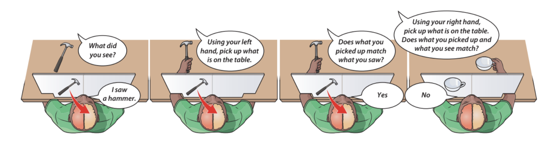
How did the other type of Sperry’s visual task work on some occasions?
Rather than saying the word or identifying the picture, participants were asked to point to an item or picture
They were shown a variety of objects or pictures including the one they had just been shown
They would then identify what they had seen using either the same hand, or the hand on the opposite side of the body.
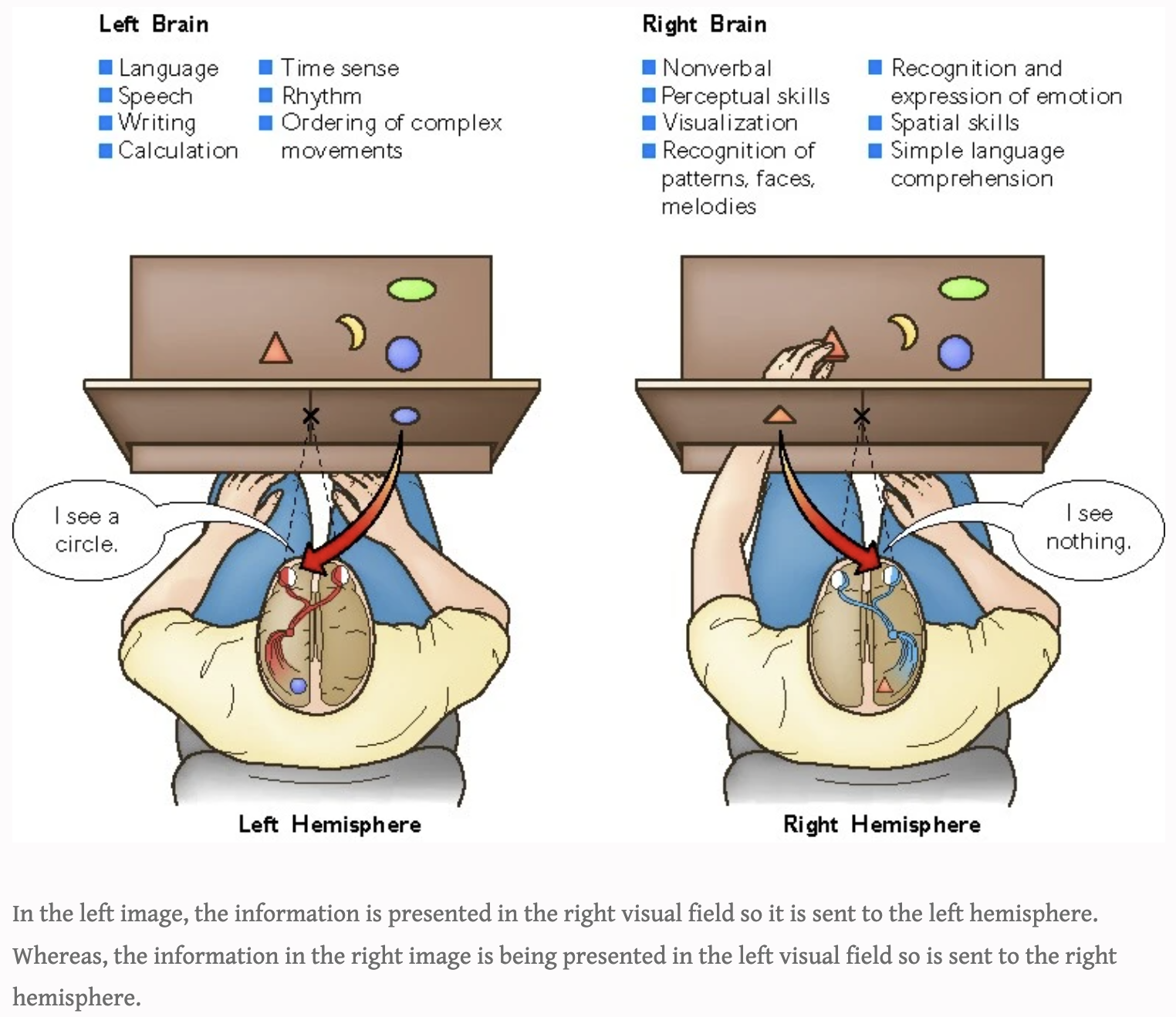
How did Sperry’s visual task work?
Participants focused on the centre of a screen
On the screen, information was presented to the left and right sides of the visual fields at the same time
2 different words or pictures were presented
1 on the left of the focal point + 1 on the right
So left side of each eye picked up 1 image- the one on the right of centre
But right side of each eye picked up the other image- the one on the left of centre
Information on the right of the visual field → passed to the left hemisphere (vice versa)
Participants were asked to say the word/s or picture/s they had seen on the screen
Variations of Sperry’s visual task
Putting unseen objects into one of the hands and asking them to identify them from touch alone
Then placing different objects in each hand and then asking them to feel for them in a large pile of different objects
In the last picture, why does it not match what the person saw?
Someone with a split-brain cannot exchange information between the 2 hemispheres
So if information goes to the right of both eyes (left hemisphere) and comes into the left hand (right hemisphere) the person cannot match the two pieces of information
Results for the tasks involving reading words or selecting objects
Words shown to the RVF → patients had no problem repeating the word to the researcher
Words shown to the LVF (sent to right hemisphere), patients had trouble saying what they had seen
Word or picture shown to the LVF (right hemisphere)→ participants had little trouble selecting an object that matched what they had seen
Word or picture shown to the RVF (left hemisphere) → participants struggled to point to the correct object
Results for the tasks for objects presented to each hand
Objects felt by right hand (passed to left hemisphere) → they could name the object
Objects felt by left hand → they found it more difficult to say what they could feel
2 different objects given to the participant (1 in each hand) → after they were asked to feel around in a pile of objects for the 2 objects → they could only identify each item with the hand that originally held it
If the opposite hand picked up the item, they could not identify it as the item they had held before.
Quasi experiment
Aims to establish a cause and effect relationship
Variables of Sperry’s experiment
IV: whether the individual had a split-brain or not
DV: individual’s performance on visual and tactile tasks.
CV: No control group → as effects of not having a split-brain were already known
Conclusion of Sperry (1968) Hemisphere Deconnection and Unity in Conscious Awareness
Each hemisphere is capable of working well without being connected to the other side
But, each hemisphere has its own memories → without a corpus callosum, it couldn’t be shared with the other side
Caused problems for some activities → supported that the right + left hemispheres have different roles
LH: better at naming items using words when they had been held by the right hand
RH: better at identifying objects by feeling for them with the left hand after previously being held by the left hand
Supports the LH controls more language abilities, but the right hemisphere controls spatial abilities
Strengths of Sperry (1968) Hemisphere Deconnection and Unity in Conscious Awareness
Reliability: gathered lots of detailed information
Standardised procedure: he designed procedures (such as the split-screen for presenting visual information) → kept the same for each participant → data gathered in a reliable way + participants’ results compared easily
Weaknesses of Sperry (1968) Hemisphere Deconnection and Unity in Conscious Awareness
Not generalisable: Sample of 11 participants → too small → few people have surgery to sever the corpus callosum so the results not useful to explain how ‘normal’ brains work
Lacks ecological validity: Lab experiment → very artificial → lacks mundane realism → you don’t look at a picture with one eye