Biology
Cells
Basics of Cellular Reproduction
Chromosomes
Chromatin
Cell Cycle
Interphase
Mitosis
Cytokinesis
Spindle
Cell Cycle Checkpoints
Internal and External Signals
Apoptosis
Cancer
Proto-Oncogenes
Tumor Suppressor Genes
Oncogenes
Chromosomal Rearrangements
BRCA1
BRCA2
RB Gene
RET Gene
Cancer Cells
University/Undergrad

Chapter 8: Cellular Reproduction
8.1 The Basics of Cellular Reproduction
Humans begin life as a single cell, but through cellular reproduction, they grow into an organism consisting of trillions of cells in less than 10 months.
Cellular reproduction continues throughout a person's life, helping to grow new tissues and repair damaged ones.
Right now, the human body is producing thousands of new red blood cells, skin cells, and cells that line the respiratory and digestive tracts.
If a person suffers a cut, cellular reproduction helps repair the injury.
Cellular reproduction is the process by which cells duplicate their contents and divide to form two new cells.
In bacteria and some protists, cellular reproduction occurs through a process called binary fission.
Binary fission is a form of asexual reproduction, which means that the new cells are identical to the original parent cell.
Sexual reproduction involves the fusion of two gametes, a sperm and an egg, to form a new organism.
Offspring produced through sexual reproduction are not identical to their parents.
The cell theory states that all cells come from preexisting cells.
A new cell cannot be created without a preexisting cell.
A new organism cannot be created without a preexisting organism.
Cellular reproduction is necessary for the production of both new cells and new organisms.
Cellular reproduction involves two important processes: growth and cell division.
During growth, a cell duplicates its contents, including the organelles and its DNA.
During cell division, the DNA and other cellular contents of the parent cell are distributed to the daughter cells.
The terms "parent cell" and "daughter cell" have nothing to do with gender; they are simply a way to designate the beginning cell and the resulting cells.
Both processes are heavily regulated to prevent runaway cellular reproduction, which can have serious consequences.
Chromosomes
DNA replication is the process by which a cell copies its DNA.
This process occurs in preparation for cell division.
Once DNA replication is complete, a full copy of all the DNA is passed on to both daughter cells.
The DNA contains all of the instructions that each cell needs to live and perform its functions.
A human cell contains 2 meters of DNA.
DNA and associated proteins are packaged into chromosomes.
Chromosomes allow DNA to be distributed to daughter cells.
Proteins and enzymes in the nucleus of the cell are responsible for packaging DNA into chromosomes and DNA replication.
Chromatin to Chromosomes
Chromatin is a complex of DNA and proteins found in eukaryotic cells.
Chromatin is organized into nucleosomes, which are bead-like structures consisting of DNA wrapped around a core of eight histone proteins.
Nucleosomes are arranged in a zigzag fashion, and then they are folded into loops for further compaction.
This looped chromatin more easily fits within the nucleus.
Just before cell division, chromatin condenses multiple times into large loops.
This produces highly compacted chromosomes, which are often 10,000 times more compact than the chromatin.
The compacted chromosomes can be viewed with a light microscope.
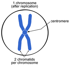
Prior to cell division, the chromosomes are duplicated.
A duplicated chromosome is composed of two identical halves, called sister chromatids.
The sister chromatids are held together at a constricted region called a centromere.
Each sister chromatid contains an identical DNA double helix.
Every species has a characteristic number of chromosomes.
Humans have 23 pairs of chromosomes, for a total of 46 chromosomes.
Sexual reproduction provides each of us with one copy of each chromosome from each parent.
8.2 The Cell Cycle: Interphase, Mitosis, and Cytokinesis
Cellular reproduction involves two steps: duplication of cell contents and cell division.
The cell cycle is an orderly sequence of stages that takes place between the time a new cell has arisen from the division of the parent cell to the point when it has given rise to two daughter cells.
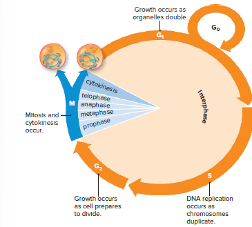
The cell cycle consists of three phases: interphase, mitosis, and cytokinesis.
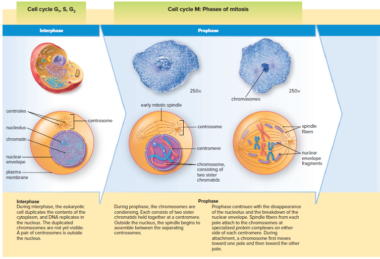
Interphase
Most of the cell cycle is spent in interphase.
The duration of interphase varies widely.
Embryonic cells complete the entire cell cycle in just a few hours.
A rapidly dividing mammalian cell, such as an adult stem cell, typically takes about 24 hours to complete the cell cycle.
A rapidly dividing mammalian cell spends 22 hours in interphase.
DNA replication occurs in the middle of interphase.
Interphase is divided into three phases: G1, S, and G2.
G1 is the phase before DNA replication.
G2 is the phase following DNA synthesis.
G stands for "growth" because the cell is metabolically active during these phases.
Protein synthesis is very much a part of these growth stages.
G1 phase:
Cell doubles its organelles and accumulates materials for DNA replication.
Cell integrates internal and external signals to decide whether to continue with the cell cycle.
G0 phase:
Some cells, like muscle cells, remain in interphase and cell division is permanently arrested.
If DNA damage occurs, many cells in G0 can reenter the cell cycle to repair the damage.
Few cell types, like nerve cells, almost never divide again once they have entered G0.
S phase is the second phase of the interphase of the cell cycle.
During S phase, DNA replication occurs, which means that the cell's DNA is copied.
At the beginning of S phase, each chromosome has one chromatid, which is a single DNA double helix.
At the end of S phase, each chromosome is composed of two sister chromatids, which are two DNA double helices that are attached at the centromere.
DNA replication produces the duplicated chromosomes, which are necessary for cell division.
G2 phase is the third phase of the interphase of the cell cycle.
During G2 phase, the cell synthesizes the proteins that will be needed for cell division, such as the protein found in microtubules.
Microtubules are part of the cytoskeleton, which is the cell's internal framework.
Microtubules play an important role in cell division, by helping to move the chromosomes around the cell.
M (Mitotic) Phase
M phase is the phase of the cell cycle in which the nucleus and cytoplasm divide.
Mitosis is the type of nuclear division that occurs during M phase.
During mitosis, the duplicated nuclear contents of the parent cell are distributed equally to the daughter cells.
The daughter nuclei are identical to the parent cell and to each other.
Each chromosome in a cell has two identical sister chromatids.
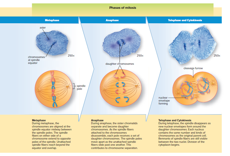
During mitosis, the sister chromatids of each chromosome separate and are now called daughter chromosomes.
The daughter nuclei produced by mitosis are genetically identical to each other and to the parent nucleus.
Most eukaryotic cells have an even number of chromosomes.
The Spindle
A spindle is used to pull the chromatids apart.
The spindle is made of microtubules that can assemble and disassemble.
The microtubules assemble to form the spindle and separate the chromatids.
The microtubules disassemble after the separation is complete.
A centrosome is the primary microtubule organizing center of a cell.
It is made up of two barrel-like structures called centrioles, and an array of microtubules called an aster.
Centrosomes duplicate at the start of the S phase of the cell cycle.
During the first part of the M phase, the centrosomes separate and move to opposite sides of the nucleus.
They form the poles of the spindle, which is used to separate the chromosomes.
Spindle fibers attach to the duplicated chromosomes and ensure their proper distribution to the daughter cells.
A centrosome will be just outside the newly formed daughter nuclei.
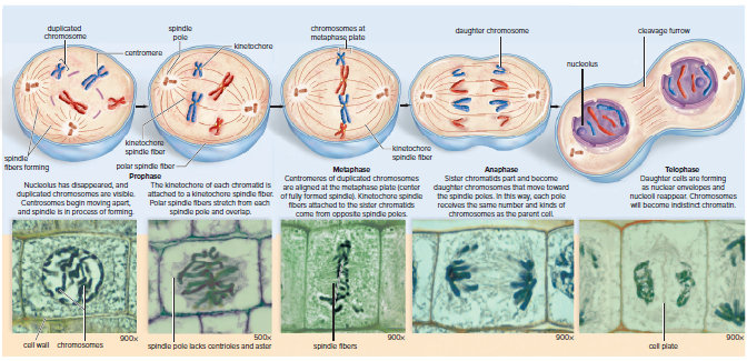
Phases of Mitosis in Animal and Plant Cells
Mitosis is a continuous process but is traditionally divided into a sequence of events.
Mitosis has four phases: prophase, metaphase, anaphase, and telophase.
During mitosis, a spindle apparatus forms and separates the sister chromatids of each duplicated chromosome.
The sister chromatids are then pulled to opposite poles of the cell, forming two daughter nuclei.
The daughter nuclei are genetically identical to the parent nucleus.
Mitosis is usually followed by cytokinesis, which is the division of the cytoplasm.
After cytokinesis, the daughter cells are genetically identical to the parent cell.
The cell cycle then repeats, with the daughter cells growing and DNA replicating in interphase.
Cytokinesis in Animal and Plant Cells
Cytokinesis follows mitosis in most cells.
If mitosis occurs, but cytokinesis doesn't, it results in a multinucleated cell.
Fungi, slime molds, and certain plant structures are multinucleated.
In animal cells, cleavage furrow begins as anaphase draws to a close.
A cleavage furrow is an indentation of the membrane between two daughter nuclei.
A band of actin filaments called the contractile ring forms a circular constriction between two daughter cells.
The contractile ring pulls the drawstring ever tighter around the middle of a balloon.
A narrow bridge between the two cells is visible during telophase.
The contractile ring continues to separate the cytoplasm until there are two independent daughter cells.
Cytokinesis in plant cells is different from animal cells due to the rigid cell wall surrounding plant cells.
Plant cells cannot undergo cytokinesis by furrowing.
Cytokinesis in plant cells involves building new plasma membranes and cell walls between daughter cells.
A small, flattened disk appears between the two daughter plant cells during cytokinesis.
The disk is composed of vesicles produced by the Golgi apparatus.
Vesicles move along microtubules to the disk region.
As more vesicles arrive and fuse, a cell plate is formed.
The cell plate is a newly formed plasma membrane that expands outward until it reaches the old plasma membrane and fuses with it.
The new membrane releases molecules that form new plant cell walls.
Cell walls are later strengthened by the addition of cellulose fibrils.
8.3 The Cell Cycle Control System
The cell cycle's control system ensures:
G1, S, G2, and M phases occur in order.
Each phase starts only when the previous phase is successfully completed.
Cell Cycle Checkpoints
Cell cycle checkpoints are control mechanisms that ensure the proper progression of the cell cycle.
There are three main checkpoints: the G1, G2, and M checkpoints.
The G1 checkpoint is especially significant because it is the point at which the cell commits to dividing.
If the cell does not pass the G1 checkpoint, it can enter G0, during which it performs its normal functions but does not divide.
The proper growth signals and the integrity of the cell's DNA are checked at the G1 checkpoint.
If the DNA is damaged, the protein p53 can stop the cycle at this checkpoint and initiate DNA repair.
If repair is not possible, the protein can cause the cell to undergo programmed cell death or apoptosis.
The cell cycle halts at the G2 checkpoint until DNA replication is verified.
This prevents initiation of M phase unless chromosomes are duplicated.
Arresting the cell cycle at this checkpoint allows time for DNA damage to be repaired.
The M checkpoint occurs during the mitotic stage.
The cycle hesitates between metaphase and anaphase to ensure proper attachment of chromosomes to the spindle.
The cell cycle does not continue until every chromosome is ready for the nuclear division process.
Internal and External Signals
The cell cycle has checkpoints that are controlled by internal and external signals.
These signals are molecules that either stimulate or inhibit cellular functions.
Cyclins are a series of internal signals that act as cellular timekeepers.
The levels of cyclins increase and decrease as the cell cycle progresses.
The appropriate cyclin must be present at the correct levels for the cell to proceed from G1 to S phase and from G2 to M phase.
Kinases are enzymes that remove phosphate from ATP and add it to another molecule.
The addition of the energized phosphate from ATP often acts as an off/on switch for cellular activities.
Kinases are active in the removal of the nuclear membrane and the condensation of the chromosomes early in prophase.
External signals such as growth factors and hormones stimulate cells to go through the cell cycle and can also stimulate tissue repair.
Even cells arrested in G0 will finish the cell cycle if stimulated by growth factors.
Epidermal growth factor (EGF) stimulates the skin to finish the cell cycle and repair damage.
Hormones act on tissues at a distance and can signal cells to divide, such as estrogen-stimulating cells lining the uterus to divide in preparation for implantation.
Contact inhibition prevents cells from overgrowing within the body and halts the cell cycle.
Mammalian cells in cell cultures divide about 70 times and then die due to senescence, which is dependent on the shortening of telomeres.
Telomeres are repeating DNA base sequences at the ends of chromosomes that ensure chromosomal stability.
Each time a cell divides, a portion of a telomere is lost, and when telomeres become too short, the cell dies by apoptosis.
Apoptosis
Apoptosis is a type of programmed cell death.
It is a natural process that occurs in all living organisms.
During apoptosis, cells go through a series of changes that lead to their death.
These changes include:
Cell rounding and loss of contact with neighboring cells
Nuclear fragmentation
Plasma membrane blebbing
Cell fragmentation
Phagocytosis by white blood cells or neighboring cells
Apoptosis is important for maintaining homeostasis in the body.
It helps to remove damaged or unwanted cells.
It also plays a role in preventing cancer.
8.4 The Cell Cycle and Cancer
Cancer is a genetic disease caused by a lack of control in the cell cycle.
The development of cancer requires several mutations, each propelling cells toward the development of a tumor.
These mutations disrupt the many redundant regulatory pathways that prevent normal cells from taking on the characteristics of cancer cells.
It often takes several years for cancer to develop, but the likelihood of cancer increases as we age.
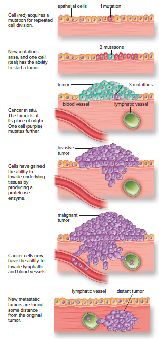
Carcinogenesis is the development of cancer.
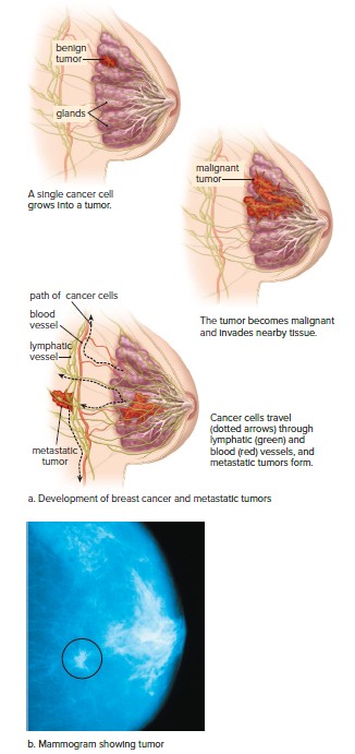
A single abnormal cell begins the process of the development of a tumor.
The most aggressive cell becomes the dominant cell of the tumor.
As additional mutations occur, the tumor cells release a growth factor, which causes neighboring blood vessels to branch into the cancerous tissue, a process called angiogenesis.
Additional mutations allow cancer cells to produce enzymes that degrade the basement membrane and invade underlying tissues.
Cancer cells are motile and can travel through the blood or lymphatic vessels to other parts of the body, where they start distant tumors, a process called metastasis.
Highly specialized cells like nerve and cardiac muscle cells rarely become cancer cells because they don't divide often.
Cells that have the ability to enter the cell cycle are more likely to become cancerous.
Fibroblasts and cells lining the cavities of the lungs, liver, uterus, and kidneys can divide when stimulated.
Adult stem cells, such as blood-forming cells in the bone marrow and basal cells of the skin and digestive tract, continue to divide throughout life.
Blood cells, intestinal cells, and skin cells need continuous division because they have a short lifespan or are regularly shed.
Proto-Oncogenes and Tumor Suppressor Genes
The cell cycle consists of interphase followed by mitosis.
Special proteins regulate the cell cycle at checkpoints.
Cancer develops due to mutations in two types of genes.
Proto-oncogenes code for proteins that promote the cell cycle and inhibit apoptosis.
Proto-oncogenes are likened to the gas pedal of a car.
Tumor suppressor genes code for proteins that inhibit the cell cycle and promote apoptosis.
Tumor suppressor genes are likened to the brakes of a car.
Tumor suppressor genes stop cells from dividing inappropriately.
Tumor suppressor genes become nonfunctional due to mutations.
Mutations in tumor suppressor genes may cause tumors to occur.
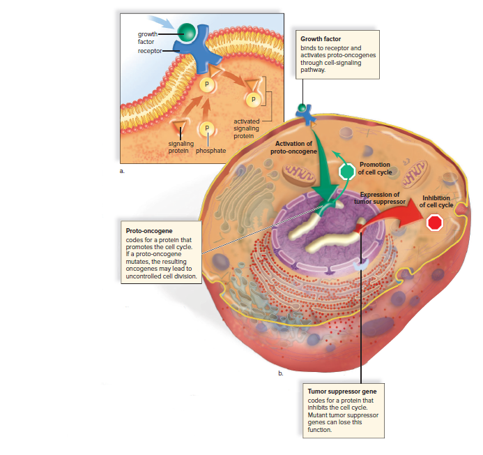
Proto-Oncogenes Become Oncogenes
Proto-oncogenes are genes that promote the cell cycle.
Mutations in proto-oncogenes can cause them to become oncogenes, which are cancer-causing genes.
Oncogenes accelerate the cell cycle, leading to uncontrolled cell growth and division.
Growth factors are chemical signals that activate a cell-signaling pathway, which then activates numerous proteins, many of which promote the cell cycle.
Some proto-oncogenes code for growth factors or for receptor proteins that receive growth factors.
When these proto-oncogenes become oncogenes, the receptor proteins are easily activated and may even be stimulated by a growth factor produced by the receiving cell.
RAS oncogenes are involved in 20–30% of human cancers.
Tumor Suppressor Genes Become Inactive
Tumor suppressor genes are genes that inhibit the cell cycle or promote apoptosis.
Mutations in tumor suppressor genes can cause them to lose their function.
Loss of function mutations in tumor suppressor genes can lead to uncontrolled cell growth and division, which can lead to cancer.
The p53 tumor suppressor gene produces a protein that checks the DNA for damage before it proceeds through the G1 checkpoint.
If the DNA is damaged, the cell is instructed to enter into G0 phase, and if the damage cannot be repaired, the cell undergoes apoptosis.
Failure of the p53 gene to perform this function allows cells with DNA damage to rapidly divide, potentially leading to cancer.
Cancer develops gradually over time.
Multiple mutations are usually required for a cell to become cancerous.
Mutations can be caused by environmental factors, such as exposure to carcinogens, or by random errors in DNA replication.
The combined effects of oncogenes and inactivated tumor suppressor genes can lead to uncontrolled cell growth and division, which is the hallmark of cancer.
Other Genetic Changes and Cancer
Absence of telomere Shortening
Telomeres are caps at the end of chromosomes that protect them from unraveling.
Each time a cell divides, telomeres shorten.
When telomeres become too short, the cell cycle stops.
Embryonic cells and certain adult cells have an enzyme called telomerase that can rebuild telomeres.
Cancer cells often have active telomerase, which allows them to divide over and over again.
Telomerase is believed to become active only after a cell has already started proliferating wildly.
Chromosomal Rearrangements
Cancer cells have unstable chromosomes, which can lead to portions of the DNA double helix being lost, duplicated, or scrambled.
Translocations, which are events where a portion of a chromosome breaks off and reattaches to another chromosome, can lead to cancer.
Translocations can disrupt genes that regulate the cell cycle, which can lead to uncontrolled cell growth.
The Philadelphia chromosome is a translocation that occurs between chromosomes 9 and 22.
The Philadelphia chromosome causes nearly 95% of cases of chronic myelogenous leukemia (CML).
A drug called imatinib (Gleevec) can successfully treat CML by inhibiting the activity of the protein coded for by BCR-ABL.
Scientists are now seeking to develop similar drugs that can inhibit the products of other oncogenes.
Cancer is usually a somatic disease, meaning that it develops only in body cells.
Some individuals may inherit a predisposition for developing some forms of cancer.
Other Cell Cycle Genes Associated with Cancer
BRCA1 and BRCA2
BRCA1 and BRCA2 are genes associated with breast cancer.
BRCA1 was identified in 1990 through DNA studies of families with a history of breast cancer.
BRCA2 was later discovered to be associated with some forms of breast cancer.
Both genes are part of mutant tumor suppressor genes.
They are inherited in an autosomal recessive manner.
Inheriting one mutated allele from either parent increases the risk of cancer only if the other allele is also mutated.
The first mutated gene is present in all cells of the body, making cancer more likely wherever the second mutation occurs, such as in the breast or ovaries.
RB Gene
The RB gene is a tumor suppressor gene.
It is associated with an eye tumor called retinoblastoma.
Retinoblastoma first appears as a white mass in the retina.
A tumor in one eye is the most common.
Mutations in both alleles are required for cancer to develop.
Children who inherit a mutated allele are more likely to have tumors in both eyes.
RET Gene
An abnormal allele of the RET gene can be inherited from a parent.
The RET gene is a proto-oncogene, which means it can become cancerous if mutated.
RET is inherited in an autosomal dominant manner, which means only one mutated allele is needed to increase a person's risk of developing thyroid cancer.
The remaining mutations necessary for thyroid cancer to develop are acquired (not inherited).
Testing for Genes Associated with Cancer
Genetic tests can detect the presence of specific alleles in many of the genes mentioned.
The advances in personal genomics are also allowing people to be screened for specific genes associated with a family history of cancer.
Persons who have inherited certain alleles of these genes may decide to have additional testing done, or sometimes elective surgery, to detect the presence of cancer.
8.5 Characteristics of Cancer
Cancers are classified based on their location.
Carcinomas are cancers of the epithelial tissue lining organs.
Sarcomas are cancers arising in muscle or connective tissue, particularly in bone or cartilage.
Leukemias are cancers of the blood.
Characteristics of Cancer Cells
Carcinogenesis is a gradual process that requires the accumulation of multiple mutations over time.
Cancer cells have abnormal nuclei, enlarged and may contain an abnormal number of chromosomes.
Gene amplification is seen much more frequently in cancer cells than in normal cells.
Cancer cells do not undergo apoptosis, preventing tumors from developing.
Cancer cells form tumors, piling on top of one another and growing in multiple layers.
Cancer cells undergo metastasis and promote angiogenesis, allowing them to invade nearby tissues and spread to other parts of the body.
Additional mutations in cancer cells allow them to secrete factors that promote angiogenesis, allowing the tumor to grow larger.
Some modes of cancer treatment are aimed at preventing angiogenesis.
The patient’s prognosis depends on whether the tumor has invaded surrounding tissues and whether there are metastatic tumors in distant parts of the body.
Cancer Treatment
Cancer treatments aim to remove the tumor or interfere with cancer cells' ability to reproduce.
Surgery is often the first line of treatment for solid tumors, especially when detected early.
Radiation therapy directs high-energy beams at the tumor to kill cancer cells within a specific area.
Chemotherapy kills cancer cells that have spread throughout the body by damaging their DNA or interfering with DNA synthesis.
Immunotherapy uses the patient's own immune system cells to target cancer cells for destruction.
Monoclonal antibodies and vaccines are being developed to act specifically on cancer cells and prime the immune system to identify and destroy them.
Prevention of Cancer
To lower the risk of developing certain cancers, people should avoid smoking, sunbathing, and excessive alcohol consumption.
Cigarette smoking is responsible for about 30% of all cancer deaths.
Smoking causes 90% of lung cancer cases among men and 80% among women.
People who smoke two or more packs of cigarettes a day have lung cancer mortality rates 15 to 25 times greater than those of nonsmokers.
Smokeless tobacco (chewing tobacco or snuff) increases the risk of cancers of the mouth, larynx, throat, and esophagus.
Sun exposure is a major factor in the development of many skin cancers, including the most dangerous type, melanoma.
The incidence of melanoma increases in people living near the equator.
Excessive exposure to radon gas in homes increases the risk of lung cancer, especially in cigarette smokers.
Heavy drinkers, especially those who also use tobacco, are more likely to develop cancers of the mouth, throat, esophagus, larynx, and liver.
Protective Diet
Obesity increases the risk of some forms of cancer by up to 40% compared to people of normal weight.
Weight loss can reduce cancer risk in obese individuals.
Increase consumption of foods rich in vitamins A and C, found in dark green, leafy vegetables, carrots, citrus fruits, and various fruits.
Vitamins A and C are antioxidants that prevent the formation of free radicals in cells, which can damage DNA.
Vitamin C also prevents the conversion of nitrates and nitrites into carcinogenic nitrosamines in the digestive tract.
Avoid salt-cured or pickled foods, which may increase the risk of stomach and esophageal cancers.
Smoked foods, such as ham and sausage, contain chemical carcinogens similar to those in tobacco smoke.
Nitrites added to processed meats and other foods can be converted to cancer-causing nitrosamines in the digestive tract.
Include vegetables from the cabbage family, such as cabbage, broccoli, brussels sprouts, kohlrabi, and cauliflower, in the diet to reduce the risk of gastrointestinal and respiratory tract cancers.