What are the 4 main functions of oral mucosa?
Protection
Sensation
Secretion
Mastication, speech and swallowing
Type of epithelium in the oral cavity
Stratified squamous epithelium to resist abrasion
Types of oral epithelium
Masticatory
Lining
Specialised
What type of oral epithelium is found on the hard palate, gingivae, dorsal surface of tongue
masticatory oral epithelium
What type of oral epithelium is found on the labial mucosa, buccal mucosa, alveolar mucosa, ventral tongue, floor of mouth, soft palate
lining oral epithelium
What type of oral epithelium is found on the gustatory mucosa of the tongue, vermillion zone/border between skin and oral mucosa
specialised oral epithelium (papillae)
Masticatory vs lining oral epithelium
Masticatory is keratinised to withstand forces generated during mastication
Lining is non-keratinised as it’s not subject to such high forces the the structures are elastic tissues which move out of the way
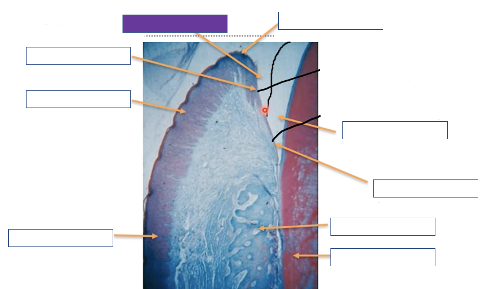
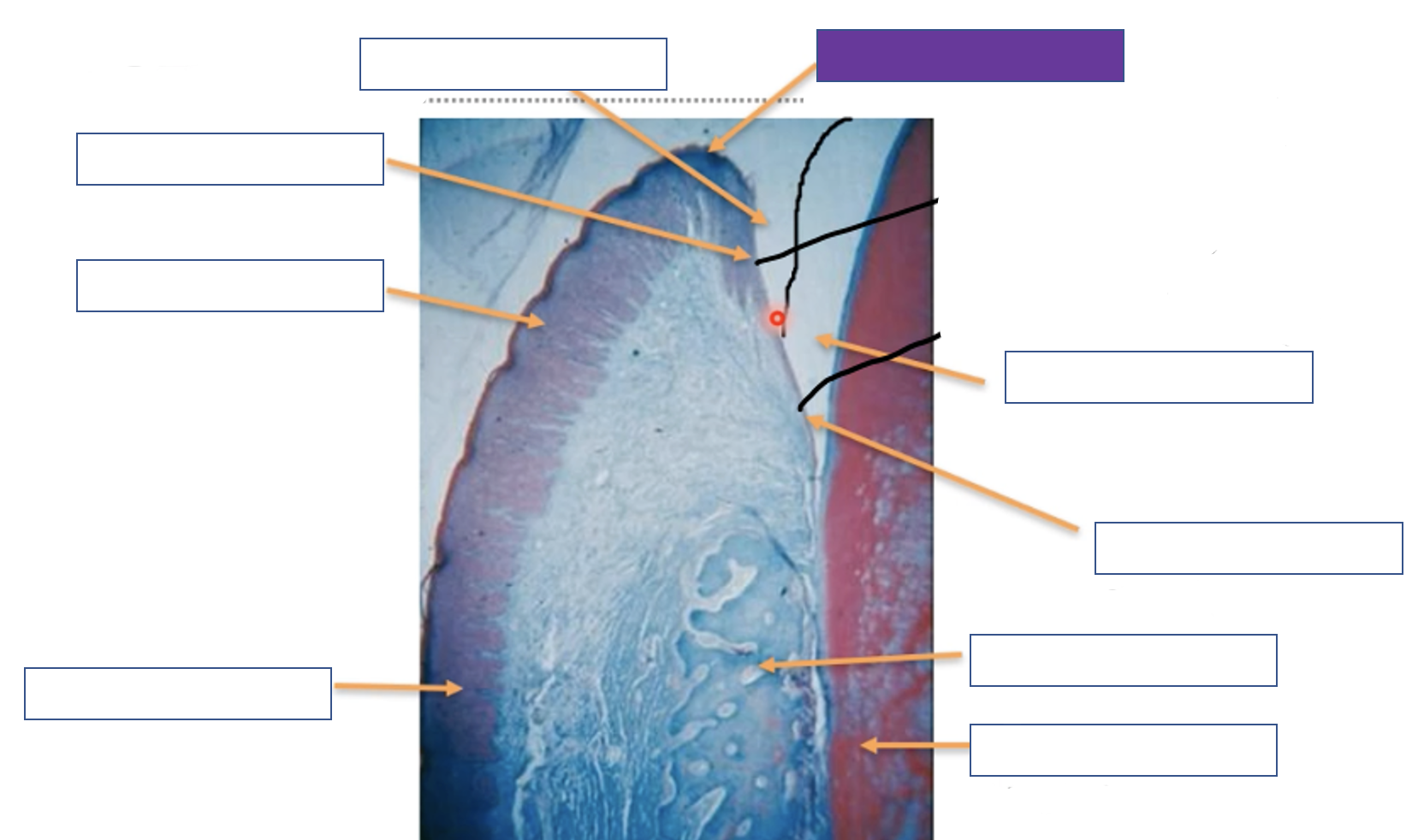
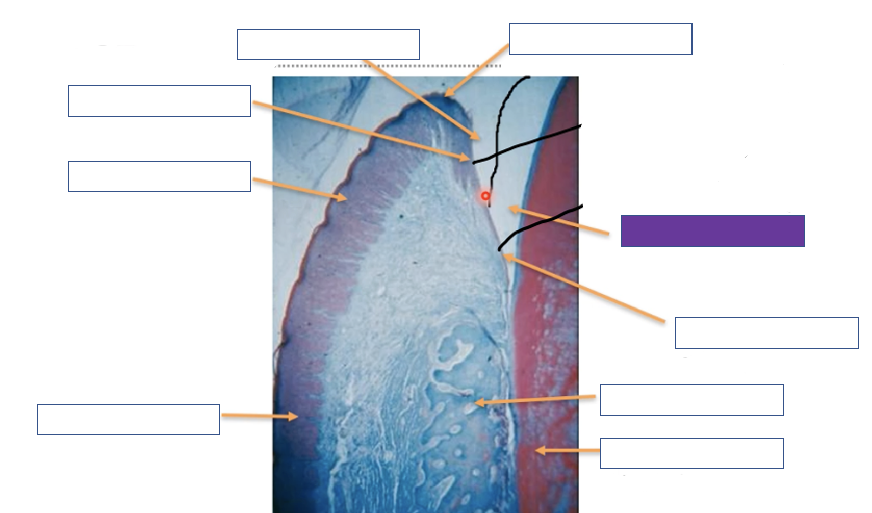
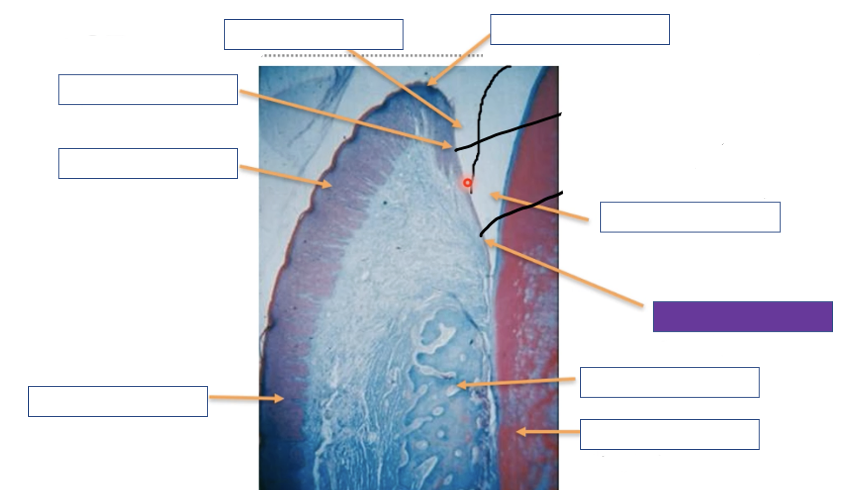
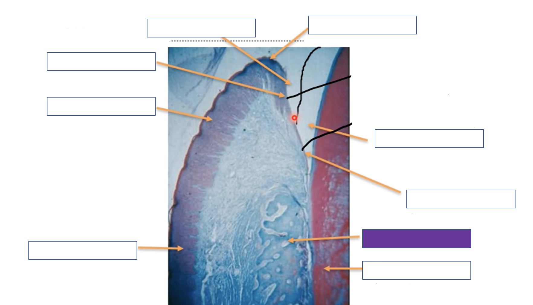
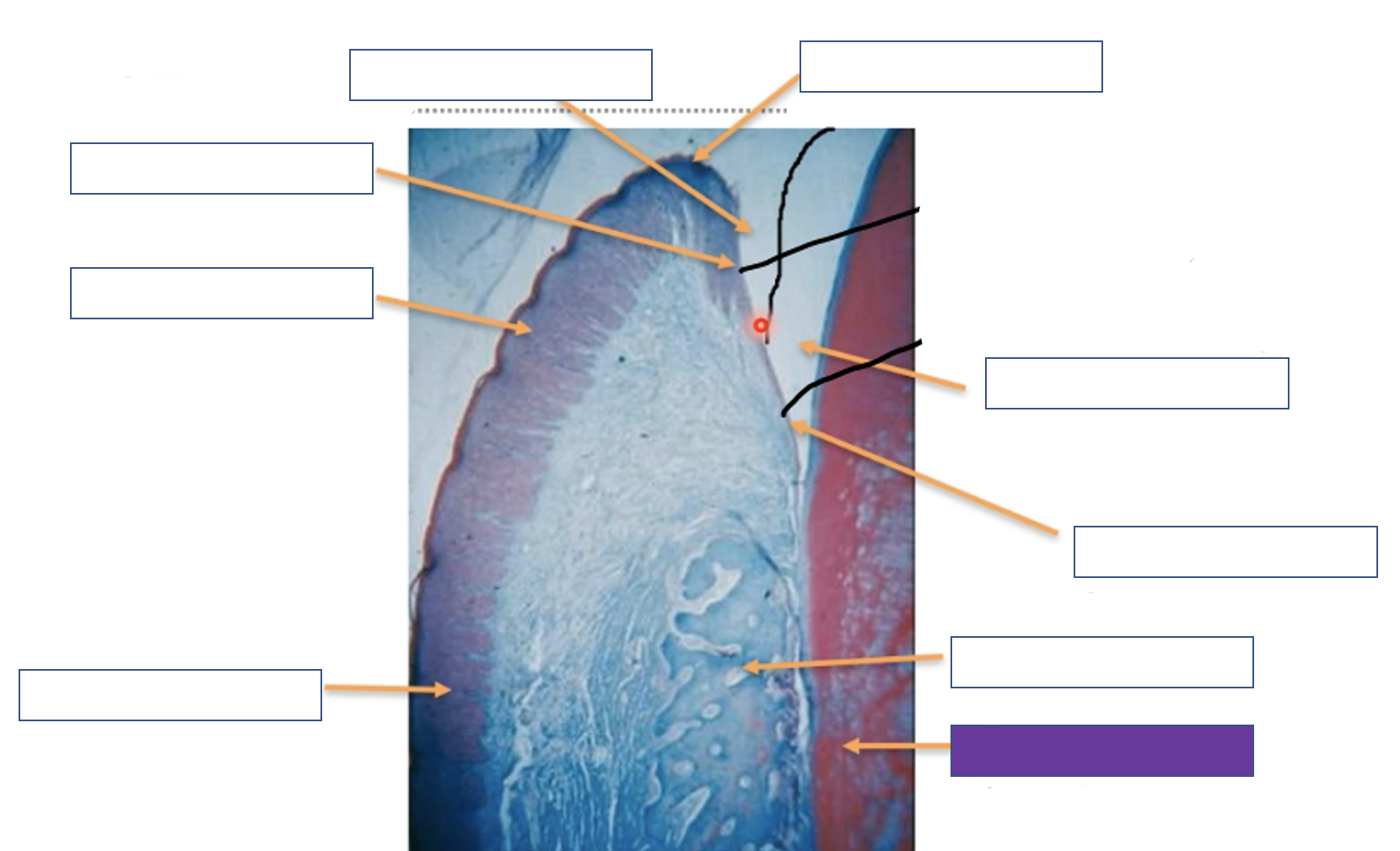
Label the 4 structures below the epithelium
Stratified squamous epithelium
Lamina propria (connective tissue)
Submucosa
Periosteum (only if over bone)
Bone/muscle
When is periosteum present
if over bone
What is found in the submucosa
fat deposits, glands, blood vessels
Name the 4 layers of keratinised oral epithelium from most to least matured
Keratinised cell layer – stratum corneum
Granular cell layer – stratum granulosum
Prickle cell layer – stratum spinosum
Basal cell layer – stratum basale
Name the 4 layers of non-keratinised epithelium
Superficial cell layer – stratum superficiale
Intermediate cell layer – stratum intermedium
Prickle cell layer – stratum spinosum
Basal cell layer – stratum basale
orthokeratinised vs parakeratinised
Orthokeratinised - no nuclei in keratinised layer
Parakeratinised - nuclei retained within keratinocytes
Function of basement membrane
Connection between epithelium and lamina propria
Site of metabolic exchange
Controls biological behaviour of epithelial cells
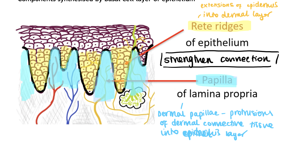
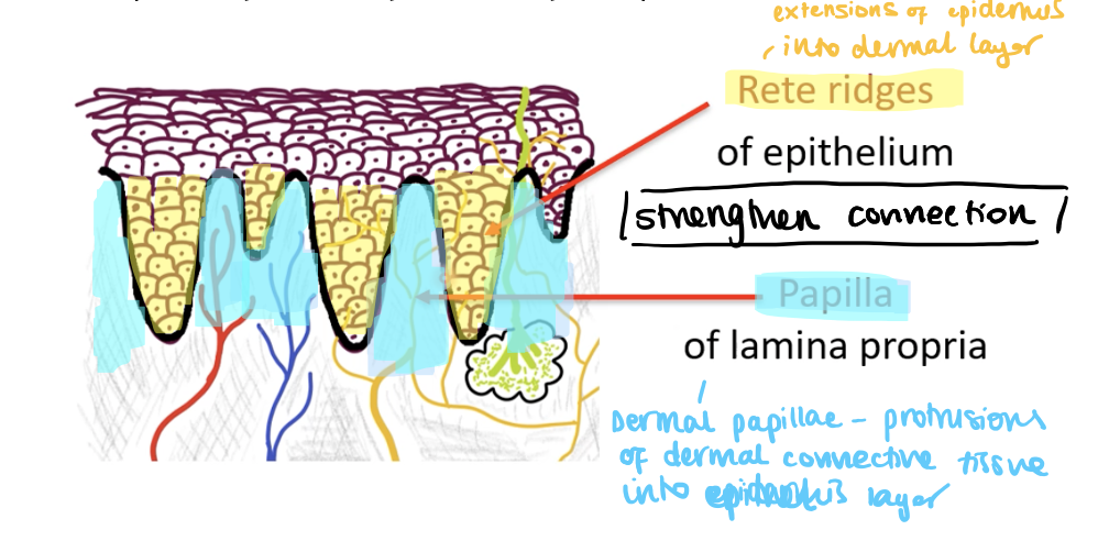
What are rete ridges of the epithelium
Extensions of the epithelial layer into the dermal layer
What are papilla of the lamina propia
Protrusions of the dermal connective tissue into the epithelial layer
What are the layers of lamina propria
Papillary layer between epithelial rete ridges – thin, loose collagen fibres
Deeper reticular layer – thick, parallel bundles collagen fibres
Function of lamina propia
Provides mechanical support and nutrition for epithelium
What is found in submucosa
Contains minor salivary glands, fat cells, blood vessels, nerves
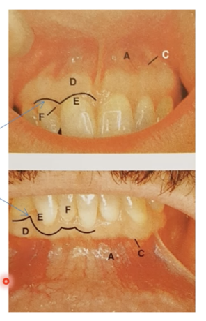
Label A - F
A - Alveolar mucosa
C - Mucogingival junction
D - Attached gingivae
E - Free gingiva
F - Interdental papilla
Texture of free vs attached gingiva
Free - smooth
Attached - textured
Keratinisation of gingiva vs alveolar mucosa
Gingiva - keratinised
Alveolar - non-keratinised
Type of mucosa on the attached gingiva
masticatory mucosa
How is attached gingiva bound to bone
directly via mucoperiosteum
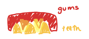
Where is interdental papilla and col found?
Triangles of the gums between teeth
how large is a healthy crevicular epithelium
0-2mm
Where is crevicular epithelium found
Unattached region between pre-gingiva and tooth (green line)
Where is junction epithelium found?
When the gingiva is connected to enamel
Why do the dark triangles appear in periodontal disease?
Interdental papilla lost
Crevicular vs junction epithelium
Crevicular has more folding at interface with underlying tissue
Crevicular has different site of keratin profile
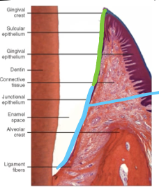
Dentogingival junction
Region where oral mucosa meets tooth surface
Principal seal between oral cavity and underlying periodontal tissues
Junction epithelium forms an epithelial collar around tooth from the cementoenamel junction to the base of the gingival sulcus
Thickness of crevicular vs junction epithelium
crevicular - 30 cells thick
junction - 1 cell thick
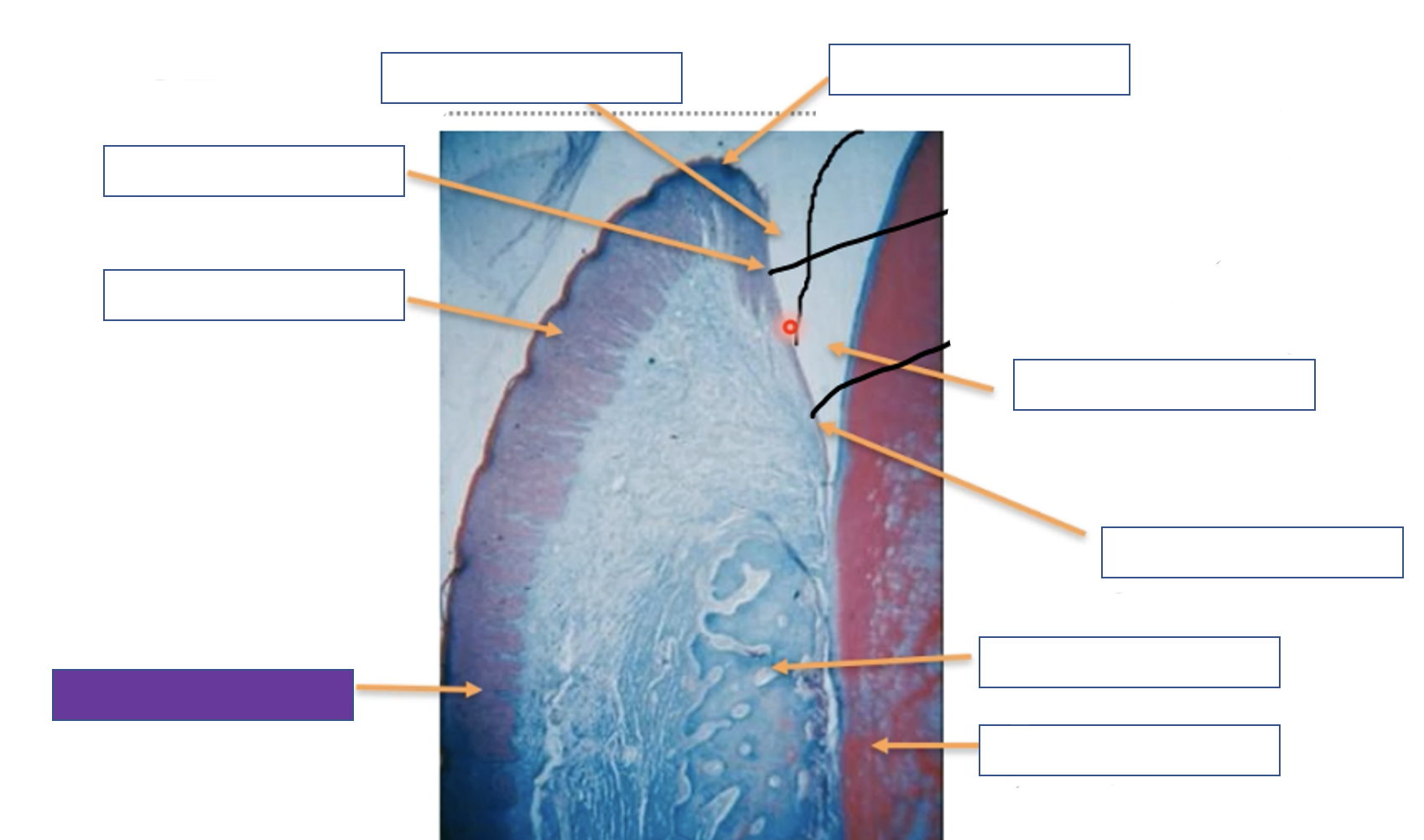
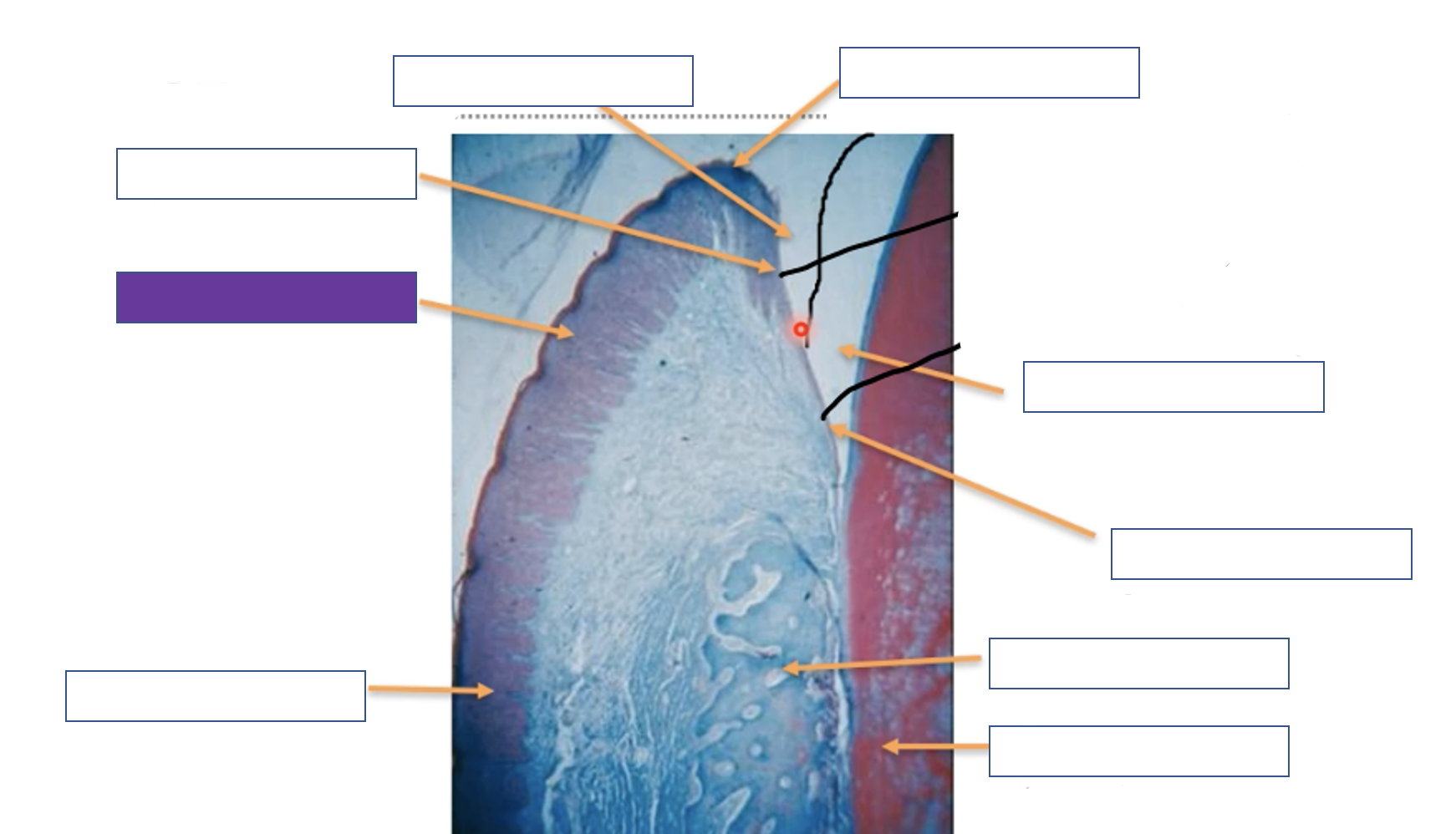
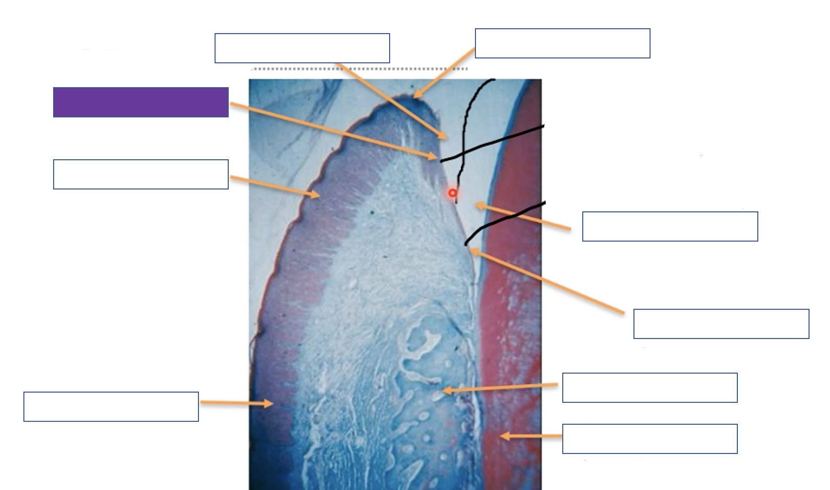
Attached gingiva
Free gingiva
Crevicular epithelium
Gingival sulcus
Gingival margin
Enamel space
Junction epithelium
Alveolar bone
Tooth (dentine)