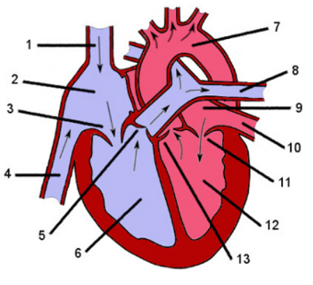
In which parts of the heart the numbers are pointing to?
Superior vena cava
Right artrium
Triscupid valve
Inferior vena cava
Pulmonary valve
Right ventrincle
Aorta
Pulmonary arteries
Left atrium
Pulmonary veins
Mitral valve
Left ventrincle
Aortic valve
What is the function of a valve in a vein?
Τo prevent reverse blood flow.
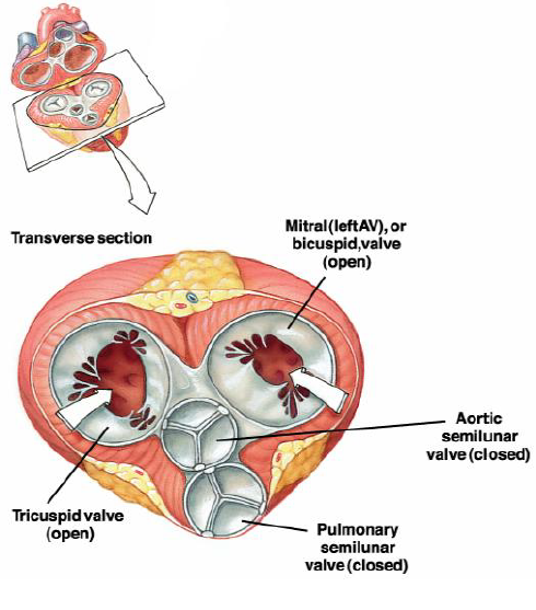
How the heart looks like when is diastoled?
Like that.
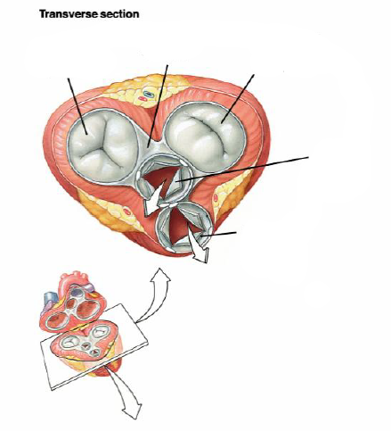
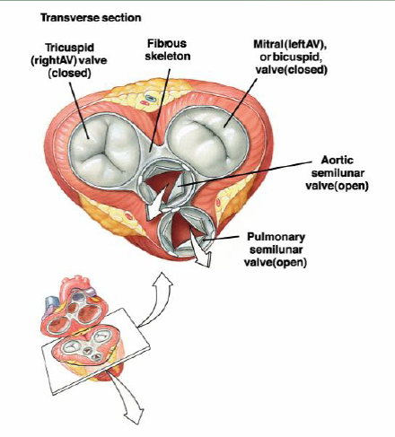
How the heart looks like when is systoled?
Like that.
Can you describe me in steps the heart cycle?
Late diastole→ Atrial systol→ Isovolumic ventricular contraction→Ventricular ejection→Isovolumic ventricular relaxation
What are the three compartments of the thoracic cavity?
2 pleural cavities and the mediastinum
Why the left ventrincle has thicker wall than the right one?
Left: pumps the blood all around the body
Right: lungs
Describe the blood flow in more details.
The oxygenated blood passes from the pulmonary veins and arrives to the left atrium
It goes from the atrial valve into the left ventricle.
When the left ventricle contracts that blood will be ejected through the aortic semilunar valve into the aorta and from there into smaller arteries.
When it arrives back from the systemic venous circulation arrives back via the superior/inferior vena cava
It will go to the right atrium from there the blood will go into the tricuspid valve into the right ventricle
When the right ventricle contracts that blood will be ejected through the pulmonary arterial semilunar valve and will go to each lung
What is the role of pericardium?
Stops the heart from over-expanding because of its tough fibrous tissue→physical protection
What is the role of endocardium?
Ensures the smooth blood flow because of the thin endothelial wall→no turbulence in the flow
What is the role of the myocardium?
The myocardium is responsible for the contractile function of the cardiac pump (contains cardiac muscle).→middle and thickest layer
What is the septum?
It seperates the 2 sides of the heart.
Which are the hemodynamic parameters of the human heart?
Heart rate: 60-80 bpm
Stroke volume (the amount of blood ejected from the ventricle with each cardiac cycle): 100 ml
Cardiac output (the product of heart rate and stroke volume): 6-7 l/min
Ejection fraction (a measurement, expressed as a percentage, of how much blood the left ventricle pumps out with each contraction): 65%
LV maximal systolic pressure: 120 mm Hg
RV maximal systolic pressure: 25 mm Hg
Ventricular end-diastolic volum 150 ml
Ventricular end-systolic volume 50 ml
Which are the main coronary arteries?
Aorta
Left main coronary artery
Circumflex coronary artery
Left anterior descending coronary artery
Right coronary artery
Which are the main cells in the heart?
Cardiomyocytes
Endothelial cells
Fibroblasts
What is the main function of cardiomyocytes?
Contractility
What is the main function of endothelial cells?
Angiogenesis
Spatial organization
Paracrine signaling
What is the main function of fibroblasts?
Transmission of Electrical Impulses
ECM Deposition
Paracrine Signaling
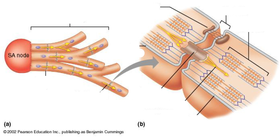
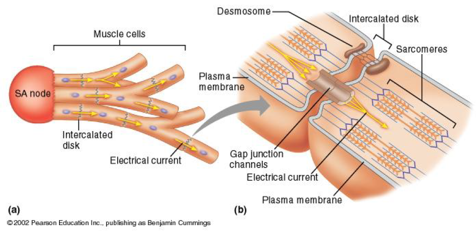
What’s the structure of myocardial muscle cells?
Branched
Single nucleus
Attached to each other by intercalated disks.
What is the excitation-contraction coupling?
E-Cc oupling links the action potential (excitation) of the muscle cell membrane (the sarcolemma) to muscular contraction.
What are the intercalated disks?
Specialized junctions that attach myocardial muscle cells to each other.
What are the steps of E-C coupling?
Depolarization of the plasma membrane and its membrane invaginations (the t-tubular system) by an action potential``
Transduction of the depolarization signal to the sarcoplasmic reticulum (SR) membrane
Activation of Ca2+ release from the SR and subsequent global elevation of intracellular [Ca2+]
Interaction of Ca2+ with contractile proteins leading to muscle contraction
return of [Ca2+] back to levels at resting conditions and muscle relaxation
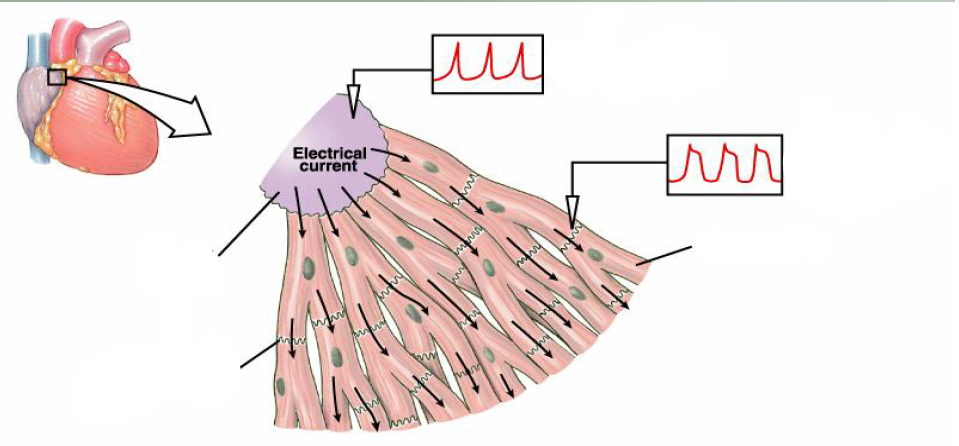
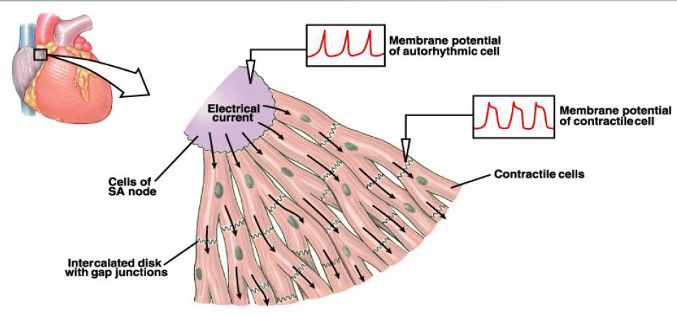
With which type of cell is generated the initial action potential?`
Sinus node cells
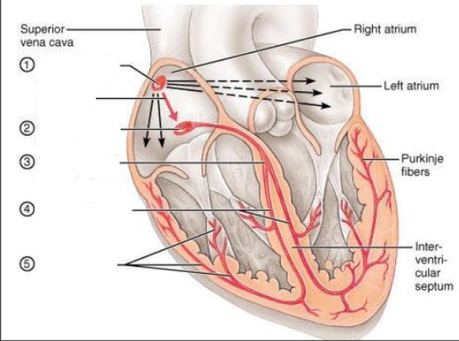
Where the arrows pointing at in this picture?
Here
How the depolarization is propagated?
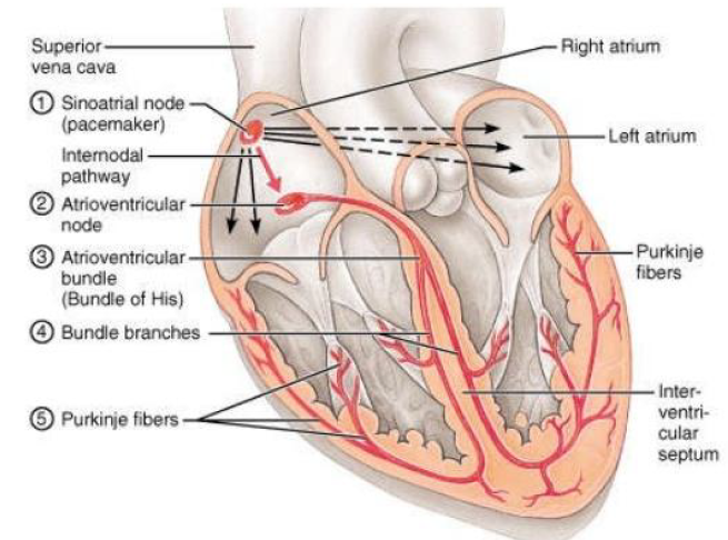
Initial action potential is generated by sinus node cells. Then it moves via gap junctions between cells.
This electrical pulse travels through the network of conducting cell.
What is the anatomy of a muscle cells?
What is the conducting system of the heart?
Network of specialized tissue that stimulates contraction.
modified cardiac myocytes.
the heart can contract without any innervation.
What is the pathway of depolarization?
Electrical activity goes rapidly to AV node via internodal pathways.
Depolarization spreads more slowly across atria. Conduction slows through AV node.
Depolarization moves rapidly through ventricular conducting system to the apex of the heart.
Depolarization wave spreads upward from the apex.
Which are the four major determinants of cardiac output?
Preload
Afterload
Myocardial contractility
Heart rate
What is the definition of preload?
Volume of blood in ventrincles at end of diastole
Which are the causes that lead to preload?
Compliance of ventrincle→end-diastolic radius→preload→myocardial end-diastolic wall stress
Total blood volume, blood volume distribution, venous return, atrial contraction→end diastolic-filling pressure→preload→myocardial end-diastolic wall stress
Total blood volume, blood volume distribution, venous return, atrial contraction→end diastolic-filling pressure→end-diastolic radius→preload→myocardial end-diastolic wall stress
Hypertrophy→myocardial wall thickness→preload→myocardial end-diastolic wall stress
What is the law that describes lenght-tension relationship?
Frank-Starling law=>Stroke volume increases preload auguments
What is the definition of afterload?
Afterload refers to the amount of resistance left ventrincle must overcome to circulate blood.
What happens if the afterload augments?
Stroke volume decreases
In which pathological conditions afterload is increased?
In hypertension and vasoconstriction
What does it mean if the afterload, is increased?
That the cardiac workload also does
In which pathological conditions preload is increased?
Hypervolemia
Regurgitation of cardiac valves
Heart failure
What is the definition of contractility?
It is the intrinsic capacity to generate force.
Which are the methods for assessment of the ventrincular and cardiomyocytes functions?
In situ: ECG, echocardiography, MRI
Isolated perfused heart –Langendorff technique, working heart.
Isolated papillary muscle.
Isolated cardiomyocytes.
Permeabilized fibers and cells.
Isolated organelles.
Methods of biochemistry and molecular biology.
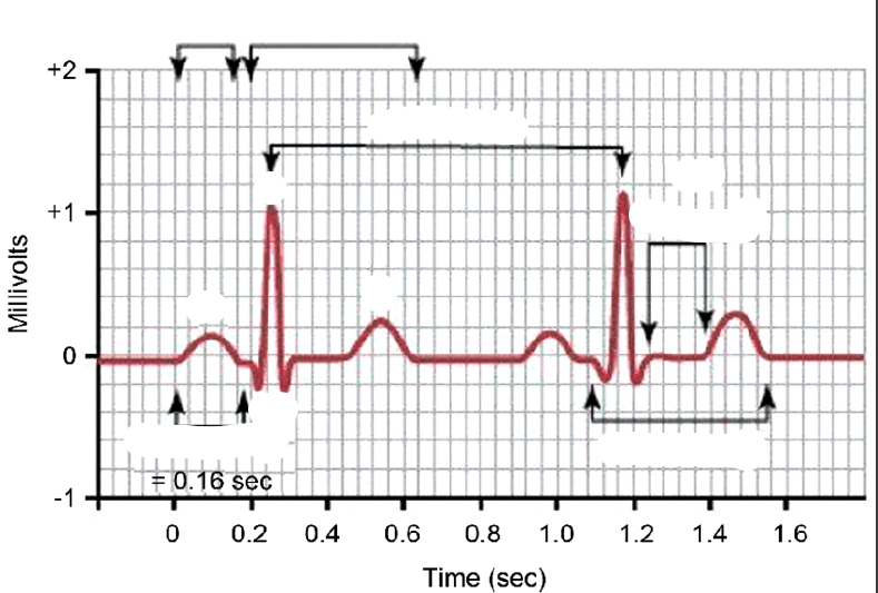
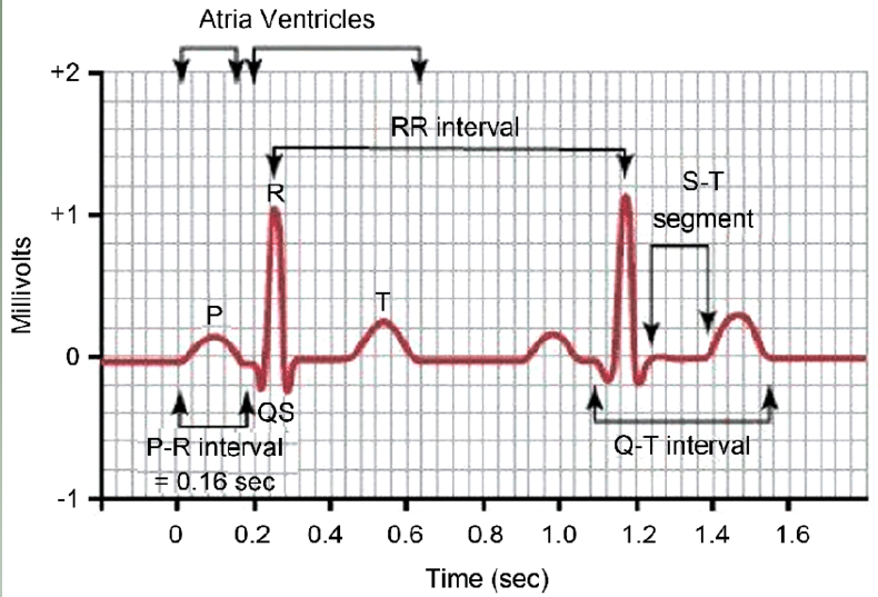
What is electrocardiogram?
It is a graph of voltage versus time of the electrical activity of the heart using electrodes placed on the skin which detect the electrical changes occured due to the cardiac muscle depolarization followed by repolarization during each cardiac cycle.
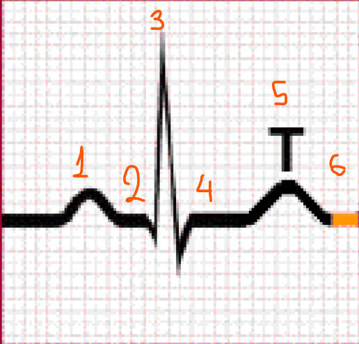
Tell me where is: P, R, T, Q, RP interval, ST segment, QT interval, PR intervla, atria and ventrincles?
What does P wave represent in a ECG?
It represents the depolarization of the atria
What does QRS compex represent in a ECG?
It represents the depolirization of the ventrincles.
What does T represent in a ECG?
It represents the repolarization of the ventrincles.
Describe briefly all the steps in a ECG?
Atria begin depolarizng
Atrial depolarization complete
Ventricular depolarization begins at apex and progresses superiorlu as atria repolarize
Ventrivular depolarization complete
Ventricular repolarization begins at apex and progresses superiority
Ventricula repolarization complete, heart is ready for the next cycle.
What abnormalities can you interpret from a ECG?
rhythm
conduction
electrical axis
myocardial mass (hypo-hypertrophy)
myocardial ischemia (a restiction in blood supply to myocardium)
What is a echocardiography?
It is a test that uses high frequency sound waves (ultrasound) to make pictures of the heart.
What is MRI?
With magnetic resonance imaging (MRI), a powerful magnetic field and radio waves are used to produce detailed images of the heart.