Looks like no one added any tags here yet for you.
What is the largest organ in the abdomen?
liver
Where is the liver located?
RUQ and extends across the midline; within the peritoneal cavity
What covers the liver?
glisson’s capsule
Where does Glisson’s capsule not cover?
bare area
What is the normal length of the liver?
12-17 cm long
Where is the measurement of the liver taken?
right lobe from diaphragm to inferior edge
What is the liver used as to view other organs?
acoustic window
What is visible through the liver?
pancreas, gallbladder, great vessels, bile duct
When would echogenicity and echotexture of the liver change?
if liver is diseased
What can degrade the quality of abdominal scan?
liver disease
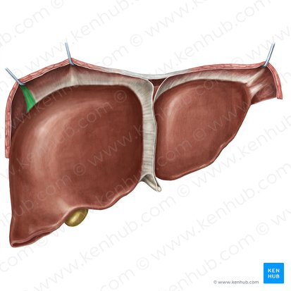
Where does the falciform attach?
sternum
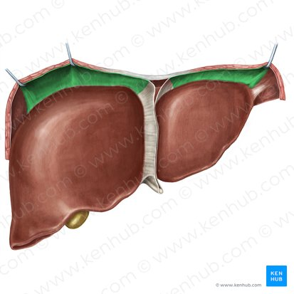
What ligament is shown?
triangular ligament
What ligament is shown?
coronary ligament
What ligament is shown?
falciform
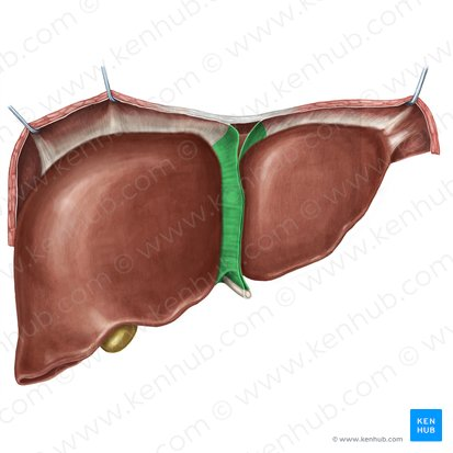
What ligament is shown?
ligamentum teres (round ligament)
How many lobes does the liver have?
3
What are the 3 lobes of the liver?
right, left, caudate
Which lobe of the liver is the smallest?
caudate
Which lobe of the liver is the largest?
right
What separates the lobes?
ligaments
What ligament separates the right and left lobe?
falciform
What ligament separates the left and caudate lobe?
lig. venosum
What is also referred to as the “grooves” of the liver
fossae
What are echogenic landmarks of the liver?
ligaments
What does the lig. teres help to locate?
left portal vein
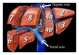
Label 1-8
Caudate
Left Lateral Superior
Left Lateral Inferior
Medial Segment
Right Anterior Inferior
Right Posterior Inferior
Right Posterior Superior
Right Anterior Superior
What does the lig. venosum help to locate?
caudate lobe & left portal vein
What does the main lobar fissure help to locate?
gallbladder & right portal vein
What does the lig. teres separate?
medial and lateral portions of the Lt lobe
What is the remnant of portal sinus?
lig. teres
How does lig. teres appear in LONG view?
echogenic streak from LPV to inf edge of liver
How does lig. teres appear in TRANS view?
echogenic spot in Lt Lobe inf to LPV
T/F: lig. teres is never seen superior to the LPV
true
What is the remnant of ductus venosus?
ligamentum venosum
What is the role of ductus venosus?
shunts blood from LPV to IVC during fetal circulation
How is the lig. venosum positioned?
posterior to Lt lobe & anterior to caudate lobe
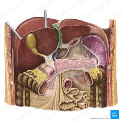
What is seen as an echogenic line joining the gallbladder and right portal vein?
main lobar fissure
What does the main lobar fissure help to locate?
gallbladder fossa
What acts as an anatomical indicator to where a gallbladder was before it was removed (post-cholecystectomy)
main lobar fissure
Where does the proper hepatic artery branch from?
common hepatic artery
What artery supplies the liver with oxygenated blood?
proper hepatic artery
Where does the PHA enter the liver?
porta hepatis
What artery is adjacent to the MPV?
PHA
Where is the portal vein formed?
at the confluence of the splenic vein and the SMV
What supplies the liver with nutrient rich blood?
portal vein
Where does the portal vein enter the liver?
porta hepatis
What is the blood/oxygen number of the liver?
80/20 split: blood
20/80 split: oxygen
Where does the MPV enter the liver?
porta hepatis
Where does the left and right portal veins supply blood to in the liver?
LPV-left lobe
RPV-right lobe
How is the liver drained?
by hepatic veins
All blood leaves the liver via the _________.
left, middle or right hepatic veins
Where do the hepatic veins drain directly into?
superior IVC
What view shows hepatic veins separated?
LONG
What view shows hepatic veins together joining the IVC as the “bunny sign”?
TRANS
What does it mean if the hepatic veins are intersegmental?
course between liver lobes
T/F: hepatic vein walls are echogenic like portal veins
false
What veins increase in caliber as they drain toward the IVC?
hepatic veins
How do the hepatic veins divide the liver?
vertically into right and left regions
How do the portal veins divide the liver?
horizontally into superior and inferior regions
How does the right hepatic vein divide the right lobe?
anterior & posterior
How does the middle hepatic vein separate the liver?
right & left lobes
How does the middle hepatic vein course?
along the main lobar fissure
How does the left hepatic vein divide the left lobe?
medial & lateral
What divides the liver into 8 segments?
Couinaud’s system
What is Couinaud’s system based on?
the course of the portal and hepatic veins
Which veins are intersegmental?
hepatic
What divides the liver into superior and inferior?
portal veins
What is #1 in Couinaud’s system?
caudate
What are the 8 segments of Couinaud’s
Caudate
Lt lateral superior
Lt lateral inferior
medial segment
Rt anterior inferior
Rt posterior inferior
Rt posterior superior
Rt anterior superior
What does hepatopedal mean?
blood flow into the liver
What means blood flow out of the liver?
hepatofugal
What flow of blood is not normal in the liver?
hepatofugal
What view shows the portal triad as “mickey mouse”?
TRANS
What view shows the portal triad as “shotgun sign” or “double barrel”?
LONG
What does the portal triad consist of?
portal vein, bile duct, hepatic artery
What does a protocol include?
required images that represent a full evaluation of an organ/tissue; images taken in LONG and TRANS; measurements of specific structures; labeled images
T/F: a protocol is repeatable
true; images are taken using anatomical landmarks found in the typical abdomen
What does a protocol include for images of pathology?
LONG & TRANS images, details in echogenicity, be measured where possible, include color and/or PW doppler images, highlight and peculiarities observed
What are some indications for a liver ultrasound?
suspected liver enlargement
hepatic or perihepatic masses
abscess
obstructive or metastatic lesions
abnormal liver function tests (abnormal labs)
known hepatic disease (cirrhosis, Hepatitis)
What are the LONG views of the liver protocol?
LONG LT LOBE Lateral
LONG LT LOBE Aorta
LONG LT LOBE Caudate lobe
LONG LT LOBE IVC
LONG RT LOBE GB & MLF
LONG RT LOBE RT KIDNEY
LONG RT LOBE Lateral
LONG RT LOBE Dome
LONG RT LOBE Inferior Border
CORONAL MPV w/out color
What are some additional liver images you may need to include?
all abnormalities with location within the liver (segment)
doppler the portal vein and branches if indicated
doppler the hepatic veins and artery if indicated