Chapter 27: The Control Systems
27.1 Nervous System
The nervous system and the endocrine system work together to regulate the activities of other systems.
Both control systems use chemical signals to respond to changes that might threaten homeostasis.
The nervous system sends a message along a nerve fiber directly to a target organ or tissue, such as skeletal or smooth muscle.
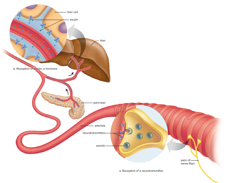
The endocrine system uses the blood vessels of the cardiovascular system to send hormones as chemical messengers to a target organ, such as the liver.
The endocrine system is slower-acting than the nervous system because it takes time for hormone molecules to move through the bloodstream to the target organ.
Hormones change the metabolism of cells, and this takes time, but the response is longer-lasting.
Cellular metabolism tends to remain the same for at least a limited period of time.
Examples of Nervous Systems
Animals have a unique characteristic of possessing a nervous system to detect stimuli in the environment and perform coordinated reactions in response to the stimuli.
The nervous system helps animals detect chemical signals that allow them to move towards a food source or away from a predator.
All animal nervous systems receive sensory input, which is processed to direct a coordinated reaction.
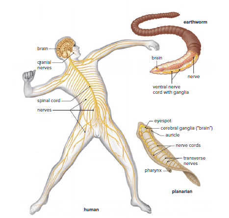
Planarians possess a ladder-like nervous system consisting of two lateral nerve cords joined together by transverse nerves.
The simple brain of planarians receives sensory information from the eyespots and sensory cells in the auricles.
The two lateral nerve cords allow a rapid transfer of information from the cerebral ganglia to the posterior end, and the transverse nerves between the nerve cords keep the movement of the two sides coordinated.
The nervous organization in planarians is a foreshadowing of the central and peripheral nervous systems found in more complex invertebrates, such as earthworms, and in vertebrates, including humans.
The Human Nervous System
The nervous system controls the muscular system and maintains homeostasis with the endocrine system in humans.
The central nervous system (CNS) includes the brain and spinal cord, while the peripheral nervous system (PNS) consists of nerves outside the CNS.
The CNS and PNS work together and are connected to each other.
The brain gives off paired cranial nerves, and the spinal cord gives off paired spinal nerves.
The human nervous system is more complex than the planarian system due to the evolution of the nervous system.
The CNS can respond to both external and internal stimuli.
Complex sense organs, such as the human eye and ear, can detect changes in the external environment.
Neurons
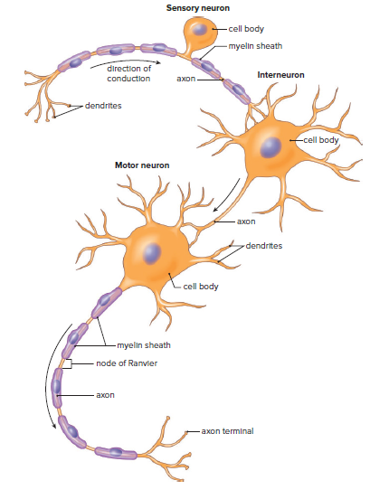
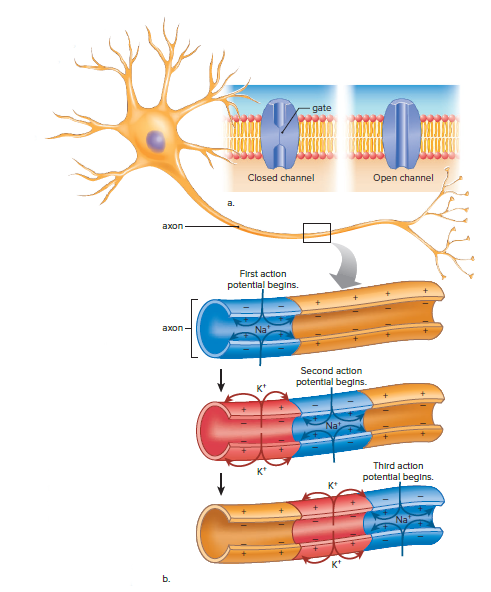
The structure of a neuron is well-suited to its function as the primary cell of the nervous system.
The cell body contains the nucleus and other organelles.
Short dendrites receive signals from sensory receptors or other neurons.
Axons conduct nerve impulses to their targets and can deliver them great distances.
Long axons are covered by a white myelin sheath formed from the membranes of tightly spiraled cells that leave gaps called nodes of Ranvier.
The nervous system has three types of neurons specific to its three functions: sensory neurons, interneurons, and motor neurons.
Sensory neurons take nerve impulses from sensory receptors to the CNS.
Interneurons take nerve impulses between various parts of the CNS.
Motor neurons take nerve impulses from the CNS to muscles or glands.
The Nerve Impulse
A nerve impulse is dependent on concentration gradients.
Neurons maintain concentration gradients through the sodium-potassium pump.
The sodium-potassium pump actively transports Na+ ions to the outside of the axon and K+ ions inside.
A charge difference exists across the axon's membrane, with the inside being negative compared to the outside.
The charge difference is due to an unequal distribution of Na+ and K+ ions across the membrane.
The charge difference plays an important role in the generation of a nerve impulse or action potential.
A nerve impulse is a rapid, short-lived, self-propagating reversal in the charge difference across the axon's membrane.
Two types of gated channel proteins in the axon's membrane are involved in a nerve impulse: one allows Na+ to pass through, and the other allows K+ to pass through.
During a nerve impulse, Na+ gates open at a particular location, and the inside of the axon becomes positive as Na+ moves from outside to inside.
Na+ gates close, and K+ gates open, causing K+ to move from inside to outside, and the charge reverses.
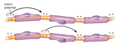
In myelinated axons, saltatory conduction occurs, which is much faster than conduction by unmyelinated axons.
Saltatory conduction is the conduction of an action potential at one node of Ranvier, causing an action potential at the next node.
The conduction of a nerve impulse is an all-or-none event, and the intensity of a message is determined by how many nerve impulses are generated within a given time span.
An axon can conduct a volley of nerve impulses because only a small number of ions are exchanged with each impulse.
During a refractory period, which follows each impulse, the sodium gates cannot open, ensuring that nerve impulses travel in only one direction and do not reverse.
The Synapse
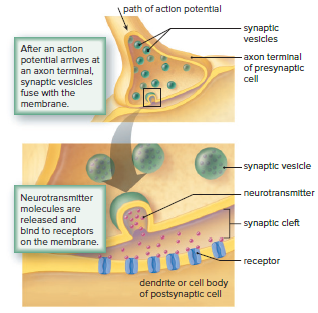
Each axon has many axon terminals.
In the CNS, a terminal of one neuron, known as the presynaptic cell, lies very close to the dendrite (or cell body) of another neuron, the postsynaptic cell.
This region of close proximity is called a synapse.
In the PNS, when the postsynaptic cell is a muscle cell, the region is called a neuromuscular junction.
A small gap exists at a synapse, and this gap is called the synaptic cleft.
Transmission across a synaptic cleft is carried out by chemical signals called neurotransmitters, which are stored in synaptic vesicles.
When nerve impulses traveling along an axon reach an axon terminal, synaptic vesicles release a neurotransmitter into the synaptic cleft.
Neurotransmitter molecules diffuse across the cleft and bind to a specific receptor protein on the postsynaptic cell.
Depending on the type of neurotransmitter and/or the type of receptor, the response of the postsynaptic cell can be toward excitation or toward inhibition.
Several dozen different neurotransmitters have been identified, a few of the more widely used neurotransmitters are acetylcholine (ACh), norepinephrine, serotonin, and gamma-aminobutyric acid (GABA).
Once a neurotransmitter has been released into a synaptic cleft and has initiated a response, the neurotransmitter is removed from the cleft.
A single neuron has many dendrites plus the cell body, and both can have synapses with many other neurons; 1,000 to 10,000 synapses per single neuron are not uncommon.
An excitatory neurotransmitter produces a potential change that drives the neuron closer to an action potential, and an inhibitory neurotransmitter produces a potential change that drives the neuron farther from an action potential.
Neurons integrate these incoming signals.
Integration is the summing up of both excitatory and inhibitory signals.
If a neuron receives many excitatory signals, chances are its axon will transmit a nerve impulse.
If a neuron receives both inhibitory and excitatory signals, the integration of these signals may prohibit the axon from firing.
Drug Abuse
Many drugs affecting the nervous system interfere with or promote neurotransmitter action.
Drugs can enhance or block neurotransmitter release, mimic action, block receptors, or interfere with removal from synaptic cleft.
Stimulants increase CNS activity; depressants decrease it.
Dopamine is believed to be responsible for mood, and new medications for drug dependence and mental illness affect its release, reception, or breakdown.
Drug abuse occurs when a person takes a drug at a harmful dose and under harmful circumstances.
Drug abusers often take more than intended and display psychological and/or physical dependence.
Physical dependence requires more of the drug for the same effect and causes withdrawal symptoms when stopped.
Cocaine
Cocaine is an alkaloid derived from the shrub Erythroxylon coca.
It is sold in powder form and as crack, a more potent extract.
Cocaine prevents the synaptic uptake of dopamine, resulting in a prolonged period of stimulation of the postsynaptic cell and a "rush" sensation.
Dopamine's epinephrine-like effects account for the state of arousal that lasts for several minutes after the rush experience.
Cocaine binge can last for days, during which the user is hyperactive, has little desire for food or sleep, but has an increased sex drive.
After the binge period, the user experiences a crash, which includes fatigue, depression, irritability, memory and concentration problems, and no interest in sex.
Cocaine causes extreme physical dependence, and continued use leads to desensitization of postsynaptic cells to dopamine, resulting in withdrawal symptoms and intense craving for cocaine.
Overdosing on cocaine can cause seizures and cardiac and respiratory arrest.
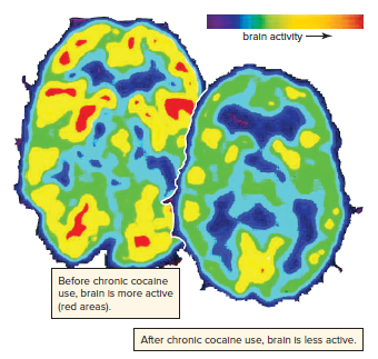
Long-term cocaine abuse may cause brain damage.
Babies born to cocaine addicts suffer withdrawal symptoms and may have neurological and developmental problems.
Methamphetamine
Methamphetamine is a synthetic drug made by adding a methyl group to amphetamine.
Over 9 million people in the United States have used methamphetamine at least once in their lifetime.
Teenagers and young adults represent approximately one-fourth of these users.
Methamphetamine is often produced from amphetamine in makeshift home laboratories.
It is available as a powder (speed) or as crystals (“crystal meth,” “glass,” or “ice”).
The crystals are smoked, and the effects are almost instantaneous and nearly as quick as when methamphetamine is snorted.
When the drug is smoked, the effects last 4–8 hours.
Methamphetamine has a structure similar to that of dopamine, and its stimulatory effect mimics that of cocaine.
Chronic use can lead to what is called an amphetamine psychosis, resulting in paranoia; auditory and visual hallucinations; self-absorption; irritability; and aggressive, erratic behavior.
Drug tolerance, dependence, and addiction are common.
Hyperthermia, convulsions, and death can occur.
Marijuana
The Indian hemp plant, Cannabis sativa, contains dried flowers, leaves, and stems that are covered by a resin rich in THC.
Cannabis and marijuana refer to either the plant or THC.
Marijuana is usually smoked in a cigarette form called a "joint."
Marijuana binds to a receptor for anandamide, a neurotransmitter that creates a feeling of peaceful contentment.
Occasional marijuana use results in mild euphoria, alterations in vision and judgment, distortions of space and time, and motor incoordination.
Heavy use can result in hallucinations, anxiety, depression, rapid flow of ideas, body image distortions, paranoid reactions, and similar psychotic symptoms.
Regular use can lead to craving and difficulty in stopping usage.
The Central Nervous System
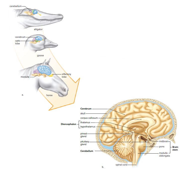
The forebrain is most prominent in humans.
The forebrain in mammals becomes the last depository for sensory information.
The forebrain carries on much of the integration for the entire nervous system before it sends out motor instructions to glands and muscles.
The spinal cord provides a means of communication between the brain and the spinal nerves, which are a part of the PNS.
Spinal nerves take messages to and from the skin, glands, and muscles in all areas of the body, except the head and face.
Long, myelinated fibers of interneurons in the spinal cord run together in bundles called tracts.
These tracts connect the spinal cord to the brain.
The left side of the brain controls the right side of the body, and vice versa.
The spinal cord is involved in reflex actions, which are programmed, built-in circuits that allow for protection and survival.
Reflex actions are present at birth and require no conscious thought to take place.
The Brain
Cerebrum
The cerebrum is the largest part of the brain.
It is divided into two hemispheres, the left and right.
The hemispheres are connected by a bundle of nerve fibers called the corpus callosum.
The cerebrum is responsible for higher-order functions such as thinking, reasoning, planning, and movement.
It also plays a role in sensory processing, emotions, and memory.
Diencephalon
The diencephalon is a small region of the brain that lies below the cerebrum.
It contains two important structures: the hypothalamus and the thalamus.
The hypothalamus is responsible for a variety of functions, including regulating body temperature, hunger, thirst, and sleep.
The thalamus is a relay station for sensory information.
It receives sensory input from the eyes, ears, skin, and other organs, and then sends it to the appropriate areas of the cerebrum for processing.
Cerebellum
The cerebellum is a small structure located at the back of the brain.
It is responsible for coordinating movement, balance, and posture.
The cerebellum receives input from the muscles, joints, and vestibular system (the inner ear).
It then uses this information to make adjustments to movement in order to maintain balance and coordination.
Brain stem
The brain stem is a small, stalk-like structure that connects the cerebrum to the spinal cord.
It contains a number of important structures, including the medulla oblongata, pons, and midbrain.
The medulla oblongata is responsible for regulating heart rate, breathing, and blood pressure.
The pons helps to coordinate movement and sleep-wake cycles.
The midbrain is responsible for vision, hearing, and motor control.
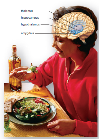
The Limbic System
The limbic system is a network that includes the diencephalon and areas of the cerebrum.
It blends higher mental functions and primitive emotions into a united whole.
It accounts for why activities such as sexual behavior and eating seem pleasurable and why mental stress can cause high blood pressure.
The hippocampus and the amygdala are two significant structures within the limbic system that are essential for learning and memory.
The hippocampus is a seahorse-shaped structure that lies deep in the temporal lobe and is well situated in the brain to make the prefrontal area aware of past experiences stored in sensory association areas.
The amygdala can cause experiences to have emotional overtones and is responsible for conditioning fear and associating danger with sensory information.
Reason can keep us from acting out strong feelings due to the connection between the frontal lobe and the limbic system.
Long-term memories are stored in bits and pieces throughout the sensory association areas of the cerebral cortex, and the hippocampus gathers this information for use by the prefrontal area of the frontal lobe when we remember.
Alzheimer's disease causes a progressive loss of memory, particularly for recent events, and clusters of abnormal tissue develop among degenerating neurons, especially in the hippocampus and amygdala.
Major research efforts are devoted to seeking a cure for Alzheimer's disease.
The Peripheral Nervous System
The peripheral nervous system (PNS) relays sensory information to the CNS for processing and motor responses to the body tissues.
Sensory information comes from sensory neurons located within sense organs or tissues of the body.
The PNS lies outside the central nervous system and contains nerves, which are bundles of axons.
Neuron cell bodies are found in the CNS or in ganglia, which are collections of cell bodies within the PNS.
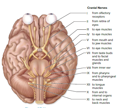
Humans have 12 pairs of cranial nerves that arise from the brain, largely concerned with the head, neck, and facial regions of the body.
The vagus nerve is a cranial nerve that has branches to the pharynx, larynx, and most of the internal organs.
Humans have 31 pairs of spinal nerves, each containing many sensory and motor axons.
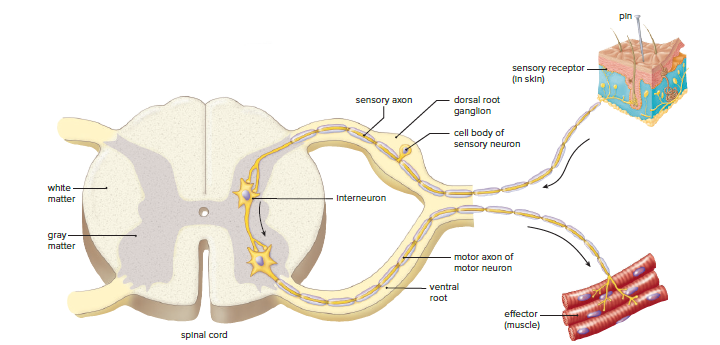
The dorsal root of a spinal nerve contains the axons of sensory neurons, which conduct impulses to the spinal cord from sensory receptors.
The cell body of a sensory neuron is in the dorsal root ganglion.
The ventral root contains the axons of motor neurons, which conduct impulses away from the cord, largely to skeletal muscles.
Each spinal nerve serves the region of the body in which it is located.
The Somatic System
Includes nerves that take information about external stimuli from sensory receptors to the CNS and motor commands away from the CNS to skeletal muscles.
Voluntary control of skeletal muscles always originates in the brain.
Involuntary responses to stimuli, called reflexes, can involve either the brain or just the spinal cord.
Flying objects cause our eyes to blink, and sharp pins cause our hands to jerk away even without our having to think about it.
Figure 27.12 illustrates the path of a reflex that involves only the spinal cord.
Sensory receptors in the skin generate nerve impulses that move along sensory axons toward the spinal cord if your hand touches a sharp pin.
Sensory neurons that enter the spinal cord pass signals on to many interneurons.
Some of these interneurons synapse with motor neurons.
The short dendrites and the cell bodies of motor neurons are in the spinal cord, but their axons leave the cord.
Nerve impulses travel along motor axons to an effector, which brings about a response to the stimulus.
In this case, a muscle contracts, so that you withdraw your hand from the pin.
Some of the interneurons involved carry nerve impulses to the brain.
Sense organs send messages to the brain that make us aware of our actions.
Your brain makes you aware of the stimulus and directs these other reactions to it.
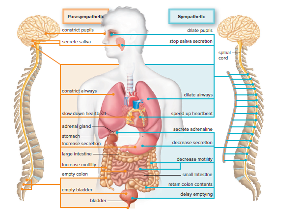
The Autonomic System
Regulates the activity of glands and cardiac and smooth muscle involuntarily.
Divided into parasympathetic and sympathetic divisions.
Reflex actions regulate blood pressure and breathing rate.
Sensory neurons send information to the CNS, and motor neurons complete the reflexes.
The parasympathetic division promotes internal responses associated with a relaxed state.
The parasympathetic division uses the acetylcholine (ACh) neurotransmitter.
The sympathetic division is important during emergency situations and is associated with "fight or flight."
Sympathetic division accelerates the heartbeat and dilates the airways.
Sympathetic division inhibits the digestive tract.
The sympathetic nervous system uses the norepinephrine neurotransmitter.
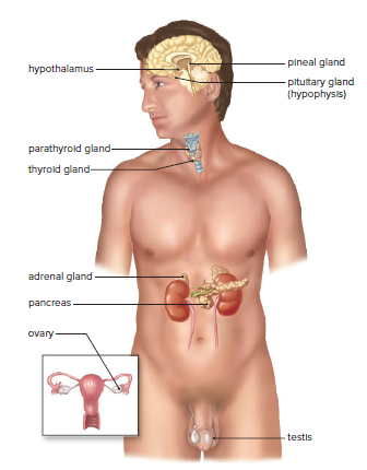
27.2 Endocrine System
Consists of glands and tissues that secrete hormones.
Hormones are chemical messengers that regulate the activity of organs, tissues, or glands.
Endocrine glands secrete hormones directly into the bloodstream.
Is contrasted with exocrine glands that have ducts and secrete products into body cavities.
The endocrine system and nervous system are involved in homeostasis.
Hormones affect blood composition and pressure, body growth, and other life processes.
Certain hormones are involved in reproductive organ maturation and function.
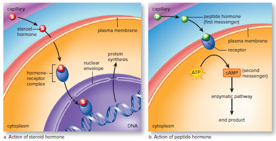
The Action of Hormones
Cells that respond to hormones have receptor proteins that bind to the hormone.
Hormones cause metabolic changes in cells.
The type of change depends on the chemical structure of the hormone.
Steroid hormones are lipids and can pass through the plasma membrane.
Hormone-receptor complex binds to DNA, and gene expression follows.
Peptide hormones can't pass through the plasma membrane.
Peptide hormones bind to a receptor protein in the plasma membrane.
Peptide hormones act as the "first messenger."
The signal transduction pathway leads to a "second messenger" that changes cell metabolism.
The enzyme pathway is activated, leading to an enzyme cascade.
Peptide hormones may be specific with regard to the types of cells they target.
Hypothalamus and Pituitary Gland
The hypothalamus is a part of the brain that helps regulate the internal environment.
It receives information about the heartbeat and body temperature.
The hypothalamus communicates with the medulla oblongata to correct any abnormalities.
The medulla oblongata contains brain centers that control the autonomic system.
The hypothalamus is a part of the endocrine system and contains specialized hormone-secreting neurons.
It controls the glandular secretions of the pituitary gland.
The pituitary gland is a small gland connected to the brain by a stalklike structure.
The pituitary gland has two portions, the anterior pituitary and the posterior pituitary, which are distinct from each other.
Anterior Pituitary
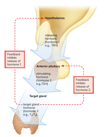
The hypothalamus controls the anterior pituitary by producing hypothalamic-releasing hormones.
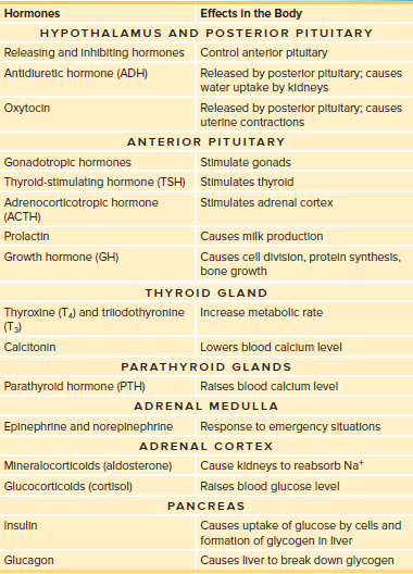
Thyroid-stimulating hormone (TSH) stimulates the thyroid to produce T3 and T4.
Adrenocorticotropic hormone (ACTH) stimulates the adrenal cortex to produce glucocorticoids.
Gonadotropic hormones (FSH and LH) stimulate the gonads to produce gametes and sex hormones.
The hypothalamic-releasing hormones are kept in balance by a three-tiered control system that uses negative-feedback mechanisms.
Prolactin (PRL) is produced during pregnancy and after childbirth, causing mammary glands to develop and produce milk, and plays a role in carbohydrate and fat metabolism.
Growth hormone (GH) promotes skeletal and muscular growth, stimulates amino acid entry into cells and protein synthesis, and underproduction leads to pituitary dwarfism, while overproduction leads to pituitary gigantism.
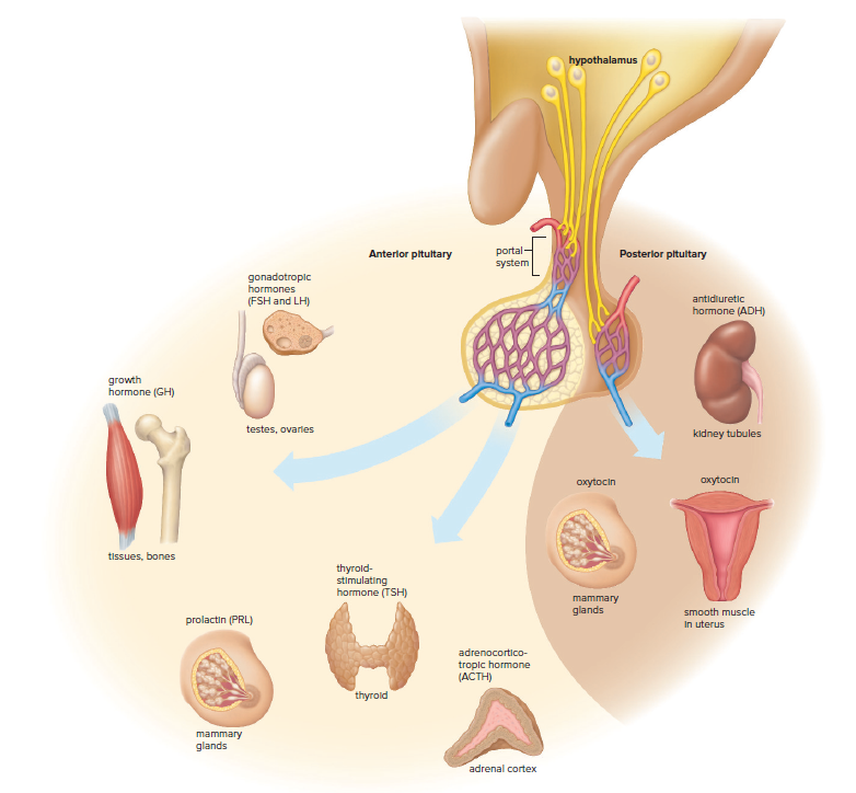
Posterior Pituitary
The hypothalamus produces two hormones: antidiuretic hormone (ADH) and oxytocin.
Hypothalamic secretory neurons' axons extend into the posterior pituitary, where hormones are stored in the axon terminals.
When the blood is too concentrated, the hypothalamus releases ADH from the posterior pituitary.
ADH causes water to be reabsorbed in the kidneys.
As the blood becomes dilute, ADH is no longer released, which is an example of negative feedback.
Negative feedback maintains homeostasis.
Oxytocin causes uterine contraction during childbirth and milk letdown when a baby is nursing.
Thyroid and Parathyroid Glands
The thyroid gland is a large gland located in the neck.
It produces three hormones: calcitonin, triiodothyronine (T3), and thyroxine (T4).
Calcitonin is involved in calcium homeostasis.
Triiodothyronine and thyroxine are produced by the thyroid gland using iodine.
The concentration of iodine in the thyroid gland can increase to as much as 25 times that in the blood.
Lack of iodine in the diet can prevent the thyroid gland from producing hormones.
Constant stimulation by the anterior pituitary can cause the thyroid to enlarge, resulting in an endemic goiter.
The use of iodized salt can help prevent endemic goiter.
Thyroid hormones increase the metabolic rate and stimulate all cells of the body to metabolize at a faster rate.
Hyperthyroidism or Graves disease can cause an overactive thyroid gland and bulging of the eyes (exophthalmos).
Removal or destruction of a portion of the thyroid by means of radioactive iodine can cure the condition.
Calcium Regulation
The thyroid gland produces calcitonin, which regulates blood calcium levels.
Calcium is important for nervous conduction, muscle contraction, and blood clotting.
Calcitonin reduces osteoclast activity and number, leading to more calcium being deposited in bone.
Negative feedback inhibits the release of calcitonin when blood calcium level returns to normal.
Low blood calcium stimulates the release of parathyroid hormone (PTH) from the parathyroid glands.
PTH promotes osteoclast activity and calcium release from bones.
PTH also promotes calcium reabsorption by the kidneys and vitamin D activation.
Vitamin D stimulates calcium absorption from the small intestine.
These effects bring the blood calcium levels back to normal, stopping PTH secretion.
Insufficient PTH production leads to tetany, which causes continuous muscle contraction due to increased nerve excitability.
Adrenal Glands
Two adrenal glands are located on top of the kidneys.
Each adrenal gland has two portions: adrenal medulla and adrenal cortex.
The adrenal medulla and adrenal cortex have no functional connection with each other.
The hypothalamus controls the activity of both portions of the adrenal glands.
The hypothalamus initiates nerve impulses that travel to the adrenal medulla, which then secretes its hormones.
The hypothalamus controls the anterior pituitary's secretion of ACTH through ACTH-releasing hormone.
ACTH stimulates the adrenal cortex.
The hypothalamus stimulates both the adrenal medulla and adrenal cortex in response to stress, including emotional and physical trauma and vigorous exercise.
Adrenal Medulla
Epinephrine (adrenaline) and norepinephrine (noradrenaline) are produced by the adrenal medulla.
They bring about rapid body changes during an emergency situation.
These hormones complement the actions of the sympathetic autonomic system.
The effects of these hormones are short-term.
Hormones produced by the adrenal cortex provide a long-term response to stress.
Adrenal Cortex
The adrenal cortex produces two major types of hormones: mineralocorticoids (e.g. aldosterone) and glucocorticoids (e.g. cortisol.)
Aldosterone regulates salt and water balance by acting on the kidneys, leading to an increase in blood volume and blood pressure.
Cortisol regulates carbohydrate, protein, and fat metabolism, leading to an increase in blood glucose levels, and acts as an anti-inflammatory agent.
The adrenal cortex also secretes small amounts of male and female sex hormones in both sexes.
Hyposecretion of adrenal cortex hormones leads to Addison's disease, which can cause bronzing of the skin, low blood pressure, dehydration, and can be fatal if left untreated.
Hypersecretion of adrenal cortex hormones leads to Cushing syndrome, which can cause diabetes mellitus, masculinization in women, and hypertension.
Excess production of adrenal male sex hormones in women can result in an increase in body hair, deepening of the voice, and beard growth.
Pancreas
The pancreas is a large gland located in the abdomen. It has two main functions:
Exocrine function: The pancreas produces digestive juices that help to break down food. These juices are released into the small intestine through a duct.
Endocrine function: The pancreas produces the hormones insulin and glucagon. Insulin helps to lower blood sugar levels, while glucagon helps to raise blood sugar levels.
Insulin
Insulin is produced when blood sugar levels are high, such as after eating.
Insulin helps to lower blood sugar levels by:
Stimulating cells to take up glucose from the blood.
Storing glucose in the liver and muscles as glycogen.
Promoting the use of glucose for energy.
Glucagon
Glucagon is produced when blood sugar levels are low, such as between meals.
Glucagon helps to raise blood sugar levels by:
Stimulating the liver to break down glycogen into glucose.
Promoting the use of fat and protein for energy.
Diabetes Mellitus
Diabetes mellitus is a hormonal disease where cells don't take up or metabolize glucose, leading to high blood glucose levels.
Type 1 diabetes is insulin-dependent and is caused by the pancreas not producing insulin due to cytotoxic T cells destroying pancreatic islets.
Type 1 diabetes requires daily insulin injections to control symptoms and prevent hypoglycemia.
Hypoglycemia symptoms include perspiration, pale skin, shallow breathing, and anxiety, which can lead to unconsciousness.
Type 2 diabetes is non-insulin-dependent and usually occurs in obese and inactive people.
Type 2 diabetes can be prevented or controlled with a low-fat and low-sugar diet and regular exercise.
Both types of diabetes can lead to blindness, kidney disease, and circulatory disorders.
Table 27.1 summarizes the major organs and glands of the endocrine system, the hormones they produce, and their effects on the body.