
Chapter 18: Blood Group Typing and Protein Profiling
18.1: Blood Group Typing
Blood Groups
Blood groups: Defined as antigen polymorphisms present on erythrocyte surfaces.
Transfusion reactions occur when an incompatible type of blood is transfused into an individual, which can lead to severe symptoms or even death.
Karl Landsteiner discovered the first blood group, known as the ABO system, in the early 1900s, while studying transfusion and transplantation.
The International Society of Blood Transfusion currently recognizes 29 blood group systems, which include hundreds of antigen polymorphisms
ABO Blood Group System
In the ABO blood group system, two types of antigens, designated A and B, give rise to four blood types:
Type A individuals have the A antigen.
Type B individuals have the B antigen.
Type AB individuals have both A and B antigens.
Type O individuals have neither A nor B antigens.
Biosynthesis of Antigens
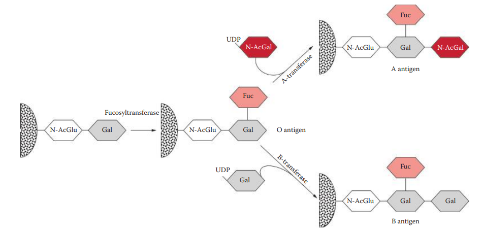
All individuals generate the O antigen, also known as the H antigen.
The O antigen is synthesized by fucosyltransferase, a fucose transferase encoded by the FUT genes, which adds a fucose on the end of a glycolipid or glycoprotein.
The specificity of this enzyme determines the ABO blood type:
The A allele produces the A-transferase, which transfers N-acetylgalactosamine to the O antigen and thus synthesizes the A antigen
The B allele produces the B-transferase, which transfers galactose to the O antigen and thus synthesizes the B antigen.
The O allele has a mutation (small deletion), which eliminates transferase activity, and no modification of the O antigen occurs.
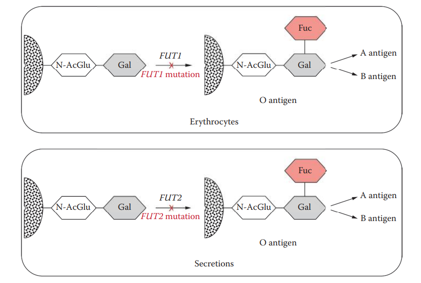
Secretors
Individuals whose A, B, and O antigens can be found in other types of bodily fluids are referred to as secretors.
Chromosome 19 contains two homologous genes:
FUT1 is expressed in tissues of mesodermal origin and is responsible for the synthesis of the O antigen in erythrocytes.
FUT2 is expressed in tissues of endodermal origin; it is responsible for the synthesis of the O antigen in secretions.
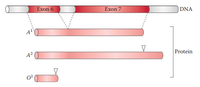
Inheritance of A and B Antigens
A and B alleles are dominant.
For AO and BO heterozygotes, the corresponding transferase synthesizes the A or B antigen.
A and B alleles are codominant in AB heterozygotes because both transferase activities are expressed.
The OO homozygote produces neither transferase activity and therefore lacks both antigens.
The inheritance of A and B alleles obeys Mendelian principles.
Blood Group System
Number | Name | Symbol | # of Antigens | Gene Names | Chromosomal Location |
|---|---|---|---|---|---|
001 | ABO | ABO | 4 | ABO | 9q34.2 |
002 | MNS | MNS | 43 | GYPA, GYPB, GYPE | 4q31.21 |
003 | P | P1 | 1 | - | 22q11.2-qter |
004 | Rh | RH | 49 | RHD, RHCE | 1p36.11 |
005 | Lutheran | LU | 20 | LU | 19q13.32 |
006 | Kell | KEL | 25 | KEL | 7q34 |
007 | Lewis | LE | 6 | FUT3 | 19p13.3 |
008 | Duffy | FY | 6 | FY | 1q23.2 |
009 | Kidd | JK | 3 | SLC41A1 | 18q12.3 |
010 | Diego | DI | 21 | SLC4A1 | 17q21.31 |
011 | Yt | YT | 2 | ACHE | 7q22.1 |
012 | Xg | XG | 2 | XG, MIC2 | Xp22.33, Yp11.3 |
013 | Scianna | SC | 5 | ERMAP | 1p34.2 |
014 | Dombrock | DO | 5 | DO | 12p12.3 |
015 | Colton | CO | 3 | AQP1 | 7p14.3 |
016 | Landsteiner-Wiener | LW | 3 | ICAM4 | 19p13.2 |
017 | Chido/Rodgers | CH/RG | 9 | C4A, C4B | 6p21.3 |
018 | H | H | 1 | FUT1 | 19q13.33 |
019 | Kx | XK | 1 | XK | Xp21.1 |
020 | Gerbich | GE | 8 | GYPC | 2q14.3 |
021 | Cromer | CROM | 12 | DAF | 1q32.2 |
022 | Knops | KN | 8 | CR1 | 1q32.2 |
023 | Indian | IN | 2 | CD44 | 11p13 |
024 | Ok | OK | 1 | BSG | 19p13.3 |
025 | Raph | RAPH | 1 | CD151 | 11p15.5 |
026 | John Milton Hagen | JMH | 1 | SEMA7A | 15q24.1 |
027 | I | I | 1 | GCNT2 | 6p24.2 |
028 | Globoside | GLOB | 1 | B3GALT3 | 3q26.1 |
029 | Gill | GIL | 1 | AQP3 | 9p13.3 |
Forensic Applications of Blood Group Typing
The application and usefulness of blood typing in forensic identification are based on the ability to group individuals into four different types using the ABO blood system, allowing individuals to be identified.
The probability that any two randomly chosen individuals have an identical blood type is very high.
The A and B antigens are very stable and can be identified in dried blood even after many years. They can also be found in semen and other bodily fluids of secretors.
Blood Group Typing Techniques
Lattes Crust Assay
It relies on the principles of Landsteiner’s experiments. It is an agglutination-based assay that utilizes the A, B, and O indicator cells to test the agglutination reaction with its corresponding naturally occurring serum antibodies in a questioned sample.
It is simple and rapid. However, one limitation is that the assay is not very sensitive and requires a large quantity of blood.
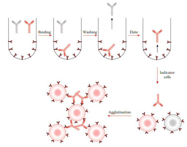
Absorption–Elution Assay
It is highly sensitive and can be used for testing dried bloodstains. This method indirectly detects the presence of antigens.
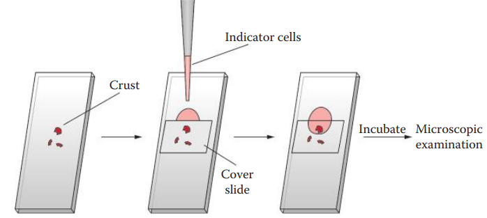
Lattes Crust Assay Procedure
Place small quantities of blood crust from a specimen on a microscopic slide and place a cover slide over the crusts. Prepare slides for A, B, and O cells separately.
Prepare cell suspensions with saline (0.85% NaCl in phosphate buffer, pH 7.4) for the A, B, and O cells separately.
Apply a few drops of the A-cell suspension and allow the cells to diffuse under the cover slip. Repeat this step for B cells and O cells.
Incubate the slides in a moisture chamber at room temperature for 2 h.
Examine results under a microscope.
18.2: Forensic Protein Profiling
Methods
Matrices Supporting Protein Electrophoresis: The matrix reduces the effects of diffusion and convection on the macromolecules.
Separation by Molecular Weight
An electrophoretic method is frequently utilized to resolve various proteins based on their molecular weights.
Native electrophoresis can be used to isolate proteins for studying the functions of proteins.
Reducing Agents
It is common to include reducing agents such as mercaptoethanol (ME), dithiothreitol (DTT), or sodium mercaptoethane sulfonate (MESNA) to denature proteins.
Reducing agents cleave the disulfide bonds of proteins.
Detergents
Detergents disrupt noncovalent interactions within the structures of native proteins.
Sodium dodecyl sulfate (SDS): A strong anion detergent that binds to most proteins in amounts proportional to the molecular weight of the protein.
Separation by Isoelectric Point
It can be used to separate proteins according to their isoelectric points (pI).
Isoelectric Points: It is the pH value at which the net electric charge of an amino acid is zero.
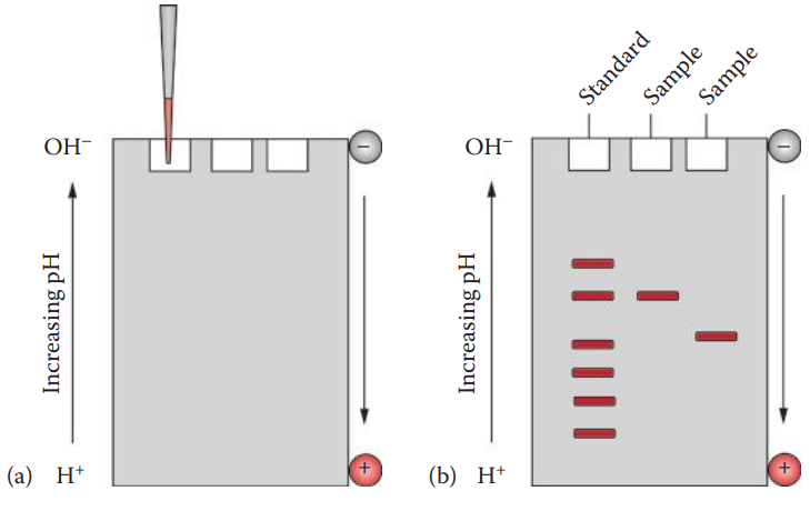
In IEF electrophoresis, a pH gradient is created in a gel between the electrodes, and a protein sample is placed in a well on the gel.
A pH gradient in the gel is established by utilizing materials such as carrier ampholytes or immobilines that are dispersed in the gel.
Carrier ampholytes are synthetic amphoteric compounds that contain multiple weak ionizable moieties acting as either acids or bases.
Erythrocyte Protein Polymorphisms
Erythrocyte Isoenzymes
Isoenzymes: Are multiple forms of an enzyme that catalyze the same reaction but differ in their amino acid sequences.
Phosphoglucomutase (PGM): An important metabolic enzyme, catalyzes the reversible conversion of glucose-1-phosphate and glucose-6-phosphate.
Hemoglobin
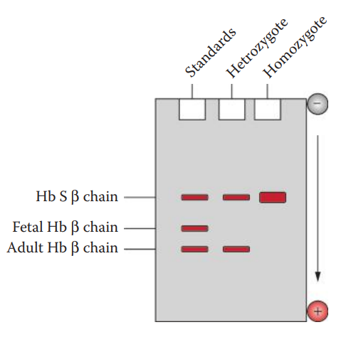
Fetal Hemoglobin: The dominant form of hemoglobin present in the fetus during gestation.
Hemoglobin S: An inherited variant of normal adult hemoglobin.
Serum Protein Polymorphisms
Haptoglobin (Hp): The most widely used of the polymorphic serum proteins in forensic biology. It is a protein that binds and transports Hb from the bloodstream to the liver for the recycling of the iron contained in the Hb.