
Introduction of Neuroscience
Analysis of the Nervous System (3 aspects)
Systems Neuroscience: investigates groups of neurons that preform a common function
Behavioral Neuroscience: looks at the interaction among systems that effect behavior (looking at the output), behaviors influenced by environment and society
Cognitive Neuroscience: looks in the fields of thinking, learning and memory
Central Nervous System: composed of the brain system and spinal cord
Peripheral Nervous System: composed of peripheral nerves, spinal and cranial nerves
Neuron = the functional unit of the nervous system
Neuron Theory: functions of the nervous system reflects the function of individual neurons, group of neurons and their connections
Bipolar Neuron: special senses of the face…found in retina, olfactory, ear (eyes & ears) , sensory info to brain
Synapses on neurons in dorsal horn
Pseudo-unipolar Neuron: sympathetic ganglion, sensory info to the brain …found in sympathetic ganglia
Multipolar Neuron: (most common neuron image) motor neurons…found in distal PNS
Synapses on skeletal muscle cells
GREY vs WHITE
Grey Matter: Ganglia, Nuclei, Cortex (information is integrated in gray matter)
(outside cortex)
White Matter: Tract, Lemniscus, Fasciculus, Column, Peduncle, Capsule (internal cortex...doing various functions) (aka the highs for information to travel)
Central Nervous System
Gila of the CNS: these cells out number neurons
Astro: help support grey matter…Others support BB
Micro-G: scavengers, remove plaques in the brain
Ependyma: Formation and movement of spinal fluid & lining of ventricles
Oligodendrocytes: myelinate nerves of the CNS
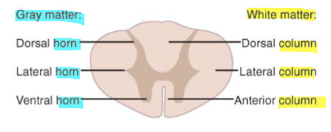
Spinal Cord
Composed of horns & columns
Information comes in through the posterior and "exits" anteriorly
Within the cross section of the spinal cord you can see what looks like the letter "H" which is composed of Grey matter (Ganglia, Nuclei, Cortex…information is integrated in gray matter), this is then divided into dorsal, lateral and ventral horns
These Horns contain cell bodies of motor neurons, interneurons and the ends of sensory neurons
Leading to these horns are columns which are composed to bundle of nerves transmitting signals
Dorsal region: contain interneurons and the ends of sensory neurons
Lateral region: autonomic cell bodies
Ventral region: bodies of motor neurons and interneurons
Columns transmit signals from the horns
 2 Main Functions of the Spinal Region:
Conveys information from the peripheral regions to the Brian
Processes Information
Brainstem:
motor and sensory fiber tracts travel through the brainstem
It is here that you find “subconscious control” to maintain equilibrium, cardiovascular activity , respiration and other functions
Composed of 3 sections: Mid-brain, Pons and Medulla Oblongata
Mid-brain: where info crosses over from one side to another. Root of cranial nerve 3 & 4
Pons: roots of cranial nerves 5,6,7,8
Medulla: roots of cranial nerves 9,10,11,12. Where we find pyramidal decussation, which is the mid-line crossing of axons
Cranial Nerves (1- 12):
Olfactory: smell
Optic: vision
Oculomotor: moves pupil of eyes up & down, raises upper eyelid, constricts pupil
Trochlear: moves pupil of eye medially/down
Trigeminal: Facial sensation, chewing sensation of TMJ
Abducens: abduct pupil of the eye
Facial: facial expression, closes eyes, tears, salivation and taste
Vestibulocochlear: sensation of head position relative to gravity and head movement, hearing
Glossopharyngeal: swallowing, salivation and taste
Vagus: regulates viscera, swallowing, speech & taste
Spinal Accessory: elevates shoulders and turns head
Hypoglossal: moves tongue
__Cerebellum (aka lil brain): __
consists of two large cerebellar hemispheres
connects to the posterior brain-stem by peduncles (large bundles of fibers)
function is to coordinate movement…smooth and accurate movements, maintain posture, movement of head in space
Cerebellar Connections:
Vestibulocerebellum (Flocculonodular Lobe): detects head position relative to gravity…coordinates with inner ear & postural reflexes to keep us upright while we sit/move.
Spinocerebellum/Spinal Cord: checks the sensory input from the spinal cord and that it matches the motor output…if not the cerebellum will correct/override
Cerebrocerebellum/ Cerebral Cortex: communicates with the cerebrum and is the final check to the movement being sent out
Cerebrum
consists of cerebral hemisphere and diencephalon
suface = cerebraal cortex
the site of cognitive thinking…so reasoning, language, non-verbal communication, intelligence and personality
The cerebrum is divided by the longitudinal fissure into 2 cerebral hemispheres.
Elevations on the surface = gyri
Grooves on the surface = sulci
The Diencephalon is composed of 4 parts
Thalamus: processes emotional and some memory information, integrates different sensations, regulates consciousness, arousal & attention…essentially processes sensory information and relays a motor response
Hypothalamus: maintains body temperature, metabolic rate and chemical composition of tissues & fluids (communicates with endocrine gland to regulate secretions)
Epithalamus: influences the secretions of other endocrine glands
Subthalamus: part of the neural circuit control movements (aids in movement control)
The Cerebral Hemispheres are each divided into 6 lobes (regions have specific functions)
Frontal
Parietal
Temporal
Occipital
Limbic
Insular
The basal ganglia in the cerebral hemispheres is composed of the: Caudate, Putamen and Globus Pallidus
Putamen & Globus Pallidus = Lenticular nucleus
Caudate & Putamen = Corpus Striatum
Subthalamic Nucleus: basal ganglia neural circuit
The Hippocampus and Amygdala: apart of the limbic system…processes emotions and memory
involved with processing emotions/emotional response
Hippocampus function = turning working memory to long lasting memory
Peripheral Nervous System
Afferent axons: carries info TOWARDS CNS
Efferent axons: carries info AWAY from CNS
2 divisions: __Somati__c vs Autonomic
Somatic: conscious about it feel it, act on it…axons, sensory nerve endings, myelin
Autonomic: nerve endings on our organs…automatically functioning, we are not conscious about entire neurons, sensory nerve endings, synapses, ganglia
Organization of the PNS
Described based on the functional components of the fibers they carry
Sensory fibers bring info to the CNS (AFFERENT)
Spinal dorsal root ganglia & Cranial Nerve Ganglia
Precise Information coming in from a specific area on the body(Soma)
Information regarding
Pain
Light Touch
Temperature
Vibration
Position in Space (Proprioception)
Motor fibers carry info towards the PNS/Muscles (EFFERENT)
Spinal Cord (spinal nerves) & Brainstem (cranial Nerves)
What we want to do.. the reaction…produces movement
Under direct voluntary control
From the ventral/anterior portion …spine to muscles
Weakness in muscles can be caused by innervation issue
All SOMATIC: "What we know we're doing"
Autonomic
Innervates hollow organs (blood vessels, glands, heart, lungs, liver, GI)
Effects smooth muscle contraction…absence of excitation = dilation
Direct innervation of some glands and nodal tissue of the heart
Parasympathetic & Sympathetic: have opposing effects on hollow organs
Sympathetic: fight or flight…pupils dilate, HR increases, airways dilate, blood directed away from GI & towards skeletal muscles, ejaculation
Parasympathetic: Rest & Digest (homeostasis) pupils constrict, HR decreases, Increase in GI blood flow and motility, blood to genitals increase
Cerebrospinal Fluid System: Ventricles and Meninges
CFS: modified filtrate of plasma, circulates from cavities in the brain to the surface of CNS into the venous blood system
4 ventricles:
pair of lateral ventricles in the cerebral hemisphere
Communicate with the 3rd along the interventricular foramina
Third ventricle along the midline of the diencephalon
Communicates/connected to the 4th by the cerebral aqueduct
Fourth ventricle deep w/in the brainstem, posterior to the pons and medulla & anterior to the cerebellum
Communicates with the spine, found in spinal cord
Compression on the ducts > creates pressure within the brain
Meninges: membranous coverings
Dura (thick)
Arachnoid (web)
Pia
Blood Supply
2 main arteries: Internal carotids & Vertebral arteries (posterior)
Vertebral fuse to create basilar > brainstem and cerebellum
Circle of Willis (anastomotic ring): supplies to the cerebral hemispheres
Anterior communicating (R-L) frontal
Posterior communicating, blood can be brought forward and back
3 majors to the Cerebral Hemispheres
Anterior - branch of the internal carotid
Middle - branch of the internal carotid
Posterior - continues from the Circle of Willis
Blood Supply to the Nerves
Nerves need blood to the Vasoneurium: contain blood supply, glial cells and supporting fascia
Present in all Peripheral Nervous Tissue