


😱AP Biology Chapter 9: The Cell Cycle
Introduction
9.1 Most cell division results in genetically identical daughter cells
9.2 The mitotic phase alternates with interphase in the cell cycle
9.3 The eukaryotic cell cycle is regulated by a molecular control system
BIG IDEAS: The continuity of life depends on a cell cycle in which genetic information from a parent cell is passed on to daughter cells. In unicellular organisms, mitosis results in transmission of a complete genome through asexual reproduction (Big Idea 3). For multicellular organisms, mitosis ensures the growth of an individual given adequate free energy (Big Idea 2).
Cells undergoing mitosis in a eukaryote go through a highly conserved and controlled process. There are three major checkpoints in the control system, found in G1, G2, and M phases in a normal cell cycle. However, there are cancer cells that do not follow the same controls. What is out of control in cancer cells? (Big Idea 3)
Unicellular organisms reproduce by cell division; multicellular organisms depend on cell division for their development from a fertilized egg and for growth and repair. Cell division is part of the cell cycle, an ordered sequence of events in the life of a cell.
Cell Division:
The reproduction of cells
How do dividing cells distribute chromosomes to daughter cells?
The functions of cell division
Cell Cycle:
An ordered sequence of events in the life of a cell, from its origin in the division of a parent cell until its own division into two
The eukaryotic cell cycle is composed of ___ and _
interphase (including G1, S, and G2 subphases); M phase (including mitosis and cytokinesis)
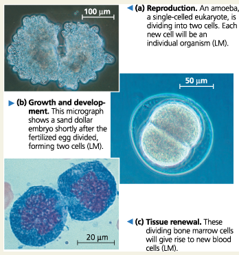
Reproduction:
An amoeba is dividing into two cells. Each new cell will be an individual organism (LM)
Amoeba:
A single-celled eukaryote
Growth and Development:
This micrograph shows a sand dollar embryo shortly after the fertilized egg divided, forming two cells (LM)
Tissue Renewal:
These dividing bone marrow cells will give rise to new blood cells
IMPORTANT VOCAB
Chromosome: A cellular structure consisting of one DNA molecule and associated protein molecules
Chromatin: The complex of DNA and proteins that make up eukaryotic chromosomes
Centromere: Most closely attached
Chromatid: Each of the strands from the chromosome (1 chromatid in a chromosome, a duplicated chromosome has two sister chromatids)
Centriole: A centrosome has a pair of centrioles
Centrosome: A structure present in the cytoplasm of animal cells that functions as a microtubule-organizing center and is important during cell division (moves away during prophase)
Concept 9.1: Most cell division results in genetically identical daughter cells
The genetic material (DNA) of a cell—its genome—is partitioned among chromosomes. Each eukaryotic chromosome consists of one DNA molecule associated with many proteins. Together, the complex of DNA and associated proteins is called chromatin. The chromatin of a chromosome exists in different states of condensation at different times. In animals, gametes have one set of chromosomes, and somatic cells have two sets.
Cells replicate their genetic material before they divide, each daughter cell receiving a copy of the DNA. Prior to cell division, chromosomes are duplicated. Each one then consists of two identical sister chromatids joined along their lengths by sister chromatid cohesion and held most tightly together at a constricted region at the centromeres. When this cohesion is broken, the chromatids separate during cell division, becoming the chromosomes of the daughter cells. Eukaryotic cell division consists of mitosis (division of the nucleus) and cytokinesis (division of the cytoplasm).
Genome:
The genetic material of an organism or virus; the complete complement of an organism’s or virus’s genes along with its noncoding nucleic acid sequences
Chromosome:
A cellular structure consisting of one DNA molecule and associated protein molecules. (In some contexts, such as genome sequencing, the term may refer to the DNA alone.)
Chromosomes in eukaryotic cells
A eukaryotic cell typically has multiple, linear chromosomes, which are located in the nucleus.
Chromosomes in prokaryotic cells
A prokaryotic cell often has a singular, circular chromosome, which is found in the nucleoid, a region that is not enclosed by a membrane
Chromatin:
The complex of DNA and proteins that makes up eukaryotic chromosomes. When the cell is not dividing, chromatin exists in its dispersed form, as a mass of very long, thin fibers that are not visible with a light microscope
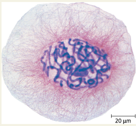
Eukaryotic chromosomes: Chromosomes (stained purple) are visible within the nucleus of this cell from an African blood lily. The thinner red threads in the surrounding cytoplasm are the cytoskeleton. The cell is preparing to divide (LM)
Somatic Cell:
Any cell in a multicellular organism except a sperm or egg or their precursors
Gamete:
A haploid reproductive cell, such as an egg or sperm. Gametes unite during sexual reproduction to produce a diploid zygote
After DNA replication, how do chromosomes and chromatin fibers change?
Chromosomes condense as a part of cell division, and each chromatin fiber becomes densely coiled and folded, making the chromosomes shorter and thicker
Sister Chromatids:
Two copies of a duplicated chromosome are attached to each other by proteins at the centromere and, sometimes, along the arms. While joined, two sister chromatids make up one chromosome. Chromatids are eventually separated during mitosis or meiosis II
Cohesins:
Protein complexes that attach chromatids
Sister Chromatid Cohesion:
When two chromatids are attached all along their lengths by cohesions; contain a centromere
Centromere:
In a duplicated chromosome, the region on each sister chromatid where it is most closely attached to its sister chromatid by proteins that bind to specific DNA sequences;
This close attachment causes a ___ in the condensed chromosome
constriction
An uncondensed, unduplicated chromosome has ___ identified by its DNA sequence
a single centromere
Chromatid Arm:
The part of a chromatid to either side of the centromere (An uncondensed, unduplicated chromosome has a single centromere and two arms)
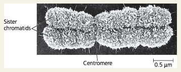
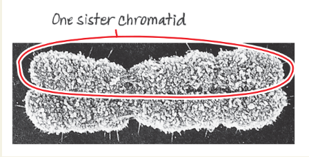
A highly condensed, duplicated human chromosome (SEM)
Circle one sister chromatid of the chromosome in this micrograph
How are chromosomes doubled?
The two sister chromatids duplicate and separate into two new nuclei, one forming at each end of the cell. Since they are separate, they are now considered individual chromosomes instead of sister chromatids
Mitosis:
The process of nuclear division in eukaryotic cells is conventionally divided into five stages. Mitosis conserves chromosome number by allocating replicated chromosomes equally to each of the daughter nuclei.
What are the stages of mitosis?
Prophase, prometaphase, metaphase, anaphase, and telophase
Cytokinesis:
The division of the cytoplasm to form two separate daughter cells immediately after mitosis, meiosis I, or meiosis II
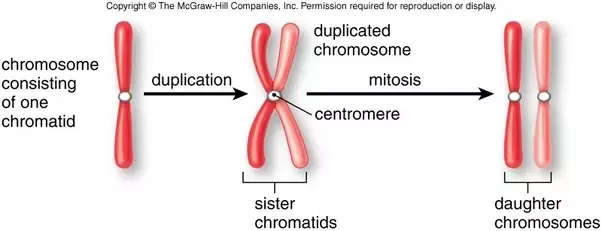
Chromosome duplication and distribution during cell division
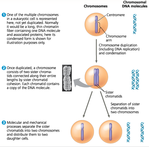
How many chromatid arms does the chromosome in step 2 have? Identify the point in the figure where one chromosome becomes two.
The chromosome has four chromatid arms. The single (duplicated) chromosome in step 2 becomes two (unduplicated) chromosomes in step 3. The duplicated chromosome in step 2 is considered one single chromosome.
How many chromosomes are drawn in each part of Figure 9.5? (Ignore the micrograph in part 2.)
1; 1; 2 2. 39; 39; 78
A chicken has 78 chromosomes in its somatic cells. How many chromosomes did the chicken inherit from each parent? How many chromosomes are in each of the chicken’s gametes? How many chromosomes will be in each somatic cell of the chicken’s offspring?
39: 39: 78
Differentiate between these terms: chromosome, chromatin, and chromatid.
The DNA of a eukaryotic cell is packaged into structures called chromosomes. Each chromosome is a long molecule of DNA, which carries hundreds to thousands of genes, with associated proteins that maintain chromosome structure and help control gene activity. This DNA-protein complex is called chromatin. The chromatin of each chromosome is long and thin when the cell is not dividing. Prior to cell division, each chromosome is duplicated, and the resulting sister chromatids are attached to each other by proteins at the centromeres and, for many species, all along their lengths (a phenomenon called sister chromatid cohesion).
Concept 9.2: The mitotic phase alternates with interphase in the cell cycle
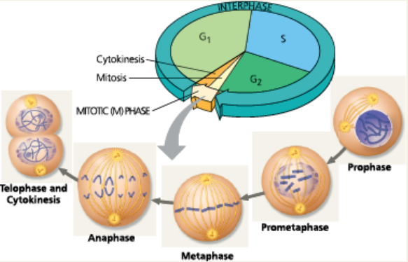
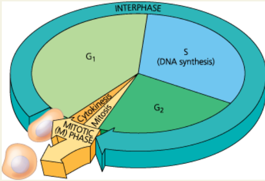
Between divisions, a cell is in interphase: the G1, S, and G2 phases. The cell grows throughout interphase, with DNA being replicated only during the synthesis (S) phase. Mitosis and cytokinesis make up the mitotic (M) phase of the cell cycle.
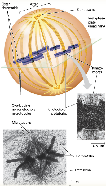
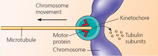
The mitotic spindle, made up of microtubules, controls chromosome movement during mitosis. In animal cells, the mitotic spindle arises from the centrosomes and includes spindle microtubules and asters. Some spindle microtubules attach to the kinetochores of chromosomes and move the chromosomes to the metaphase plate. After sister chromatids separate, motor proteins move them along kinetochore microtubules toward opposite ends of the cell. The cell elongates when motor proteins push nonkinetochore microtubules from opposite poles away from each other.
Mitosis is usually followed by cytokinesis. Animal cells carry out cytokinesis by cleavage, and plant cells form a cell plate.
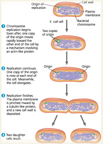
During binary fission in bacteria, the chromosome replicates and the daughter chromosomes actively move apart. Some of the proteins involved in bacterial binary fission are related to eukaryotic actin and tubulin. Since prokaryotes preceded eukaryotes by more than a billion years, it is likely that mitosis evolved from prokaryotic cell division.
Mitotic (M) Phase:
The phase of the cell cycle that includes mitosis and cytokinesis
Interphase:
The period in the cell cycle when the cell is not dividing. During interphase, cellular metabolic activity is high, chromosomes and organelles are duplicated, and cell size may increase
Interphase often accounts for about ___ of the cell cycle
90%
G1 Phase:
The portion of interphase before DNA synthesis begins (the first gap, or growth phase)
S Phase:
The portion of interphase during which DNA is replicated (the synthesis phase)
G2 Phase:
The portion of interphase after DNA synthesis occurs (the second gap, or growth phase)
During all three subphases of interphase, in fact, a cell grows by ___
producing proteins and cytoplasmic organelles such as mitochondria and endoplasmic reticulum
Duplication of the chromosomes, crucial for the eventual division of the cell, occurs entirely during the ___
S phase
The Cell Cycle: In a dividing cell, the mitotic (M) phase alternates with interphase, a growth period. The first part of interphase (G1) is followed by the S phase, when the chromosomes duplicate; G2 is the last part of interphase. In the M phase, mitosis distributes the daughter chromosomes to daughter nuclei, and cytokinesis divides the cytoplasm, producing two daughter cells
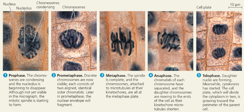
Prophase:
The chromatin condensed into discrete chromosomes visible with a light microscope, the mitotic spindle begins to form, and the nucleolus disappears but the nucleus remains intact (the first stage of mitosis)
Prometaphase:
The nuclear envelope fragments and the spindle microtubules attach to the kinetochores of the chromosomes (the second stage of mitosis)
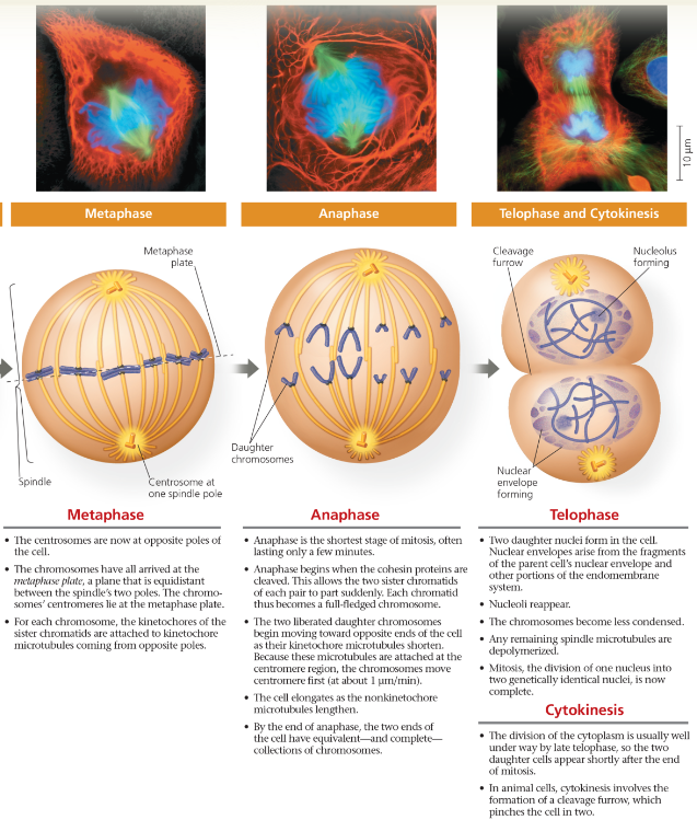
Metaphase:
The spindle is complete and the chromosomes, attached to microtubules at the kinetochores, are all aligned at the metaphase plate (the third stage of mitosis)
Anaphase:
The chromatids of each chromosome have separated and the daughter chromosomes are moving to the poles of the cell (the fourth stage of mitosis)
Telophase:
Daughter nuclei are forming and cytokinesis has typically begun (the fifth and final stage of mitosis)
Exploring Mitosis in an Animal Cell
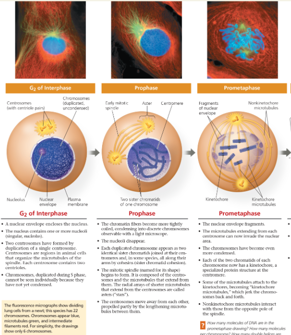
Exploring Mitosis in an Animal Cell part 2
How many molecules of DNA are in the prometaphase drawing? How many molecules per chromosome? How many double helices are there per chromosome? Per chromatid?
12; 2; 2; 1
Mitotic Spindle:
An assemblage of microtubules and associated proteins that are involved in the movement of chromosomes during mitosis
The spindle microtubules ___ by incorporating more subunits of the protein tubulin and shorten _ by losing subunits
elongate (polymerize); shorten (depolymerize)
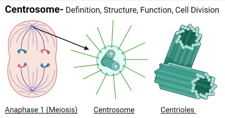
Centrosome:
A structure present in the cytoplasm of animal cells that functions as a microtubule-organizing center and is important during cell division
A centrosome has ___ centrioles
two
In animal cells, the assembly of spindle microtubules starts at the ___
centrosome
Aster:
A radial array of short microtubules that extends from each centrosome toward the plasma membrane in an animal cell undergoing mitosis
Kinetochore:
A structure of proteins attached to the centrosome that links each sister chromatid to the mitotic spindle
The kinetochore acts as a ___ that attaches the motor of the spindle to the cargo that it moves—the chromosome
coupling device
Explain the consequences of a chromosome’s kinetochores being “captured” by microtubules
The chromosome begins to move toward the pole from which those microtubules extend. However, this movement is checked as soon as microtubules from the opposite pole attach to the kinetochore on the other chromatid. What happens next is like a tug-of-war that ends in a draw. The chromosome moves first in one direction and then in the other, back and forth, finally settling midway between the two ends of the cell
Metaphase Plate:
An imaginary structure located at a plane midway between the two poles of a cell in metaphase on which the centromeres of all the duplicated chromosomes are located
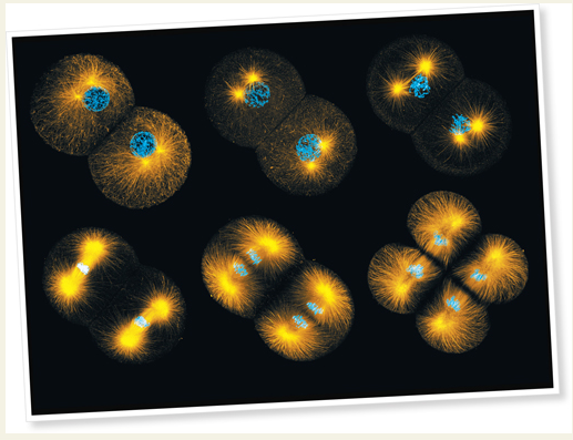
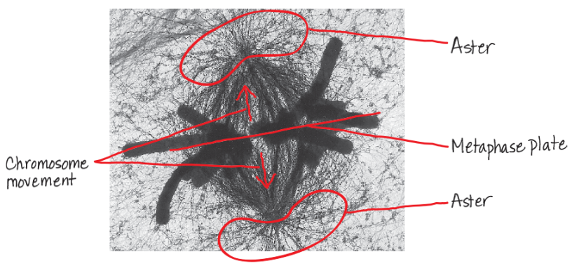
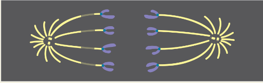
The mitotic spindle at metaphase: The kinetochores of each chromosome’s two sister chromatids face in opposite directions. Here, each kinetochore is attached to a cluster of kinetochore microtubules extending from the nearest centrosome. Nonkinetochore microtubules overlap at the metaphase plate (TEMs).
On the lower micrograph, draw a line indicating the position of the metaphase plate. Circle an aster. Draw arrows indicating the directions of chromosome movement once anaphase begins.
Gary Borisy and colleagues at the University of Wisconsin wanted to determine whether kinetochore microtubules depolymerize at the kinetochore end or the pole end as chromosomes move toward the poles during mitosis. First they labeled the microtubules of a pig kidney cell in early anaphase with a yellow fluorescent dye. (Nonkinetochore microtubules are not shown.)
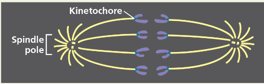
Then they marked a region of the kinetochore microtubules between one spindle pole and the chromosomes by using a laser to eliminate the fluorescence from that region, while leaving the microtubules intact (see below). As anaphase proceeded, they monitored the changes in microtubule length on either side of the mark.
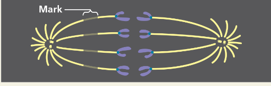
As the chromosomes moved poleward, the microtubule segments on the kinetochore side of the mark shortened, while those on the spindle pole side stayed the same length.
During anaphase in this cell type, chromosome movement is correlated with kinetochore microtubules shortening at their kinetochore ends and not at their spindle pole ends. This experiment supports the hypothesis that during anaphase, a chromosome is walked along a microtubule as the microtubule depolymerizes at its kinetochore end, releasing tubulin subunits.
If this experiment had been done on a cell type in which “reeling in” at the poles was the main cause of chromosome movement, how would the mark have moved relative to the poles? How would the microtubule segment lengths have changed?
The mark would have moved toward the nearer pole. The lengths of fluorescent microtubules between that pole and the mark would have decreased, while the lengths between the chromosomes and the mark would have remained the same.
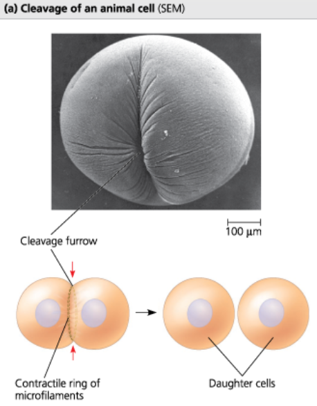
Cleavage:
(1) The process of cytokinesis in animal cells, characterized by pinching of the plasma membrane (2) The succession of rapid cell division without significant growth during early embryonic development that converts the zygote to a ball of cells
Cleavage Furrow:
The first sign of cleavage in an animal cell; a shallow groove around the cell in the cell surface near the old metaphase plate
Cytokinesis in animal cells
Cytokinesis in plant cells
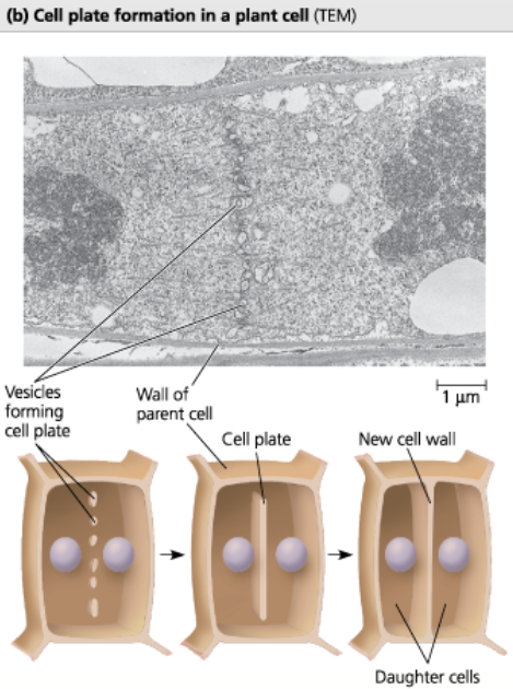
Cell Plate:
A membrane-bounded, flattened sac located at the midline of a dividing plant cell, inside which the new cell wall forms during cytokinesis
Mitosis in a plant cell: These light micrographs show mitosis in cells of an onion root
Binary Fission:
A method of asexual reproduction in single-celled organisms in which the cell grows to roughly double its size and then divides into two cells.
In prokaryotes, binary fission does not involve ___, but in single-celled eukaryotes that undergo binary fission, it is part of the process
mitosis
Origin of Replication:
The site where the replication of a DNA molecule begins, consisting of a specific sequence of nucleotides
Bacterial cell division by binary fission: The bacterium E. coli, shown here, has a single, circular chromosome
Describe the evolution of mitosis
Given that prokaryotes preceded eukaryotes on Earth by more than a billion years, we might hypothesize that mitosis evolved from simpler prokaryotic mechanisms of cell reproduction. The fact that some of the proteins involved in bacterial binary fission are related to eukaryotic proteins that function in mitosis supports that hypothesis.
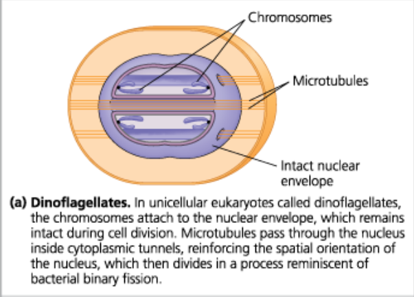
Mechanisms of cell division: Some unicellular eukaryotes existing today have mechanisms of cell division that may resemble intermediate steps in the evolution of mitosis (Dinoflagellates)
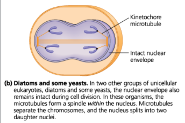
Mechanisms of cell division: Some unicellular eukaryotes existing today have mechanisms of cell division that may resemble intermediate steps in the evolution of mitosis (Diatoms and some yeasts)
How many chromosomes are shown in the drawing in Figure 9.8? Are they duplicated? How many chromatids are shown?
6 chromosomes; they are duplicated; 12 chromatids
Compare cytokinesis in animal cells and plant cells
Following mitosis, cytokinesis results in two genetically identical daughter cells in both plant cells and animal cells. However, the mechanism of dividing the cytoplasm is different in animals and plants. In an animal cell, cytokinesis occurs by cleavage, which divides the parent cell in two with a contractile ring of actin filaments. In a plant cell, a cell plate forms in the middle of the cell and grows until its membrane fuses with the plasma membrane of the parent cell. A new cell wall grows inside the cell plate.
During which stages of the cell cycle does a chromosome consist of two identical chromatids?
From the end of S phase in interphase through the end of metaphase in mitosis
Compare the roles of tubulin and actin during eukaryotic cell division with the roles of tubulin-like and actin-like proteins during bacterial binary fission
During eukaryotic cell division, tubulin is involved in spindle formation and chromosome movement, while actin functions during cytokinesis. In bacterial binary fission, it’s the opposite: Tubulin-like molecules are thought to act in daughter cell separation, and actin-like molecules are thought to move the daughter bacterial chromosomes to opposite ends of the cell.
In which of the three subphases of interphase and the stages of mitosis do chromosomes exist as single DNA molecules?
Chromosomes exist as single DNA molecules in G1 of interphase and in anaphase and telophase of mitosis. During S phase, DNA replication produces sister chromatids, which persist during G2 of interphase and through prophase, prometaphase, and metaphase of mitosis.
Concept 9.3: The eukaryotic cell cycle is regulated by a molecular control system
Signaling molecules present in the cytoplasm regulate progress through the cell cycle.
The cell cycle control system is molecularly based; key regulatory proteins are cyclins and kinases. The cell cycle clock has specific checkpoints where the cell cycle stops until a go-ahead signal is received; important checkpoints occur in the G1, G2, and M phases. Cell culture has enabled researchers to study the molecular details of cell division. Both internal signals and external signals control the cell cycle checkpoints via signal transduction pathways. Most cells exhibit density-dependent inhibition of cell division as well as anchorage dependence.
Cancer cells elude normal cell cycle regulation and divide unchecked, forming tumors. Malignant tumors invade nearby tissues and can undergo metastasis, exporting cancer cells to other sites, where they may form secondary tumors. Recent cell cycle and cell-signaling research, and new techniques for sequencing DNA, have led to improved cancer treatments.
The cell cycle is driven by ___ present in the _
specific signaling molecules; cytoplasm
Do molecular signals in the cytoplasm regulate the cell cycle?
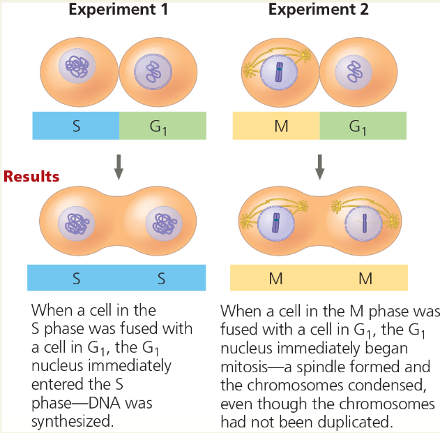
Experiment: Researchers at the University of Colorado wondered whether a cell’s progression through the cell cycle is controlled by cytoplasmic molecules. They induced cultured mammalian cells at different phases of the cell cycle to fuse
Conclusion: The results of fusing a G1 cell with a cell in the S or M phase of the cell cycle suggest that molecules present in the cytoplasm during the S or M phase control the progression to those phases
If the progression of phases did not depend on cytoplasmic molecules and each phase began when the previous one was complete, how would the results have differed?
In both cases, the G1 nucleus would have remained in G1 until the time it normally would have entered the S phase. Chromosome condensation and spindle formation would not have occurred until the S and G2 phases had been completed.
Cell Cycle Control System:
A cyclically operating set of molecules in the eukaryotic cell that both triggers and coordinates key events in the cell cycle
Mechanical analogy for the cell cycle control system: In this diagram of the cell cycle, the flat “stepping stones” around the perimeter represent sequential events. Like the control device of an automatic washer, the cell cycle control system proceeds on its own, driven by a built-in clock. However, the system is subject to internal and external regulation at various checkpoints; three important checkpoints are shown (red).
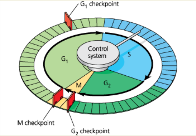
Checkpoint:
A control point in the cell cycle where stop and go-ahead signals can regulate the cycle
If a cell receives a go-ahead signal at the G1 checkpoint, it will usually ___. If it does not receive a go-ahead signal at that point, it may exit the cycle, switching into _
complete the G1, S, G2, and M phases and divide; a nondividing states called the G0 phase
G0 Phase:
A nondividing state occupied by cells that have left the cell cycle, sometimes reversibly
Two Important Checkpoints: At certain checkpoints in the cell cycle (red gates), cells do different things depending on the signals they receive. Events of the (a) G1 and (b) M checkpoints are shown. In part (b), the G2 checkpoint has already been passed by the cell.
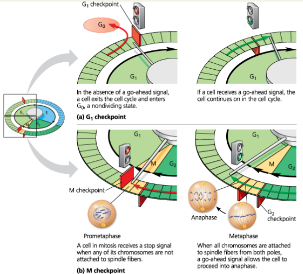
What might be the result if the cell ignored either checkpoint and progressed through the cell cycle?
The cell would divide under conditions where it was inappropriate to do so. If the daughter cells and their descendants also ignored either of the checkpoints and divided, there would soon be an abnormal mass of cells. (This type of inappropriate cell division can contribute to the development of cancer.)
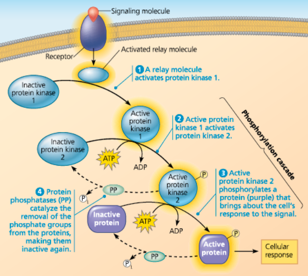
Cyclins:
A protein that can regulate the cell cycle at the molecular level
Kinase:
Enzymes that activate or inactivate other proteins by phosphorylating them
A Phosphorylation Cascade
Anaphase, the separation of sister chromatids, does not begin until all the chromosomes are ___
properly attached to the spindle at the metaphase plate
Growth Factor:
(1) A protein that must be present in the extracellular environment (culture medium or animal body) for the growth and normal development of certain types o cells. (2) A local regulator that acts on nearby cells to stimulate cell proliferation and differentiation
Platelet-Derived Growth Factor (PDGF):
A growth factor made by blood cell fragments called platelets is required to divide cultures of fibroblasts
The effect of PDGF on cell division
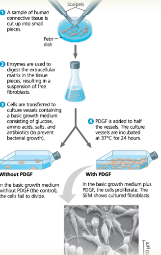
PDGF signals cells by binding to a cell-surface receptor that then becomes phosphorylated, activating it so that it sends a signal. If you added a chemical that blocked phosphorylation, how would the results differ?
The cells in the vessel with PDGF would not be able to respond to the growth factor signal and thus would not divide. The culture would resemble that without the added PDGF.
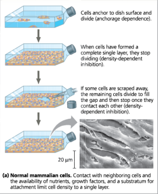
Density-Dependent Inhibition:
The phenomenon observed in normal animal cells that causes them to stop dividing when they come into contact with one another
Density-dependent inhibition and anchorage dependence of cell division (cells shown are disproportionately large): Normal Mammalian Cells
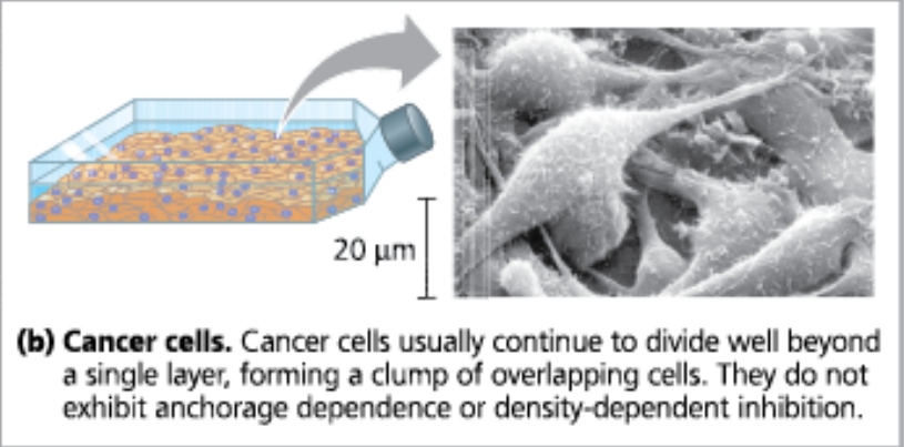
Density-dependent inhibition and anchorage dependence of cell division (cells shown are disproportionately large): Cancer Cells
Anchorage Dependence:
The requirement that a cell must be attached to a substratum in order to initiate a cell division
Cancer cells do not need ___
growth factors in their culture medium
Transformation:
(1) The process by which a cell in culture acquires the ability to divide indefinitely, similar to the division of cancer cells. (2) A change in genotype and phenotype due to the assimilation of external DNA by a cell. When the external DNA is from a member of a different species, transformation results in horizontal gene transfer
Apoptosis:
A programmed cell death
Benign Tumor:
A mass of abnormal cells with specific genetic and cellular changes such that the cells are not capable of surviving at a new site and generally remain at the site of the tumor’s origin.
Malignant Tumor:
A cancerous tumor contains cells that have significant genetic and cellular changes and are capable of invading and surviving in new sites.
How are benign and malignant tumors different?
Most benign tumors do not cause serious problems and can be removed by surgery. Malignant tumors are transformed cells that spread to tissues impairing the functions of organs; it shows cancer
The growth and metastasis of a malignant breast tumor: A series of genetic and cellular changes contribute to a tumor becoming malignant (cancerous). The cells of malignant tumors grow in an uncontrolled way and can spread to neighboring tissues and, via lymph and blood vessels, to other parts of the body (metastasis)
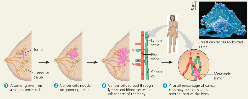
Metastasis:
The spread of cancer cells to locations distant from their original site
How does radiation affect cancer cells?
A tumor that appears to be localized may be treated with high-energy radiation, which damages DNA in cancer cells much more than DNA in normal cells, apparently because the majority of cancer cells have lost the ability to repair such damage
How does chemotherapy affect cancer cells?
To treat known or suspected metastatic tumors, chemotherapy is used, in which drugs that are toxic to actively dividing cells are administered through the circulatory system
What are some other effects of chemotherapy?
It interferes will the cell cycle, so they may stop actively dividing. Also, it can affect intestinal cells, hair follicle cells, and immune system cells leading to nausea, hair loss, and susceptibility to infection respectively.
Interpreting Histograms
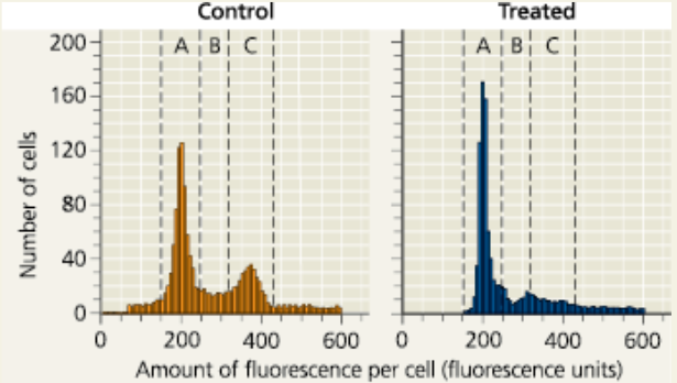
In Figure 9.14, why do the nuclei resulting from experiment 2 contain different amounts of DNA?
The nucleus on the right was originally in the G1 phase; therefore, it had not yet duplicated its chromosomes. The nucleus on the left was in the M phase, so it had already duplicated its chromosomes.
What phase are most of your body cells in?
Most body cells are in a nondividing state called G0
Compare and contrast a benign tumor and a malignant tumor
Both types of tumors consist of abnormal cells, but their characteristics are different. A benign tumor stays at the original site and can usually be surgically removed; the cells have some genetic and cellular changes from normal, non-tumor cells. Cancer cells from a malignant tumor have more significant genetic and cellular changes, can spread from the original site by metastasis, and may impair the functions of one or more organs
What would happen if you performed the experiment in Figure 9.17 with cancer cells?
The cells might divide even in the absence of PDGF. In addition, they would not stop when the surface of the culture vessel was covered; they would continue to divide, piling on top of one another
Explain the significance of the G1 and M checkpoints and the go-ahead signals involved in the cell cycle control system
Checkpoints allow cellular surveillance mechanisms to determine whether the cell is prepared to go to the next stage. Internal and external signals move a cell past these checkpoints. The G1 checkpoint, called the “restriction point” in mammalian cells, determines whether a cell will complete the cell cycle and divide or switch into the G0 phase. The signals to pass this checkpoint often are external—such as growth factors. Regulation of the cell cycle is carried out by a molecular system, including proteins called cyclins and other proteins that are kinases. The signal to pass the M phase checkpoint is not activated until all chromosomes are attached to kinetochore fibers and are aligned at the metaphase plate. Only then will sister chromatid separation occur.