


Anatomy
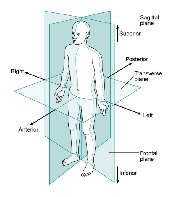
Important Terminology
Inferior - below or further away from the head
Superior - above or nearer to the head
Proximal - nearer to where a limb attaches to the body
Distal - further away from where a limb attaches to the body
Posterior – behind or nearer to the back
Anterior - the front or nearer to the front
Internal - located inside or further away from the surface
External - located on or near the surface
Lateral - further away from the midline of the body
Medial - closer to the midline of the body
Skeletal System
Axial Skeleton (80 bones)
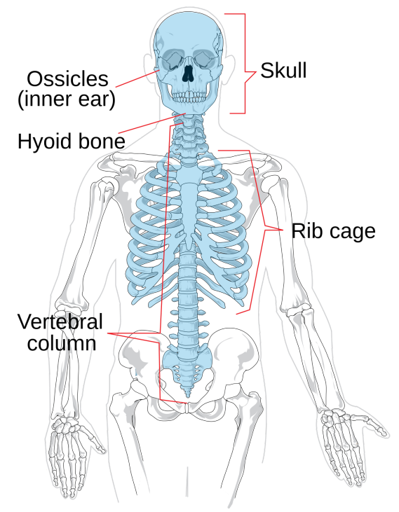
Skull
Protects the brain, forms the orbit of the eyes, attachment to muscles, and structure to the face.
Ribs/Thoracic Cage
Protects and supports the internal organs of the body such as the heart and lungs and some of the abdominal organs like kidneys and liver.
12 pairs of ribs
1-7 → true ribs
Directly attached to the sternum
8-10 → false ribs
Indirectly attached to sternum
11-12 → floating ribs
Not attached to the sternum
Sternum
A flat bone that starts at the bottom of the throat and runs to about halfway down the centre of the chest.
Vertebral Column (33)
Supports the spinal cord and supports the head. It provides articulation sites for ribs and innominate bones of the pelvic girdle. It is also responsible for the flexibility of the back.
Cervical Vertebrae (7)
Smallest vertebrae
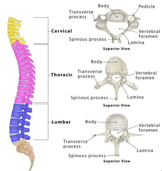
More movement than thoracic and lumbar vertebrae
Thoracic Vertebrae (12)
Restricts movement
Ribs are attached to the side of each vertebrae
Lumbar Vertebrae (5)
The biggest and strongest of the vertebrae
Plays a major role in weight-bearing
Sacral Vertebrae (5)
Transmits weight from body to pelvis and legs
Coccygeal Vertebrae (4)
The bone at the end of the spinal column that is composed of four vertebrae combined into one bone
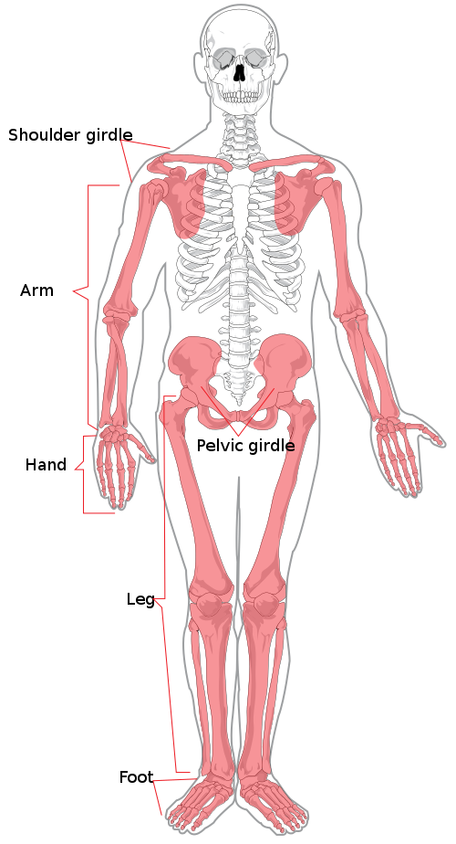
Appendicular Skeleton (126 bones)
Pectoral (Shoulder) Girdle
Functions to anchor and support the upper limbs serve as an important attachment site for many muscles that help to move the arms.
Pelvic (Hip) Girdle
Supports and protects the soft vital organs of the abdominal cavity, transfers the weight of the upper axial skeleton to lower appendicular parts, especially during body movement, and provides attachment to the lower limbs.
Upper Extremity/Arms (humerus, ulna, radius, carpal bones, metacarpals, and phalanges)
Helps in the hand movement to perform various activities, and helps the shoulder to perform a wide range of motions.
Lower Extremity/Legs (femur, tibia, fibula, tarsal bones, metatarsals, and phalanges)
Weight-bearing bones that support the entire structure of the body while walking, jumping, or running.
Types of Bones:
Long bones usually have a long cylindrical shaft and are enlarged at both ends; can be large or small, but the length is always greater than the width; most important bones for movement.
They include the femur, metatarsals, and clavicle
Short bones are small and cube-shaped, and they usually articulate with more than one other bone.
Short bones include the carpals of the hand and tarsals of the foot.
Flat bones usually have curved surfaces and vary from being quite thick to very thin; provide protection, and the broad surfaces also provide a large area for muscle attachment.
Flat bones include the sternum, scapula, ribs, and pelvis
Irregular bones have specialized shapes and functions.
Irregular bones include the vertebrae, sacrum, and coccyx.
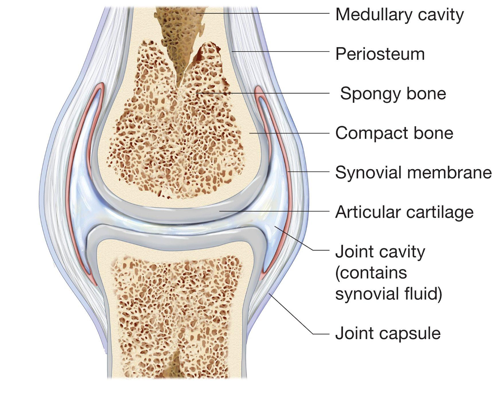
Parts of Long Bone
Epiphysis: Two end partitions of a long bone, each covered by articular cartilage.
Diaphysis: Compact part of a long bone; a long shaft covered by a periosteum membrane. Important for protection.
Periosteum: Membrane of a long bone for protection.
Spongy Bone: A type of bone tissue found at the ends of long bones and in the middle of other bones such as the vertebrae. It is lighter and less dense than compact bone; and contains red bone marrow, which is responsible for producing blood cells.
Articular Cartilage: Smooth, white tissue that covers the ends of bones where they come together to form joints, helps to reduce friction, and absorbs shock.
Bone Marrow: Soft fatty substance in the cavities of bones, in which blood cells are produced; RED → produces blood cells and platelets. Yellow marrow → stores fat
Compact Bone: The external layer of the bone that is very dense, filled with passageways for nerves, blood vessels, and the lymphatic system.
Marrow Cavity: Space within the diaphysis where yellow marrow is stored for white cell production.
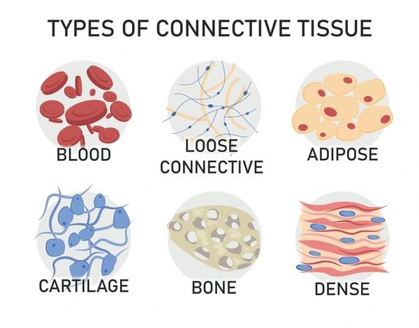
Connective Tissue
Functions:
to join bodily structures like bones and muscles to one another or hold tissues like muscles, tendons, or even organs in their proper place in the body.
gives reinforcement to joints, strengthening and supporting the articulations between bones.
transports nutrients and metabolic by-products between the bloodstream and the tissues to which it adheres.
Structure:
made up of proteins like collagen, elastin, and intercellular fluid.
the form can range from a thin sheet to a dense rope of fibers.
Joints
Also known as an articulation; where two or more bones come into contact or articulate with each other.
Different Types of Joints
Fixed Joints
Very stable, with no observable movement, bones are joined by strong fibers called sutures.
Cartilaginous Joints
Allows slight movement, the ends of the joint are covered with white pads of fibrocartilage, which act as shock absorbers.
Synovial Joints
The most common type of joint that allows a wide range of movement and is subdivided according to movement possibilities, is characterized by the presence of a joint capsule and cavity lined with a synovial membrane.
Synovial Joints
Features
Ligament
Structure: A band of strong fibrous connective material.
Function: Joins bone to bone, and provides stability.
Pads of fat
Found between capsule, bone, or muscle.
Increases joint stability, acts as a shock absorber, and reduces friction.
Meniscus Tough
Flexible discs of fibrocartilage.
Improves fit between the bone ends, increases stability, and reduces wear and tear to joint surfaces.
Bursae Fluid
A filled sac is found between the tendon and bone.
Reduces friction, found in body areas of high stress.
Articular Cartilage
Smooth and spongy covers of the end of bones
Prevents friction between articulating bones
Synovial Fluid
Slippery fluid that fills the joint capsule.
Reduce friction, nourish cartilage, and get rid of waste from the joint.
Layered Joint Cavity
Outer layer – tough and fibrous
Inner layer – synovial membrane covers all internal surfaces
Strengthen joint, secrete synovial fluid
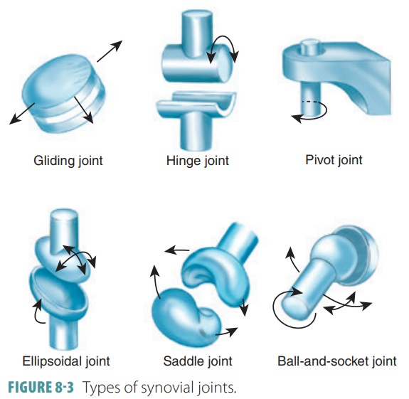
Types
Gliding
Usually flat or slightly curved, slide across each other, with the least amount of movement.
Hinge
The articular surfaces have been fused so movement in one direction, joined by ligaments, movement is only allowed in one plane (extension/flexion).
Pivot
The rounded surface of one bone that rolls around a ring formed by bone and ligament.
Condyloid
A ball-shaped bone that fits into a cup.
Saddle
Saddle-shaped bone that fits into a bone shaped like the legs, and can move up, down, side to side.
Ball and Socket
A sphere-shaped bone that fits into a rounded cavity, covered in cartilage to prevent friction and a high range of movements.
Muscular System
Origin: the attachment of a muscle tendon to a stationary bone, usually the most proximal attachment Insertion: the attachment of a muscle tendon to a moveable, usually the most distal attachment
Characteristics:
Contractility: The ability of the muscle to contract and generate a force when it is stimulated by a nerve
Extensibility: The ability to extend before its normal resting state.
Elasticity: The muscle's ability to return to its original resting length.
Atrophy: Muscle wastage, lack of physical activity, poor nutrition, and disease.
Hypertrophy: Growth and increase in the size of the muscle, most commonly as a result of weight training.
Nerve Stimuli: A nerve that sends a signal for the muscle to contract.
Fed by capillaries: Gaseous exchange that occurs in the capillaries so oxygen can be delivered to the muscles.
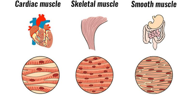
Types of Muscles:
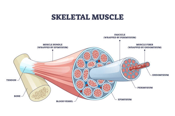
Skeletal muscle: Under voluntary control, has a striated appearance, has tendons that attach the muscle to the bone, and the main function is to move the skeleton.
Cardiac muscle: Under involuntary control, striated, heart muscle.
Smooth muscle: Lines the walls of the blood vessels and hollow organs such as the stomach or intestines, involuntary control, not striated.
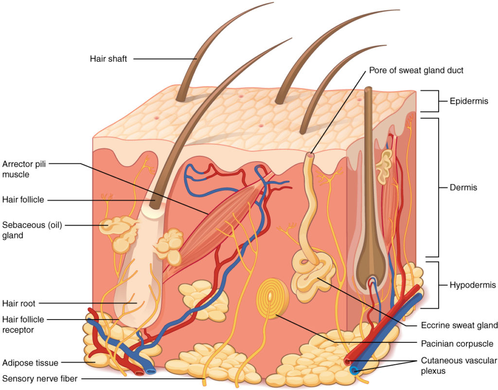
Integumentary System
Functions:
Regulation of Body Temperature: if it is cold, hairs on the skin will stand up and blood flow in the capillaries is decreased.
If it is warm, hair muscle relaxes so heat can escape; Also, sweat is secreted which cools us down.
Protection and Immunity: The skins form a physical barrier through specialized cells of the immune system.
These cells detect bacteria and viruses, and they are called antibodies.
Sensation: Sensation is a feeling that is localized on the skin’s surface.
They are processed through receptors in the dermis.
Excretion: Sweat glands remove waste such as urea, uric acid, and ammonia and help regulate body temperature when overheating.
Synthesis of Vitamin D: From the sunlight, we need vitamin D to aid with calcium, iron, magnesium phosphate, and zinc absorption through the liver and kidney.
The epidermal cells convert ultraviolet rays into vitamin D.
Nervous System
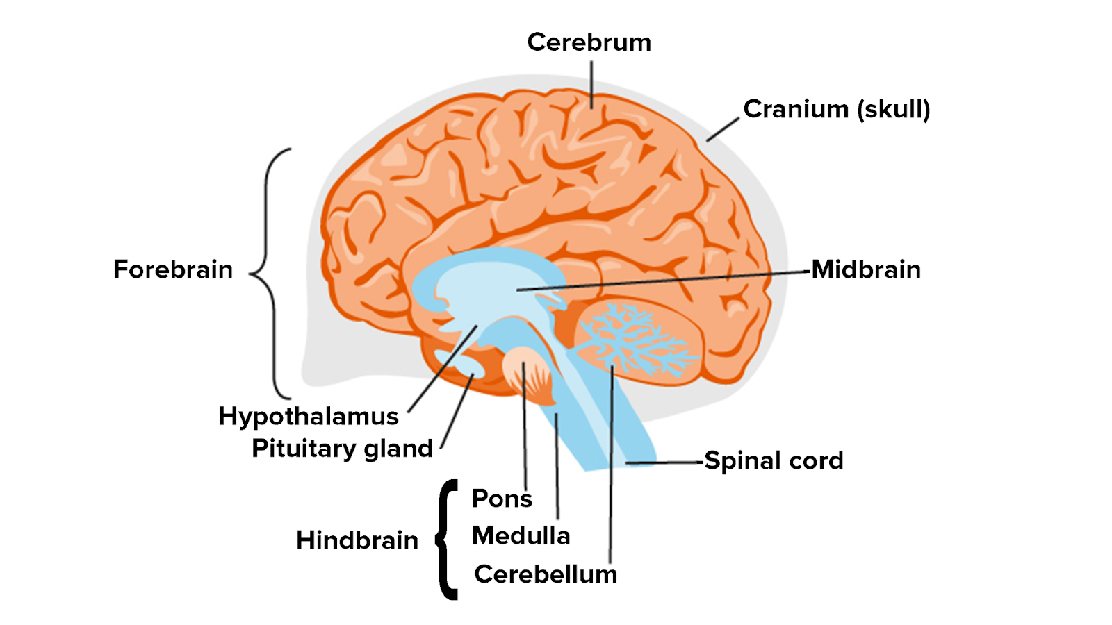
Functions of Parts of the Brain:
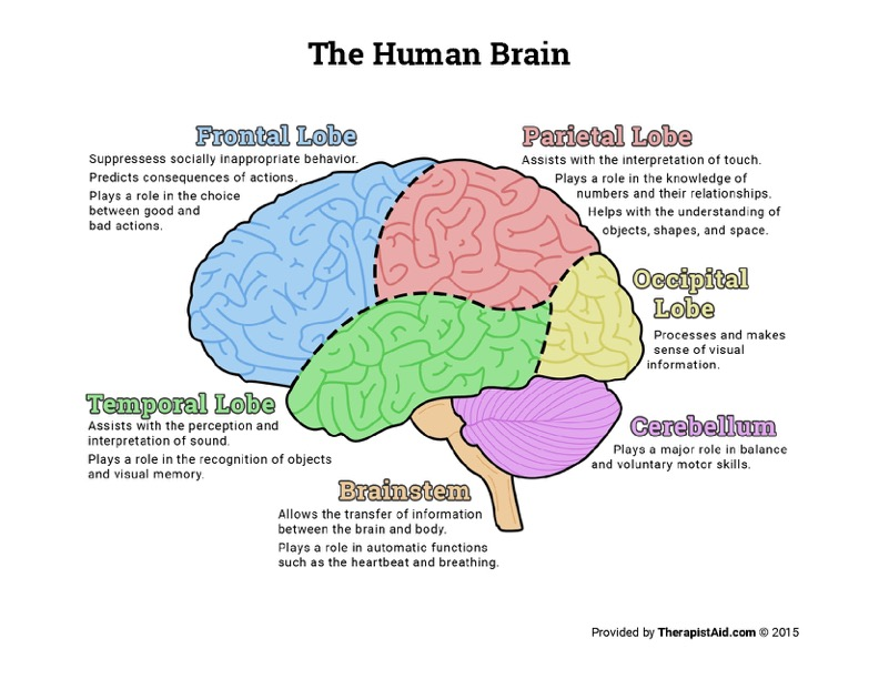
Brain Stem
Location: Posterior part of the brain linking to the top of the spinal cord.
Control center for the regulation of cardiac and respiratory function, consciousness, and sleep cycle.
Vehicle for sensory information.
Made up of the medullary oblongata, pons, and midbrain.
Medullary Oblongata: Centre for respiration and circulation; regulates breathing, heart, and blood vessel function.
Pons: Links brainstem to spinal cord.
Midbrain: Links brain to spinal cord.
Thalamus
Relays motor and sensory signals from the cerebral cortex.
Involved in cognition, pain, temperature, pressure, and sensation in general.
Hypothalamus
Controls the autonomic nervous system (ANS) and helps to maintain your internal balance.
Regulates heart rate, blood pressure, the pituitary gland, body temperature, appetite, thirst, fluid and electrolyte balance and circadian rhythms.
Cerebrum
The largest part of the brain
Responsible for high-level brain functions
Ex. thinking, language, emotion, and motivation
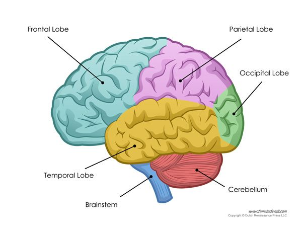
Cerebellum
Top of the brain stem
Receives information from the sensory system
Responsible for:
Coordinating movements
Regulating balance and posture
Allowing skilled motor activities to be carried out
Frontal Lobe
Directly behind the forehead
Largest lobe in the human brain
The most common region of injury
Primary Function:
Behavior and Emotional Control Centre
Important for voluntary movement, expressive language, and managing higher-level executive functions ]→ cognitive skills
Controls:
Personality/Emotions
Intelligence
Attention/Concentration
Judgment
Body movement
Problem-solving
Speech
Damage or injury can cause:
Loss of movement (paralysis)
Repetition of a single thought
Unable to focus on tasks
Mood swings/Irritability/Impulsiveness
Changes in social behavior and personality
Difficulty problem solving
Difficulty with language – unable to get words out (aphasia)
Parietal Lobe
Near the back and top of the head
Informs about objects in our external environments through touch and the position and movement of body parts.
Responsible for integrating sensory input, and the construction of a spatial system to represent the world around us.
Controls:
Sense of touch, pain, and temperature
Distinguishing size, shape, and color
Spatial perception
Visual perception
Damage or injury can cause:
Difficulty drawing objects
Difficulty distinguishing left from right
Spatial disorientation and navigation difficulties
Problems reading
Lack of awareness of certain body parts or surrounding space
Inability to focus visual attention
Difficulty with complex movement
Occipital Lobe
At the back of the head
Controls:
Vision
Responsible for visual perception including color, form, and motion.
Damage or injury can cause:
Difficulty locating objects in the environment
Difficulty identifying colors
Production of hallucinations
Visual illusions – inaccurately seeing objects
Word blindness
Difficulty reading and writing
Temporal Lobe
Behind the ears and is the second largest lobe
Controls:
Speech (understanding language)
Memory
Hearing
Sequencing
Organization
Process auditory information and encode memory.
Plays an important role in processing affect/emotions, language, and certain aspects of visual perception.
The dominant temporal lobe (left side for most) → helps to understand language. learning and remembering verbal information.
The non-dominant → learning and remembering non-verbal information.
Damage or injury can cause:
Difficulty understanding spoken words
Disturbance with selective attention
Difficulty identifying and categorizing objects
Impaired factual and long-term memory
Persistent talking
Difficulty recognising faces
Increased or decreased interest in sexual behavior
Emotional Disturbance
Limbic Lobe
Top of the brain stem and under the cerebral cortex
Controls:
Emotional processing
Behavior
Motivation
Long term memory
It is involved in many emotions and motivations, especially those related to survival.
Processing emotions such as fear, anger
Emotions related to sexual behavior
Blood Supply
The brain needs oxygen + nutrients.
Cerebral Arteries: Posterior supply (basilar artery) in the cerebellum and the anterior supply in the cerebrum.
Communicating Arteries: Surround the pituitary gland and make up the ‘Circle of Willis’ – this allows the brain to receive blood and nutrients from either the carotid or vertebral arteries.
Carotid Artery
Internal
Origin: Subclavian artery
Supply: Blood to the cerebrum.
Anterior supply to the brain ascends to three branches and reduces the risk of circulation interruption as there are two supplies.
External
Origin: Bifurcation of the common carotid artery
Branches: Split into 5 arteries e.g., facial artery, occipital artery
Supply: Blood to the face, scalp, base of the skull, and neck.
Vertebral Artery
Origin: Branches of the 1st part of the subclavian artery.
Course: Ascends posterior to the internal carotid artery in the transverse foramina of the cervical vertebrae branches.
numerous small branches
radicular/spinal branches
Posterior inferior cerebellar artery (PICA)
Termination: Combines with the contralateral vertebral artery to form the basilar artery.
Blood-Brain Barrier
It protects the brain from foreign substances that could injure it.
Protects it from hormones and neurotransmitters.
Maintains a constant environment for the brain (homeostasis).
The highly selective barrier separates circulating blood from the brain’s extracellular fluids in the Central Nervous System.
Energy
The brain’s main sources of energy are glucose and oxygen → travel from the blood to the brain cells.
Glucose and oxygen help make ATP within the brain through the process of aerobic respiration.
Adenosine triphosphate (ATP): nucleotide which is vital for brain function because it enhances the delivery of nutrients and oxygen to the brain and stimulates the removal of waste products such as glucose and oxygen.
Glucose
Glucose is a simple carbohydrate that provides fuel for the brain.
Glucose travels into the brain cells from the blood through the process of diffusion.
The supply of glucose is continuous because carbohydrate storage is limited.
The energy from glucose is crucial for communication activity inside the brain, as well as for maintaining memory function.
Oxygen
Used by the brain to perform its functions.
Needed for brain growth and healing.
The brain requires 3x as much oxygen as the muscles.
Supplied to the brain cells through the blood via diffusion.
Supply is always continuous.
The effect of low glucose or oxygen levels:
Without a constant supply of glucose and oxygen, the brain is unable to function properly. If blood entering the brain is low on either glucose or oxygen, can suffer from:
Mental confusion
Dizziness
Convulsions
Loss of consciousness