Chapter 17: The Cell Cycle
Overview of the Cell Cycle
The cell cycle is a highly regulated process that ensures proper cell growth, DNA replication, and cell division, leading to the production of two daughter cells with identical genetic material.
The cell cycle is the series of events that occur in a cell leading to its division and the production of two daughter cells.
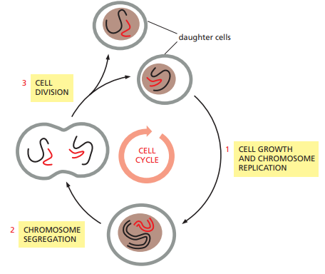
It consists of distinct phases, including interphase and mitotic phase.
Interphase
Interphase is the longest phase of the cell cycle, where the cell grows, carries out its normal functions, and replicates its DNA.
Interphase is further divided into three stages: G1 (Gap 1), S (Synthesis), and G2 (Gap 2).
G1 phase is characterized by cell growth, protein synthesis, and preparation for DNA replication.
S phase involves DNA replication, where the genetic material is duplicated, resulting in two identical copies of the genome.
G2 phase is a period of growth and preparation for cell division, during which the cell checks the accuracy of DNA replication and repairs any errors.
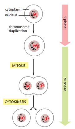
Mitotic Phase
The mitotic phase (M phase) is the shortest phase and involves the actual division of the cell into two daughter cells.
The M phase is subdivided into four stages: prophase, metaphase, anaphase, and telophase.
During prophase, the chromatin condenses into visible chromosomes, the nuclear envelope breaks down, and the mitotic spindle begins to form.
In metaphase, the chromosomes align at the equator of the cell, facilitated by microtubules attached to the kinetochores.
Anaphase is characterized by the separation of sister chromatids, which are pulled to opposite poles of the cell by the shortening of microtubules.
Telophase marks the end of mitosis, where the nuclear envelope reforms around the separated chromosomes, and the cell prepares for cytokinesis.
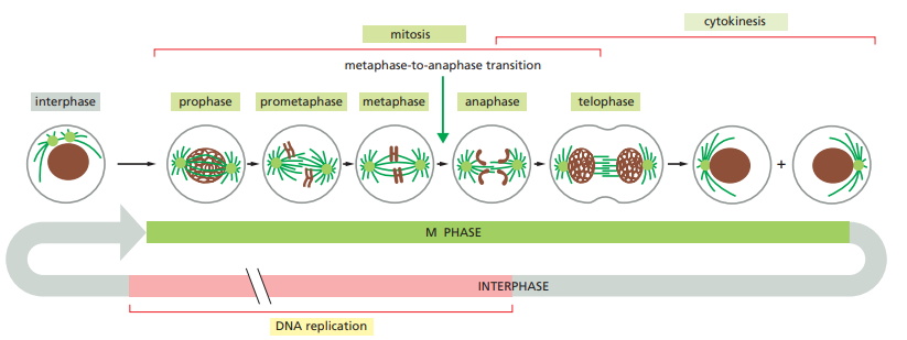
Cytokinesis
Cytokinesis is the final stage of the cell cycle, where the cytoplasm divides, and two daughter cells are formed.
Cytokinesis differs in animal and plant cells, with animal cells undergoing cell cleavage through the formation of a contractile ring, while plant cells form a cell plate to divide the cytoplasm.
The completion of cytokinesis marks the end of one cell cycle and the beginning of the next, resulting in the formation of two genetically identical daughter cells.
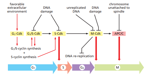
Cell-Cycle Control System
The cell-cycle control system is a complex network of molecular checkpoints and regulatory mechanisms that ensure the orderly progression of the cell cycle.
It functions to monitor and regulate the timing and coordination of cell-cycle events, including DNA replication, chromosome segregation, and cell division.
The cell-cycle control system consists of various proteins, enzymes, and signaling pathways that interact to regulate the cell cycle.
Key components of the control system include cyclins, cyclin-dependent kinases (CDKs), and checkpoints.
Cyclins are proteins that fluctuate in concentration during the cell cycle and bind to specific CDKs to activate them.
CDKs are enzymes that regulate the progression of the cell cycle by phosphorylating target proteins involved in cell-cycle events.
Checkpoints are critical control points that monitor the integrity of DNA, ensure proper replication and repair, and verify accurate chromosome alignment.
Checkpoints include the G1 checkpoint (restriction point), G2 checkpoint, and spindle assembly checkpoint (SAC) during mitosis.
At each checkpoint, the cell-cycle control system assesses the conditions and signals from the environment and other cellular processes before deciding whether to proceed or halt the cell cycle.
If the cell-cycle control system detects DNA damage, replication errors, or other abnormalities, it can activate DNA repair mechanisms or induce cell-cycle arrest to allow time for repair or prevent the transmission of damaged DNA.
Failure of the cell-cycle control system can lead to uncontrolled cell growth, genomic instability, and the development of cancer.
Abnormalities or mutations in cell-cycle control genes, such as tumor suppressor genes (e.g., p53) or oncogenes (e.g., cyclins, CDKs), can disrupt the normal functioning of the control system.
Studying the cell-cycle control system provides insights into the mechanisms underlying cell division, DNA replication, and cell-cycle regulation, with implications for understanding diseases like cancer and potential targets for therapeutic interventions.
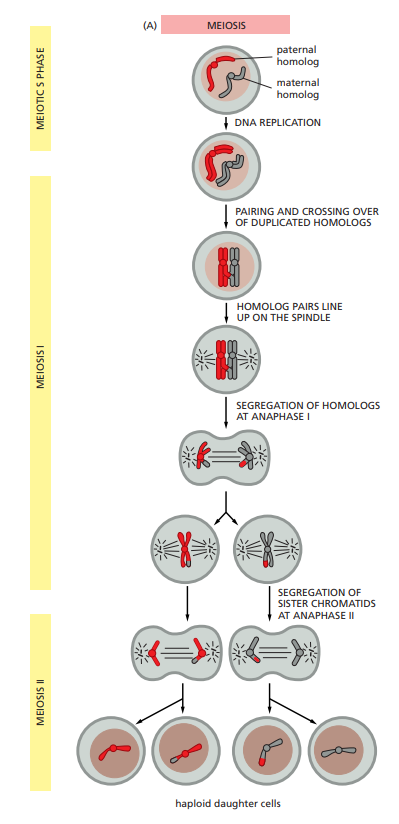
Meiosis
Meiosis is a specialized form of cell division that occurs in sexually reproducing organisms.
It involves the division of a diploid (2n) cell into four haploid (n) daughter cells, resulting in the production of gametes (sperm and eggs).
Meiosis consists of two successive divisions, known as meiosis I and meiosis II.
Meiosis I is the reduction division, where homologous chromosomes pair up and exchange genetic material through a process called crossing over.
Crossing over leads to genetic recombination and promotes genetic diversity among offspring.
During meiosis I, homologous chromosomes separate, resulting in the formation of two haploid cells with duplicated chromosomes (consisting of sister chromatids).
Meiosis II is similar to mitosis, where the sister chromatids of each chromosome separate, resulting in the formation of four haploid daughter cells.
The four daughter cells produced at the end of meiosis are genetically distinct due to independent assortment of chromosomes during meiosis I and the random segregation of sister chromatids during meiosis II.
Meiosis I
Meiosis I is the first division of meiosis and consists of several distinct steps, including prophase I, metaphase I, anaphase I, and telophase I.
Prophase I is the longest phase of meiosis I and can be further divided into five sub-stages: leptotene, zygotene, pachytene, diplotene, and diakinesis.
During leptotene, the chromosomes condense, becoming visible as thread-like structures, and the nuclear envelope starts to break down.
In zygotene, homologous chromosomes pair up and form structures called synaptonemal complexes. This pairing is called synapsis.
Pachytene is characterized by the crossing over of genetic material between non-sister chromatids of homologous chromosomes. This genetic exchange increases genetic diversity.
Diplotene is marked by the separation of homologous chromosomes but with the physical connection points known as chiasmata still present. This is when the synaptonemal complexes dissolve partially.
During diakinesis, the chromosomes further condense, and the nuclear envelope completely disintegrates. The spindle apparatus starts to form.
Metaphase I is the stage where homologous chromosome pairs line up along the equator of the cell, called the metaphase plate.
The orientation of the chromosomes is random, which contributes to genetic diversity through a process called independent assortment.
Anaphase I follows metaphase I, and it involves the separation of homologous chromosomes, with one member of each pair moving towards opposite poles of the cell.
Telophase I marks the end of meiosis I, where the chromosomes arrive at the poles and start decondensing. Nuclear envelopes may form around each group of chromosomes, and cytokinesis typically occurs, resulting in the formation of two haploid daughter cells.
Meiosis II
Meiosis II is the second division of meiosis and follows meiosis I. It is similar to a mitotic division, consisting of four stages: prophase II, metaphase II, anaphase II, and telophase II.
Prophase II initiates with the condensation of the chromosomes, and the nuclear envelope disintegrates.
The centrosomes, containing the centrioles, duplicate and move towards opposite poles of the cell, forming the spindle apparatus.
Metaphase II is the stage where individual chromosomes line up along the equator of the cell, called the metaphase plate.
Unlike meiosis I, the chromosomes align individually in metaphase II, without homologous pairs.
Anaphase II follows metaphase II and is characterized by the separation of sister chromatids. The connections between the sister chromatids break, and each chromatid is pulled towards opposite poles of the cell.
Telophase II marks the end of meiosis II, where the chromosomes arrive at the poles and start decondensing.
Nuclear envelopes may form around each set of chromosomes, and the spindle apparatus disintegrates.
Cytokinesis typically occurs, resulting in the division of the cytoplasm, and the formation of four haploid daughter cells.
Each daughter cell produced at the end of meiosis II contains a single set of chromosomes, making them genetically distinct from one another and the parent cell.
Meiosis II completes the process of meiosis, resulting in the production of four haploid daughter cells from the original diploid (2n) parent cell.