


Chapter 17: Identification of Urine, Sweat, Fecal Matter, and Vomitus
17.1: Identification of Urine
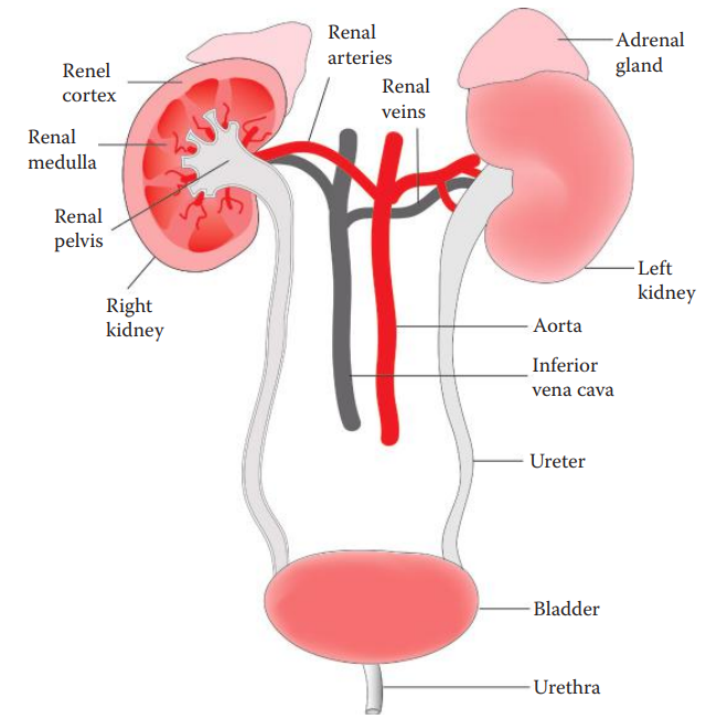
Urine Formation
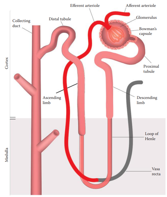
Nephron: The basic structural and functional unit of the kidney.
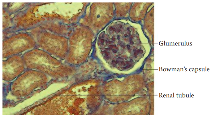
Each nephron is composed of a glomerulus, a Bowman’s capsule, and a renal tubule.
Glomerulus: It is formed by a network of capillaries and is surrounded by a Bowman’s capsule.
Filtration is the first step in urine formation.
As blood flows through the glomeruli, much of its fluid, except cells and large molecules, is filtered through the capillaries into the Bowman’s capsule.
Glomerular filtrate: The preliminary form of urine, consists of water, salts glucose, and waste products such as urea.
The filtrate is then passed through the renal tubule where reabsorption occurs.
During the reabsorption process, water, glucose, nutrients, and ions such as sodium are reabsorbed back into the blood.
Secretion: It is the last process of urine formation. It occurs at the distal and the collecting tubules of the nephron where ions ammonia, and certain metabolites are secreted from the blood into the lumen of the renal tubule to be eliminated in the urine.
Urine: An aqueous solution consisting largely of water and waste products.
Urea: The most abundant waste product in urine, resulting from the elimination of ammonia that is produced from the metabolic process of amino acids.
The average urea concentration in human urine is approximately 9 g/L.
Presumptive Assays
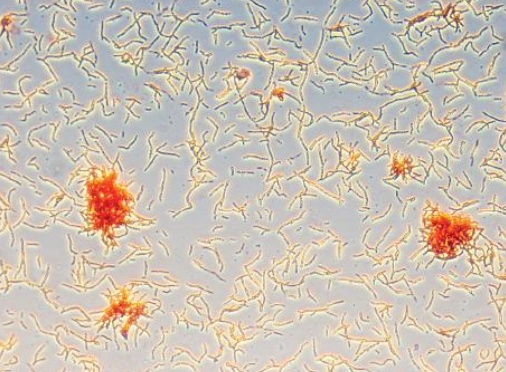
In forensic identification, urine stains can be located by visual examination based on the characteristic yellow color of urine and the detection of the distinctive odor of urine stains.
Under alternative light sources, urine stains emit a fluorescent light that facilitates the locating of urine stains in clothing and bedding.
Chemical analysis can be carried out to detect the major inorganic anions in urine such as phosphate and sulfate as well as the major organic compounds in urine such as urea, creatinine, and uric acid.
Identification of Urea
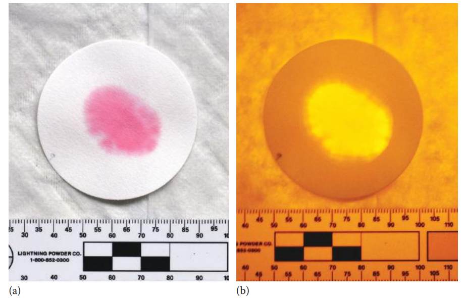
The para-dimethylaminocinnamaldehyde (DMAC) assay is simple and rapid and is the most commonly used presumptive assay for the forensic identification of urine stains.
In the colorimetric method, a portion of a stain (~1 cm2) is cut and is extracted with 1 mL of distilled water. The extraction is transferred onto a piece of filter paper and is allowed to dry.
One drop of 0.1% DMAC solution is then added to the filter paper. DMAC reacts specifically with urea, if present, producing a pink-colored product.
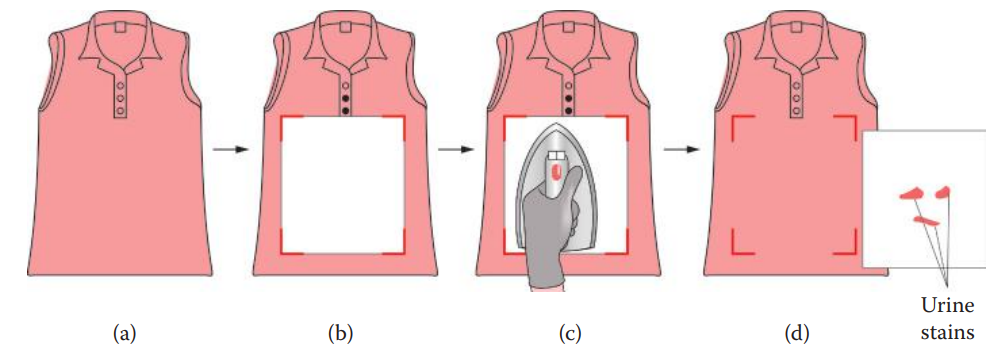
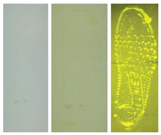
The fluorometric method is useful for locating urine stains on large pieces of evidence such as clothing and bedding.
This method can detect patterns of urine stains which can be useful in crime scene reconstructions.
In the fluorometric method, the evidence to be examined, such as a garment, is covered by a sheet of filter paper that has been preabsorbed with the DMAC solution. The evidence and the filter are then wrapped together in a sheet of aluminum foil and are left overnight in a press, ensuring that the evidence is in close contact with the filter paper.
Using a light source at 473–548 nm, the DMAC-treated urine stain fluoresces. The fluoresced stain is best observed with a 549 nm filter.
However, colored fabrics interfere with the assay since dyes and pigments can inhibit the fluorescence.
Identification of Creatinine
Creatinine is produced during normal muscle cell metabolism.
During this metabolism, phosphocreatine, an energy-storing molecule in muscle cells, breaks down to form creatine.
Creatine is then metabolized to creatinine, which is released from the muscle cells into the blood. Serum creatinine is largely filtrated by the renal glomeruli.
A small amount of creatinine is secreted by the renal distal tubules. The amount of creatinine excreted in urine is proportional to the muscle mass of an individual.
The creatinine present in urine can be detected using the Jaffe color test.
In this test, picric acid is used to convert creatinine, under alkaline conditions, to form creatinine picrate, which is a bright red product.
Confirmative Assays
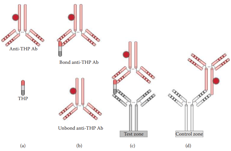
Identification of Tamm–Horsfall Protein
Tamm–Horsfall protein (THP): Also known as uromodulin, is the most abundant protein in urine, and accounts for 40% of the urine proteins.
THP is exclusively synthesized in the epithelial cells of Henle’s loop. THP is secreted from the apical plasma membrane of the epithelial cells into the lumen.
THP can be detected using an ELISA and RSID-Urine.
RSID-Urine: It utilizes a polyclonal rabbit antibody that is specific to THP. This test is rapid and simple and thus can be used as a screening test in laboratories and as a field test at crime scenes to identify urine.
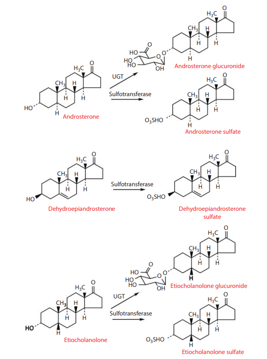
Identification of 17-Ketosteroids
Androsterone: A steroid hormone with a weak potency of testosterone.
DHEA: A metabolic intermediate in the biosynthesis of the gonadal steroids. It also has a potential function as a steroid hormone.
Etiocholanolone: A metabolite of testosterone.
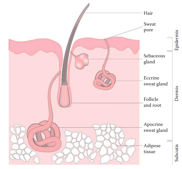
17.2: Identification of Sweat
Biology of Perspiration
Eccrine sweat glands: Are distributed almost all over the body and are controlled by the sympathetic nervous system. It plays a role in regulating body temperature.
Apocrine sweat glands: Are associated with hair follicles, are usually restricted to the underarm and genital areas and are controlled by emotional stress.
Sweat Identification Assays
Sweat evidence has been analyzed using presumptive assays such as elemental analysis using scanning electron microscopes coupled with energy dispersive x-ray spectroscopy in the detection of lactic acid.
Raman microspectroscopy is potentially useful for the identification of sweat for forensic purposes, which is based largely on the profiles of lactate, lactic acid, urea, and single amino acids in urine.
Dermcidin: Identified as a potential biomarker of human sweat. It belongs to a class of human antimicrobial peptides of the innate immune defense system and plays an important role in protecting epithelial barriers from infections.
The detection of dermcidin in sweat stains can be performed using ELISA assays utilizing antibodies specific to human dermcidin.
Dermcidin is encoded by the DCD gene. Its mRNA can be detected using reverse transcription polymerase chain reaction (RT-PCR) assays that can detect DCD mRNA in 10 ìL of sweat sample.
17.3: Identification of Fecal Matter
Fecal Formation
Feces: Are a type of waste matter that is the direct result of food that has been processed by the digestive system.
Human feces contain undigested foodstuffs, sloughed intestinal epithelial cells, intestinal bacteria, bile pigments, electrolytes, and water.
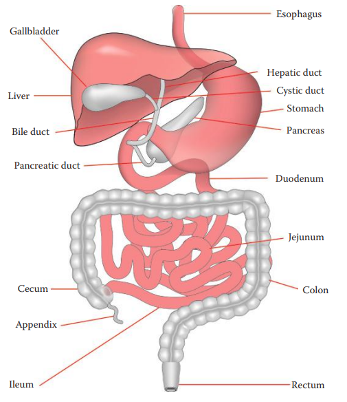
Feces are formed in the intestines during the last phase of digestion. Feces first enter the colon in liquid form.
Most of the nutrients are absorbed on the surface area of the small intestine. In the large intestine, water, sodium, and chloride are absorbed on the surface of the lumen.
Food stays for approximately 2–6 h in the stomach. It takes an additional 3–5 h to travel through the small intestine and 12–24 h to travel through the large intestine.
Fecal Matter Identification Assays
Macroscopic and Microscopic Examination
The color and odor of human feces are useful characteristics for fecal identification.
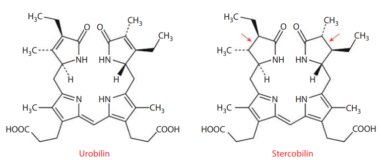
The normal brown color of feces primarily results from the presence of urobilinoids, which are heme catabolic by-products.
The characteristic odor of feces is caused by the metabolic by-products of the intestinal bacterial flora.
Indole, skatole, and hydrogen sulfide are the compounds that are responsible for the odor of feces.
Urobilinoids Tests
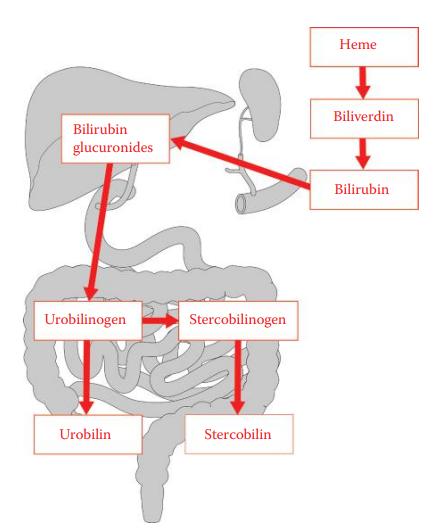
Urobilinoids: These are generated from the degradation of heme and are excreted into feces.
Erythrocytes are continuously undergoing hemolysis in which erythrocytes are naturally broken down and are usually processed in the reticuloendothelial system of the spleen.
Hemoglobin is released daily during the hemolysis process and is degraded into heme, globin, and iron.
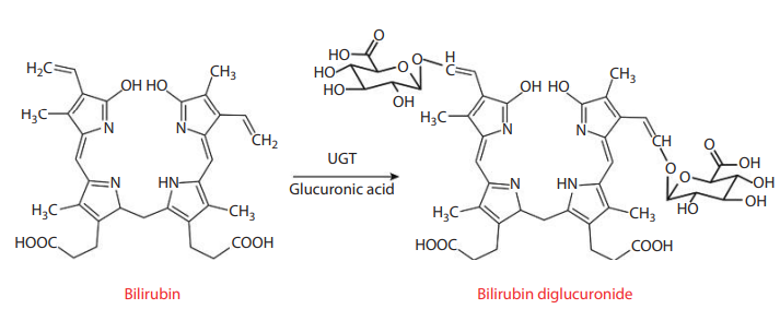
In the peripheral tissues, heme undergoes catabolism to form bilirubin. Bilirubin is further converted to urobilinogen in the intestine. A portion of urobilinogen is reduced to stercobilinogen.
In the Schlesinger test, a sample is mixed with saturated zinc acetate in an ethanol solution to form aurobilinoid–zinc chelation complex that emits a characteristic green fluorescence under ultraviolet light.
The Edelman test is a variation of the Schlesinger test. A sample is treated with a mercuric salt solution to yield a pink-colored compound. Further treatment with a zinc salt produces fluorescence.
17.4: Identification of Vomitus
Biology of Gastric Fluid
Gastric fluid can be found in stains derived from stomach wounds.
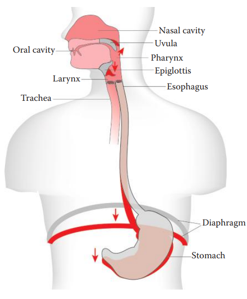
Vomiting: The forceful expulsion of the contents of the stomach through the mouth. It is usually preceded by salivation, sweating, and the sensation of nausea.
Vomiting usually begins with a deep inhalation and the closure of the glottis.
Subsequent contractions of the diaphragm and the abdominal muscles compress the stomach.
The gastric contents are then forced upward through the relaxed sphincters and the esophagus and are expelled through the mouth.
Since the glottis is closed, vomitus usually does not enter the respiratory tract.
Although the uvula is usually raised to close the nasal cavity, vomitus sometimes enters the nose.
The stomach stores ingested food until it can be emptied into the small intestines.
When food enters the stomach, hydrochloric acid is secreted in large quantities, which facilitates the initial degradation of proteins.
The stomach also secretes mucus that lubricates the gastric surface to protect the epithelium from acidic environments.
Hormones regulate acid secretion and gastric movement.
A number of enzymes are secreted into the gastric fluid, including lipase, which plays a role in lipid hydrolysis, and gelatinase, which can hydrolyze gelatin.
The stomach also secretes pepsinogens, which are enzyme precursors, into the gastric fluid. In the stomach, pepsinogens are activated by hydrochloric acid into pepsin, which is largely responsible for the digestion of proteins.
Vomitus Identification Assays
Vomitus is highly acidic and tends to be malodorous.
Fresh blood in the vomit is usually bright red and suggests bleeding due to injuries, while dark red blood clots suggest bleeding in the stomach due to pathological conditions such as an ulcer.
The presence of gastric fluid in vomitus can also be identified by the detection of pepsins secreted from the stomach. This identification test is based on the proteolytic activity of pepsins.
Pepsins are endopeptidases that cleave primarily on peptide bonds in the middle of the protein.
Aromatic amino acids such as tryptophan, phenylalanine, and tyrosine, are the preferred targeted amino acids for the cleavage reaction by pepsins.
In the pepsin-proteolytic assay, fibrin blue is used as a substrate for pepsin.
Fibrin Blue: An insoluble protein–dye complex that is colorless.
In the presence of vomitus, pepsin cleaves fibrin blue and releases a chromophore that is soluble in water and exhibits a blue color.
In the assay, a fibrin blue–containing agarose gel is utilized. The sample from vomitus is loaded onto the gel plate. After incubation, a blue ring around the sample can be observed as a result of the enzymatic reactivity of pepsin.
This method can determine the pepsin content of fresh and aged forensic samples.