
1. Intro to Imaging & Digital Image Processing
Defining Imaging
Modality: The difference between different bioimaging methods and machines (ex: CT and MRI are different ______)
Four necessary components of a modality: source (illumination), camera (detector), digitizer (frame grabber), imaging processing unit
Imaging processing unit (hardware and/or software)
acquisition (takes in the data and understands it)
preprocessing (combines information from multiple points)
segmentation (identifying components, facial recognition)
& more
An image is a 2D representation of a physical quantity as rendered by an imaging modality
X-ray attenuation (projection x-ray yor CT)
Proton density (MRI)
Acoustic reflectivity (ultrasound)
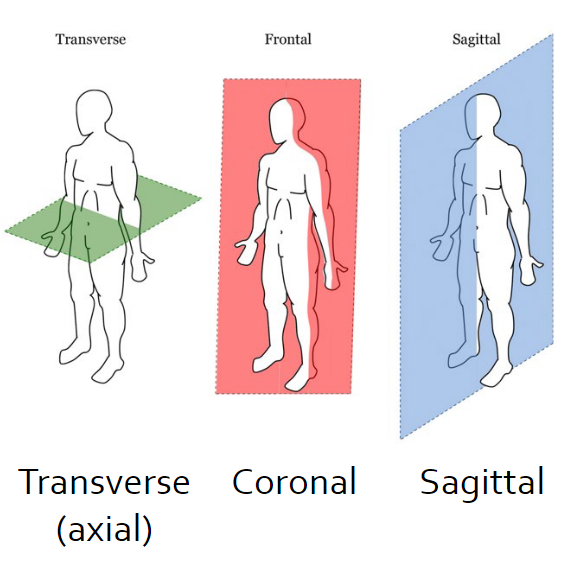
An image represents a “finite-thickness” plane within a volume of interest
Types of imaging
Anatomical: Imaging that represents structure/composition of objects (e.g. CT imaging)
Functional: Imaging that represents function/physiology of an organ (e.g. PET scans)
Projection: Imaging that shoes a single planar representation (e.g. x-ray)
Tomographic: Imaging that shows cross-sectional representation (e.g. CT imagings)
Imaging mechanisms
Transmission: The imaging mechanism by which information comes from what travels through the body (e.g. x-ray)
Reflection: Transmission: The imaging mechanism by which information that comes from what reflects back from the body (e.g. ultrasound)
Modality Comparison
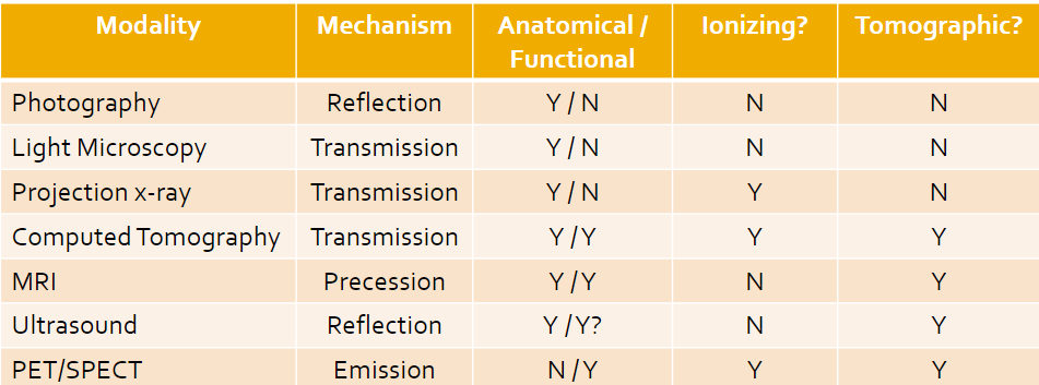
Ionizing is when the energy we work with is higher than others, and electrons in the atoms can bump up to unsafe levels; this is something we want to avoid (can lead to cancer)
Digital Imaging
Digital images are digital files saved on a computer
2D arrays of “picture elements” called pixels
Voxels are for 3D elements
Image size = width x height
Real-world image size (or FOV) is (Ncolumn x pixel width) x (Nrow x pixel height)
Resolution: Number of pixels per square inch
Image Pixels
Addressed with x,y coordinates
Top left corner is (1, 1)
(coumn, row)
Storage type
Pixel values depend on the storage type
Grayscale images are NOT called black and white
8-Bit: Greyscale images with values from 0 to 2^8 minus 1
16-Bit: Greyscale images with values from 0 to 2^16 minus 1
Color images: each pixel can have 4 values
1 value per pixel – e.g. indexed image
3 values per pixel e.g. 3x1 bytes – R_G_B, 3x2 bytes – R_G_B, ...
4 values per pixel RGB e.g. 4x1 byte – R_G_B_Alpha, ...
Image Processing
Enhancement/restoration of image info for human reading
Segmentation
Characterization
Representation of images for machine analysis
Visualization
Processing of image data for storage
Processing of image data for transmission
Matlab
Load the image and info
imread()
iminfo()
Display image
imtool()
imshow()
imagesec()
image()
imshowpair()
Perform needed operation
Display and evaluate results
Save resulting image
imwrite()