
CHAPTER 1
The structure and functions of the skeleton
Muscular skeletal system : The name used to describe the muscular skeletal system and the muscular system working together.
There are 206 bones in the human body.
There are 4 different types of bones
Long bones – to enable gross (large) movements
Short bones – to enable finer controlled movements
Flat bones – often large and protect vital organs
Irregular bones - specifically shaped to protect
Articulating bones : Bones that meet at a joint to enable movement
Shoulder : scapula, clavicle, humerus
Elbow : humerus, radius, ulna
Hip : pelvis, femur
Knee : femur, patella, fibula, tibia
Ankle : tibia, fibula, talus
Functions of the skeleton
The skeleton is rigid supporting the framework of bones inside the body, to which all the soft tissues and organs are attached.
Together the bones and muscles form a machine which can perform many different tasks.
There are 6 main functions of the skeleton
Support – without support we would be unable to move and be a mass of soft tissue.
Eg: our vertebrae supports the head
Protection – The hard nature of the bones means the skeleton can protect the more delicate parts of the body (vital organs). This reduces the chance of injury allowing players to continue to train and play sports.
Eg: our ribcage protects our lungs, cranium protects the soft tissue of the brain
Movement - The skeleton is joined to allow us to move when the muscles attached to them contract and pull on the bone
Eg: The bones and joints work with muscles to enable us to walk, jog and sprint
Shape and structure – Without the skeleton we would not be able to move. The skeleton provides something for the muscles to attach to
Eg: the bones in the legs support the body, the vertebrae supports the head
Produce blood cells – Red and white bloods cells are made of bone marrow which is found at the ends of the femur and the humerus and also at the ribs ,sternum, pelvis and vertebrae. Red blood cells are important for aerobic activity as they help oxygen transportation around the body. White blood cells are fight off infections, and platelets help blood to clot following an injury
Storage of minerals – Bones act as reservoirs for vital minerals such as calcium and phosphorus. These are essential for major body functions. Their role in physical activity is linked to the general health of the athlete, which affects sporting performance.
Joints
A joint is where two or more bones meet and muscles act together to cause movement
The human skeleton is jointed to allow movement
Muscular contraction causes the bones to move about the joints
Synovial joints are otherwise known as freely-movable joints
These are the largest group of joints found in the body, eg: hips, shoulders, elbow, ankle and knees
Types of synovial joints -
Ball and socket joints – These are the most moveable joints in the body. These can move away from the body, back towards the body and can also rotate.
Eg: Hips and shoulders
Hinge joints – Hinge joints can only move towards or away from the body like a hinge on a door.
Eg: Knee, ankle, elbow
Movement at synovial joints – Different types of synovial joints allow different kinds of movement…
Extension : Straightening or extending a limb
Eg: the arm can be extended at the elbow
Flexion : Bending or flexing a limb
Eg: the leg can be flexed at the knee
Abduction – Moving a limb away/towards from the centre line of the body.
Eg : the leg can be moved away from the centre of the body at the hip
Adduction – Moving a limb towards the centre line of the body
Eg : the arm can be moved towards the centre line of the body at the shoulder.
Rotation – A circular movement around a joint.
Circumduction : Movement of a bone or a limb in a circular pattern a combination of flexion, extension, adduction and abduction
Eg : the shoulder when swimming butterfly
Plantar Flexion : movement at the ankle joint that point the toes
Eg : in the ankle
Dorsi-flexion : movement at the ankle that flexes the foot upwards
The structure and function of the muscular system
Tendons
A tendon is connective tissue that attaches muscle to bone
The role of a tendon is to transfer the effort created by a contracting muscle to the bone, resulting in movement of that bone
Muscles at the 5 main joints -
Shoulder - Deltoid, Trapezius, Pectorals, Latissimus dorsi, Biceps, Triceps, Rotator cuff
Elbow – Biceps, Triceps
Hip - Gluteals, Hip flexors
Knee – Quadriceps, Hamstrings
Ankle – Gastrocnemius, Tibialis anterior
Antagonistic Pairs
Muscles can only PULL not push and are arranged in pairs on either side of joints
These pairings of muscles are known as antagonistic pairs
The muscle that tenses is the agonist (prime mover)
The muscle that eccentrically contracts is the antagonist
Muscles pull by contracting, they cannot push to produce the opposite movement
These muscles make up obvious antagonistic pairs
Biceps and triceps - (acting at the elbow to create flexion and extension)
Hip flexors and gluteals - (acting at the hip to create flexion and extension)
Hamstring group and quadriceps groups- (acting at the knee to create flexion and extension)
Tibialis anterior and gastrocnemius (acting at the ankle to create dorsiflexion and plantar flexion)
Types of Muscular Contraction
Isometric – Do not create movement, the muscle neither shortens nor lengthens it remains the same Eg : To support a weight in a stationary position/ To hold the body in a particular position eg handstand, plank, arabesque
Isotonic – These create movement. The muscle length changes when it contracts, resulting in limb movement
Concentric – When the muscle contracts and shortens
Eccentric – When the muscle contracts and lengthens
The structure and function of the cardio-respiratory system
The respiratory system – This brings oxygen into the body so it can be used to produce energy and enable activity. it then gets rid of carbon dioxide a waste product which is produced in the muscles during exercise.
Key terms
Cardio-respiratory- The name used to describe the respiratory system and the cardiovascular system working together
Circulatory system – Heart, blood vessels, and blood
Respiratory System – Lungs and airways
Functions – Enable the body to breathe, pump blood and oxygen around the body
Gaseous exchange – The process where oxygen from the air in the alveoli moves into the blood in the capillaries, while carbon dioxide moves from the blood in the capillaries into the air in the alveoli.
Haemoglobin - The protein found in red blood cells that transports oxygen as oxyhaemoglobin and carbon dioxide around the body
Oxyhaemoglobin - A chemical formed when haemoglobin bonds to oxygen
Alveoli - Small air sacks in the lungs where gas exchange takes place. Very thin, one cell thick, provides a moist and extremely large surface area for gaseous exchange to occur. Numerous capillaries run across the alveoli, ensuring a large blood supply to the area
Capillaries - A network of microscopic blood vessels. They are only one cell thick.
Diffusion Pathway - The distance travelled during diffusion. The diffusion pathway is short in gaseous exchange
Oxygen that has been breathed in passes through the alveoli and into the red blood cells in the capillaries
In the capillaries the oxygen combines with haemoglobin to form oxyhaemoglobin and is then carried around the body
At the same time haemoglobin carries carbon dioxide from the body to the capillaries
The carbon dioxide in the capillaries passes through the alveoli and is breathed out
Mechanics of breathing
Inhalation
The diaphragm contracts. It flattens and moves downwards
The intercostal muscles contract, raising the ribs and pushing out the sternum, making the chest cavity larger
The lungs increase in size and the air pressure inside the lungs is reduced
The air pressure outside the body is now higher than inside the body. Air travels from areas of high concentration to areas of low concentration, so air is pulled into the lungs
Exhalation
The diaphragm relaxes. It moves back up into a dome shape.
The intercostal muscles relax, lowering the ribs and dropping the sternum, making the chest cavity smaller.
The lungs reduce in size and the air pressure inside the lungs is increased.
The air pressure outside the body is now lower than the air pressure inside the body. Air travels from areas of high concentration to areas of low concentration, so air leaves the lungs.
What happens to your breathing during exercise?
The lungs expand and contract more as more air is inhaled to supply more oxygen to the working muscles and more air is exhaled to remove the increased amount of carbon dioxide produced by the working muscles.
More muscles are involved in breathing when you exercise. The sternocleidomastoid, paired muscles in the side of the neck, assist in raising the sternum when you inspire. The abdominals pull the rib cage down more quickly, forcing air out quickly, when you expire.
Spirometer Trace
A spirometer is a piece of equipment that measures the air capacity of the lungs
It is a way of recording and drawing these volumes. The pattern of the trace will change as the amount of air you inspire and expire changes as a result of exercise.
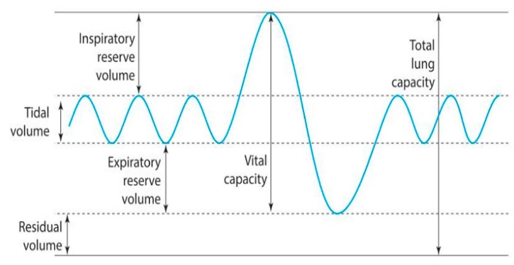 A spirometer measures the 5 following volumes associated with the lungs
Tidal volume – The normal amount of air inhaled or exhaled per breath
Expiratory reserve volume – The amount of air that can be forced out after tidal volume (after normal expiration)
Inspiratory reserve volume – The amount of air that can be forced in after tidal volume (a normal inspiration)
Residual volume - The amount of air that remains in the lungs after maximal expiration
Structure of the heart
KEY TERMS
Pulse – the rhythmic throbbing that you can feel as your arteries pump blood around the body. You can measure your heart rate using your pulse.
Backflow - the flowing backwards of blood for. Valves in the veins prevent back flow.
Diastole – the phase of the heartbeat when the chambers of the heart relax and filled with blood.
Systole – the phase of the heart between the chambers of the heart contract and empty of blood; when blood is ejected from the heart.
Cardiac cycle – one cycle of diastole and systole is called the cardiac cycle.
Blood pressure – the pressure that blood is under. The systolic reading measures the pressure the blood is under when the heart contracts. The dear stolice reading measures the pressure the blood is under when the heart relaxes
Blood Vessels
Arteries
Thick muscular walls with a small internal diameter - carry oxygenated blood away from the heart under high pressure
Arteries do not have valves
Your pulse is located in your arteries as they pulse as blood is carried through them
Can vasodilate and vasoconstrict
Veins
Thinner walls with a larger internal diameter - blood pressure is low in the veins
Veins carry deoxygenated blood back to the heart
Veins contain valves that open due to the pressure of the blood flow and close to make sure that the blood does not flow backwards so there is no back-flow
Capillaries –
Microscopic blood vessels that link the arteries to the veins
walls are very thin just one cell thick - tell out oxygen and carbon dioxide to pass through them during gaseous exchange
 deoxygenated blood becomes oxygenated at the capillaries
Pathway of blood
Deoxygenated blood enters the right atrium from the superior vena cava and inferior vena cava
It then passes into the right atrium, through a valve and into the right ventricle.
The pulmonary artery transports deoxygenated blood to the lungs.
Gaseous exchange occurs, resulting in oxygenated blood.
The pulmonary vein transports oxygenated blood from the lungs to the left atrium.
It then passes through a valve to the left ventricle.
Oxygenated blood is ejected from the heart and is transported to the body via the aorta.
Cardiovascular system
The cardiovascular system carries blood around the blood.
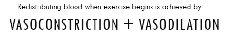
How is blood redistributed during exercise?
During exercise the body alters its priorities. When the body is resting most of the blood is directed towards the organs, but during exercise most of the blood is directed towards the voluntary muscles. This ensures that voluntary muscles are able to work aerobically, which is the most efficient. Redistributing blood when exercise begins is achieved by vasodilation and vasoconstriction, which changes the internal diameter of the arteries that supply the body with blood. Vasoconstriction is the narrowing of the internal diameter of a blood vessel to restrict the volume of the blood travelling through it. This is so less blood is delivered to inactive areas. Vasodilation is when the internal diameter of a blood vessel widens to increase the volume of blood travelling through it. The arteries dilate more during exercise so more blood is delivered to active areas therefore increasing their oxygen supply
Cardiac Cycle
DIASTOLE | SYSTOLE |
The chambers of the heart RELAX and FILL with blood. | When the chambers of the heart CONTRACT and empty, when blood is EJECTED from the heart. |
One cycle of diastole and systole is called the CARDIAC CYCLE |
Blood pressure
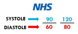 Two readings are taken when blood pressure is measured.
The pressure that the blood is under when contracting and ejecting the blood from the heart – SYSTOLIC PRESSURE.
The pressure that the blood is under when the heart relaxes (and fills with blood) – DIASTOLIC PRESSURE.
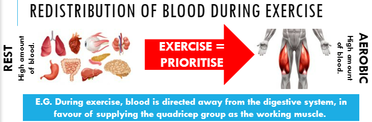 Redistribution of blood during exercise
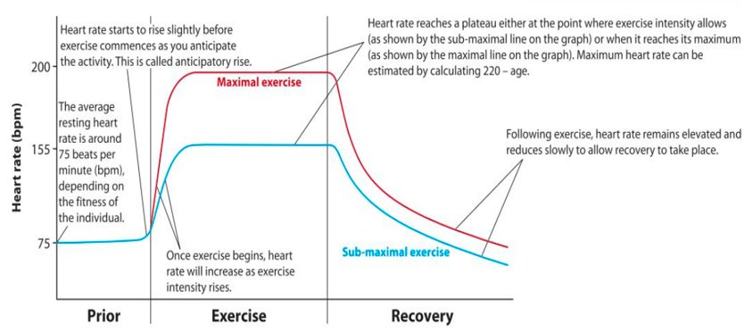
Heart Rate
The number of times the heart beats in one minute
Units = beats per minute (bpm)
Resting heart rate = 60-80 bpm
Maximum heart rate = 220 – your age =
Stroke volume
The amount of blood pumped out of the left ventricle each beat
Unit = Milliliters (ml)
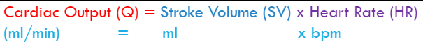 Cardiac output
Aerobic and anaerobic exercise
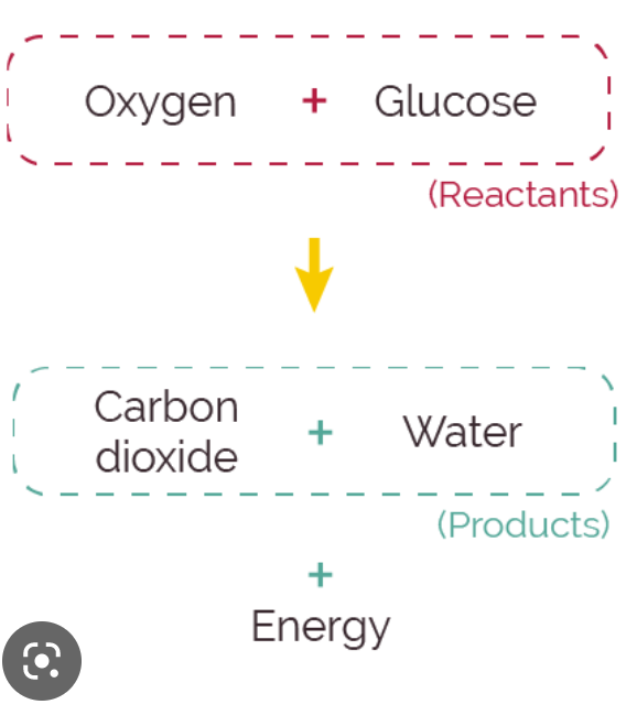 Aerobic exercise – Working at a low to moderate intensity so then the body has time to use oxygen for energy production and can work for a long amount of time
Aerobic exercise takes place in the presence of oxygen
When exercise is over a long period of time, not too fast and steady, the heart can supply all the oxygen the working muscles need
When working aerobically the energy comes from carbohydrates
These carbs are converted into glucose and oxygen
Intensity – The amount of energy needed to complete an activity. Working at a high intensity requires a large amount of energy. Working at a low intensity requires less energy
Sporting examples include – long distance running, endurance cycling, long distance swimming
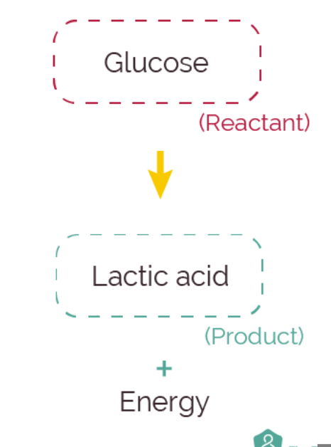 Anaerobic exercise - Working for short periods of time at a high intensity without oxygen for energy production
Takes place in the absence of enough oxygen
The heart and lungs cant supply enough blood and therefore oxygen to the working muscles
Glucose is converted to energy without the presence of oxygen
Sporting examples include – short distance sprinting (100m,200m), short distance swimming (25m,30m)
Excess Post-exercise Oxygen Consumption (EPOC) – The amount of oxygen needed to recover after exercise. It is characterised by an increased breathing after exercise
The recovery process
Cool down
Gradually reduces the intensity of the exercise
Helps maintain an elevated breathing rate/ heart rate
Ensures blood continues to flow quickly to the muscles
Replenishes working muscles with oxygen
Helps the body convert lactic acid produced anaerobic exercise into glucose, carbon dioxide and water so your not stiff
Manipulation of diet
Drink water
Drink water to rehydrate to replace fluids lost during exercise (sweat)
How much water you need to drink depends on the intensity levels and duration of the exercise that was done
Air temperature, humidity, and altitude effect how much water you need
Carbohydrate loading
Carbohydrate loading increases the amount of carbs eaten which maximises the amount of glucose in the body
More carbs = more glucose (body can meet better demands of the performance)
Limits the length and severity of the recovery period
Boosts performance
Timing of protein intake
Important for power athletes to increase strength and speed
Consuming protein after training
Provides the body with nutrients it needs to heal tears quickly and build muscle
Protein helps repair small tears that occur (hypertrophy)
Ice baths or massages
Immediately after exercise
Helps prevent DOMS (Delayed Onset Muscle Soreness)
The muscle soreness you feel a day after intense exercise
The effects of exercise
Immediate effects
Heart rate increases – as your heart works harder to deliver oxygen to the working muscles
Breathe more deeply and frequently – as your body delivers more oxygen to working muscles
Feel hotter – body temperature increases
Sweat and skin will redden – this occurs as part of the body’s temperature control system
Short term effects (24-36 hours)
Feel nauseous
Feeling light headed
Muscle ache/cramps
DOMS (if exercise was intense)
Feel tired/ fatigued
Fatigue – Physical fatigue is a feeling of extreme or severe tiredness due to a build-up of lactic acid in the muscles or working for a long period of time
The long term effects of exercise (occurs after months or years)
Stamina will improve – this us your ability to exercise
Muscles will increase in size and produce greater strength – when a muscle is strained, small tears are created. As they heal, they become thicker. This is called hypertrophy
Heart will increase in size. This is called cardiac hypertrophy and your cardiac output increases. Your heart is able to deliver more blood and more oxygen to your working muscles, removing more carbon dioxide and other waste products like lactic acid
Resting heart rate is lower (Bradycardia) – This is if you have a resting heart rate under 60 bpm
Your body will change shape (for the better) – Exercise helps keep body weight down
Improvements in specific components of fitness – increase in strength, muscular endurance, cardiovascular endurance, improvement in speed and flexibility
Hypertrophy – the enlargement of an organ or tissue caused by an increase in the size of its cells . When a muscle is trained, small tears are created. As they heal, they become thicker and increase in size