


Chapter 12: The Nervous System and Nervous Tissue
The Central and Peripheral Nervous Systems

The central nervous system (CNS) is the brain and spinal cord, and the peripheral nervous system (PNS) is everything else.
A glial cell is one of a variety of cells that provide a framework of tissue that supports the neurons and their activities.
Neurons are cells and therefore have a soma, or cell body, but they also have extensions of the cell; each extension is generally referred to as a process.
There is one important process that every neuron has called an axon, which is the fiber that connects a neuron with its target.
Dendrites are responsible for receiving most of the input from other neurons.
These two regions within nervous system structures are often referred to as gray matter (the regions with many cell bodies and dendrites) or white matter (the regions with many axons).
But white matter is white because axons are insulated by a lipid-rich substance called myelin.
A localized collection of neuron cell bodies in the CNS is referred to as a nucleus.
In the PNS, a cluster of neuron cell bodies is referred to as a ganglion.
A bundle of axons, or fibers, found in the CNS is called a tract whereas the same thing in the PNS would be called a nerve.
Basic Functions
The nervous system can be divided into regions that are responsible for sensation (sensory functions) and for the response (motor functions).
Some regions of the nervous system are termed integration or association areas.
Sensation
The first major function of the nervous system is sensation—receiving information about the environment to gain input about what is happening outside the body (or, sometimes, within the body).
The sensory functions of the nervous system register the presence of a change from homeostasis or a particular event in the environment, known as a stimulus.
Response
The nervous system produces a response on the basis of the stimuli perceived by sensory structures.
An obvious response would be the movement of muscles, such as withdrawing a hand from a hot stove, but there are broader uses of the term.
The nervous system can cause the contraction of all three types of muscle tissue.
Responses also include the neural control of glands in the body as well, such as the production and secretion of sweat by the eccrine and merocrine sweat glands found in the skin to lower body temperature.
Integration
Stimuli that are received by sensory structures are communicated to the nervous system where that information is processed.
This is called integration.
Controlling the Body
The somatic nervous system (SNS) is responsible for conscious perception and voluntary motor responses.
The autonomic nervous system (ANS) is responsible for involuntary control of the body, usually for the sake of homeostasis (regulation of the internal environment).
The enteric nervous system (ENS) is responsible for controlling the smooth muscle and glandular tissue in your digestive system.
Parts of a Neuron
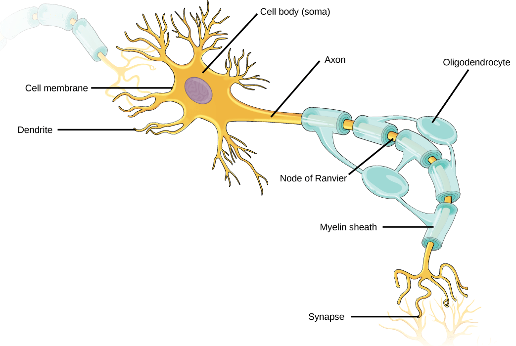
The other processes of the neuron are dendrites, which receive information from other neurons at specialized areas of contact called synapses.
Where the axon emerges from the cell body, there is a special region referred to as the axon hillock.
Within the axon hillock, the cytoplasm changes to a solution of limited components called axoplasm.
Because the axon hillock represents the beginning of the axon, it is also referred to as the initial segment.
Each gap is called a node of Ranvier and is important to the way that electrical signals travel down the axon.
The length of the axon between each gap, which is wrapped in myelin, is referred to as an axon segment.
At the end of the axon is the axon terminal, where there are usually several branches extending toward the target cell, each of which ends in an enlargement called a synaptic end bulb.
Types of Neurons

Unipolar cells have only one process emerging from the cell.
Bipolar cells have two processes, which extend from each end of the cell body, opposite to each other.
Multipolar neurons are all of the neurons that are not unipolar or bipolar.
Glial Cells

Glial cells, or neuroglia or simply glia, are the other type of cell found in nervous tissue.
They are considered to be supporting cells, and many functions are directed at helping neurons complete their function for communication.
The name glia comes from the Greek word that means “glue,” and was coined by the German pathologist Rudolph Virchow, who wrote in 1856: “This connective substance, which is in the brain, the spinal cord, and the special sense nerves, is a kind of glue (neuroglia) in which the nervous elements are planted.”
Glial Cells of the CNS
One cell providing support to neurons of the CNS is the astrocyte, so named because it appears to be star-shaped under the microscope (astro- = “star”).
Those processes extend to interact with neurons, blood vessels, or the connective tissue covering the CNS that is called the pia mater.
Some ways in which they support neurons in the central nervous system are by maintaining the concentration of chemicals in the extracellular space, removing excess signaling molecules, reacting to tissue damage, and contributing to the blood-brain barrier (BBB).
Also found in CNS tissue is the oligodendrocyte, sometimes called just “oligo,” which is the glial cell type that insulates axons in the CNS. The name means “cell of a few branches” (oligo- = “few”; dendro- = “branches”; -cyte = “cell”).
Microglia are, as the name implies, smaller than most of the other glial cells.
The ependymal cell is a glial cell that filters blood to make cerebrospinal fluid (CSF), the fluid that circulates through the CNS.
Ependymal cells line each ventricle, one of four central cavities that are remnants of the hollow center of the neural tube formed during the embryonic development of the brain.
The choroid plexus is a specialized structure in the ventricles where ependymal cells come in contact with blood vessels and filter and absorbcomponents of the blood to produce cerebrospinal fluid.
Glial Cells of the PNS
One of the two types of glial cells found in the PNS is the satellite cell.
The second type of glial cell is the Schwann cell, which insulate axons with myelin in the periphery.
Myelin
Myelin is a lipid-rich sheath that surrounds the axon and by doing so creates a myelin sheath that facilitates the transmission of electrical signals along the axon.
Myelin sheaths can extend for one or two millimeters, depending on the diameter of the axon.
The Function of Nervous Tissue
Found in the skin of your fingers or toes is a type of sensory receptor that is sensitive to temperature, called a thermoreceptor.
The amount of change is dependent on the strength of the stimulus (how hot the water is). This is called a graded potential.
The voltage at which such a signal is generated is called the threshold, and the resulting electrical signal is called an action potential.
The action potential travels—a process known as propagation—along the axon from the axon hillock to the axon terminals and into the synaptic end bulbs. When this signal reaches the end bulbs, it causes the release of a signaling molecule called a neurotransmitter.
The target of this neuron is another neuron in the thalamus of the brain, the part of the CNS that acts as a relay for sensory information.
The thalamus then sends the sensory information to the cerebral cortex, the outermost layer of gray matter in the brain, where conscious perception of that water temperature begins.
The upper motor neuron is in this region, called the precentral gyrus of the frontal cortex, which has an axon that extends all the way down the spinal cord.
At the level of the spinal cord at which this axon makes a synapse, a graded potential occurs in the cell membrane of a lower motor neuron.
Electrically Active Cell Membranes
Channels for anions (negative ions) will have positively charged side chains in the pore. This is called electrochemical exclusion, meaning that the channel pore is charge-specific.
Because of the surrounding water molecules, larger pores are not ideal for smaller ions because the water molecules will interact, by hydrogen bonds, more readily than the amino acid side chains. This is called size exclusion.
Some ion channels are selective for charge but not necessarily for size, and thus are called a nonspecific channel.
Some are opened by certain events, meaning the channels are gated.
A ligand-gated channel opens because a signaling molecule, a ligand, binds to the extracellular region of the channel.This type of channel is also known as an ionotropic receptor because when the ligand, known as a neurotransmitter in the nervous system, binds to the protein, ions cross the membrane changing its charge
A mechanically gated channel opens because of a physical distortion of the cell membrane.
A voltage-gated channel is a channel that responds to changes in the electrical properties of the membrane in which it is embedded.
A leakage channel is randomly gated, meaning that it opens and closes at random, hence the reference to leaking.
The Membrane Potential
The electrical state of the cell membrane can have several variations. These are all variations in the membrane potential.
A potential is a distribution of charge across the cell membrane, measured in millivolts (mV).
With the ions distributed across the membrane at these concentrations, the difference in charge is measured at -70 mV, the value described as the resting membrane potential.
The Action Potential
Resting membrane potential describes the steady state of the cell, which is a dynamic process that is balanced by ion leakage and ion pumping.
The resting potential is the state of the membrane at a voltage of -70 mV, so the sodium cation entering the cell will cause it to become less negative.
This is known as depolarization, meaning the membrane potential moves toward zero.
Repolarization, meaning that the membrane voltage moves back toward the -70 mV value of the resting membrane potential.
One is the activation gate, which opens when the membrane potential crosses -55 mV. The other gate is the inactivation gate, which closes after a specific period of time—on the order of a fraction of a millisecond.
While an action potential is in progress, another one cannot be initiated.
That effect is referred to as the refractory period.
There are two phases of the refractory period: the absolute refractory period and the relative refractory period.
Propagation of the Action Potential
Propagation along an unmyelinated axon is referred to as continuous conduction; along the length of a myelinated axon, it is saltatory conduction.
Much as water runs faster in a wide river than in a narrow creek, Na+ -based depolarization spreads faster down a wide axon than down a narrow one.
This concept is known as resistance and is generally true for electrical wires or plumbing, just as it is true for axons, although the specific conditions are different at the scales of electrons or ions versus water in a river.
Graded Potentials
Graded potentials can be of two sorts, either they are depolarizing or hyperpolarizing.
Types of Graded Potentials
For the unipolar cells of sensory neurons—both those with free nerve endings and those within encapsulations—graded potentials develop in the dendrites that influence the generation of an action potential in the axon of the same cell. This is called a generator potential.
For other sensory receptor cells, such as taste cells or photoreceptors of the retina, graded potentials in their membranes result in the release of neurotransmitters at synapses with sensory neurons. This is called a receptor potential.
A postsynaptic potential (PSP) is the graded potential in the dendrites of a neuron that is receiving synapses from other cells.
Depolarization in a postsynaptic potential is called an excitatory postsynaptic potential (EPSP) because it causes the membrane potential to move toward threshold.
Hyperpolarization in a postsynaptic potential is an inhibitory postsynaptic potential (IPSP) because it causes the membrane potential to move away from threshold.
Summation
All types of graded potentials will result in small changes of either depolarization or hyperpolarization in the voltage of a membrane.
These changes can lead to the neuron reaching threshold if the changes add together, or summate.
Spatial summation is related to associating the activity of multiple inputs to a neuron with each other.
Temporal summation is the relationship of multiple action potentials from a single cell resulting in a significant change in the membrane potential.
Synapses
In a chemical synapse, a chemical signal—namely, a neurotransmitter—is released from one cell and it affects the other cell.
In an electrical synapse, there is a direct connection between the two cells so that ions can pass directly from one cell to the next.
Neurotransmitter Systems
The first group, which is a neurotransmitter system of its own, is the cholinergic system. It is the system based on acetylcholine.
The cholinergic system has two types of receptors, the nicotinic receptor is found in the NMJ as well as other synapses.There is also an acetylcholine receptor known as the muscarinic receptor.
Another class of neurotransmitter is the biogenic amine, a group of neurotransmitters that are enzymatically made from amino acids.
A neuropeptide is a neurotransmitter molecule made up of chains of amino acids connected by peptide bonds.
A metabotropic receptor involves a complex of proteins that result in metabolic changes within the cell.
An effector protein is an enzyme that catalyzes the generation of a new molecule, which acts as the intracellular mediator of the signal.