Looks like no one added any tags here yet for you.
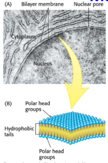
What are membranes?
sheetlike structures, two molecules thick, that form closed boundaries.
What are membranes composed of?
They are composed of lipids and proteins, either of which can be decorated with carbohydrates.
What are membrane lipids and what do they form?
small amphipathic molecules that form closed bimolecular sheets preventing the movement of polar or charged molecules.
What is the role of proteins in cell membranes?
Proteins help make membranes permeable and facilitate movement of molecules and information across the cell membrane.
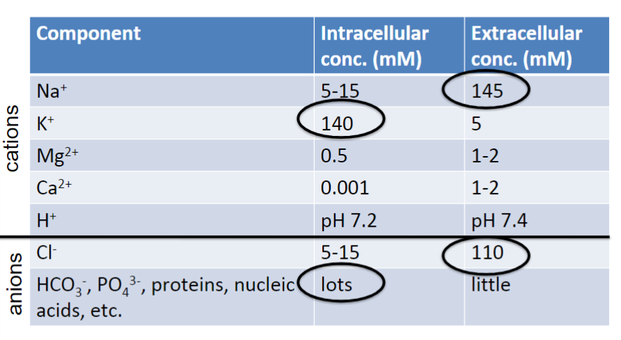
How are most cell membranes electrically polarized?
Most cell membranes are electrically polarized with the inside being negative.
Name three membrane facts.
Membranes are noncovalent assemblies.
Membranes are asymmetric in that the outer surface is always different from the inner surface.
Membranes are fluid structures.
What forms lipid bilayers in aqueous solutions?
Phospholipids and glycolipids (composed of two lipid sheets)
What powers the formation of membranes?
The hydrophobic effect.
the hydrophobic tails interact with one another by Van
der Waals attractive forces
favor close packing of the tails.
What stabilizes lipid bilayers by burying hydrophobic groups out of contact with water?
The energy gained from hydrophobic interactions.
1. A hydrophobic chain in water forces the formation of a cage of water around it.
2. When several hydrophobic regions cluster in a bilayer, the surface area exposed to water decreases, and the water molecules in the cage are released, accompanied by a gain in entropy. That drives the formation of water.
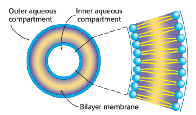
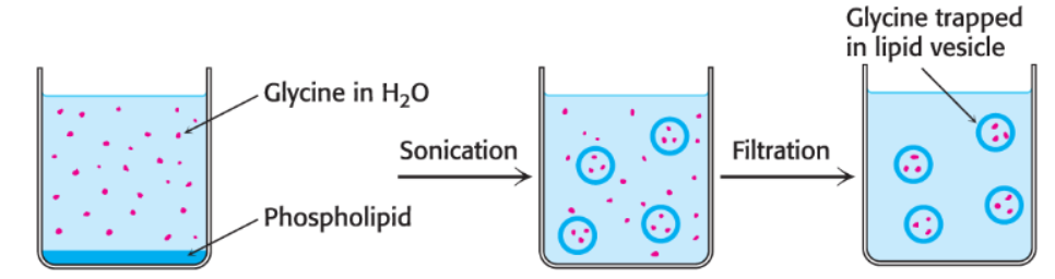
What are liposomes?
aqueous compartments enclosed by a lipid membrane, used in drug delivery and cosmetic products (formed by sonicating mixture of phospholipids in aqueous solution)
What is an advantage of drug delivery by liposomes compared to systemic drugs?
Liposomes deliver more targeted drugs, reducing exposure of the body to potentially toxic drugs.
What are lipid bilayers impermeable to?
ions and most polar molecules (especially large and water soluble)
What determines the ability of small molecules to cross a membrane?
it’s a function of its hydrophobicity
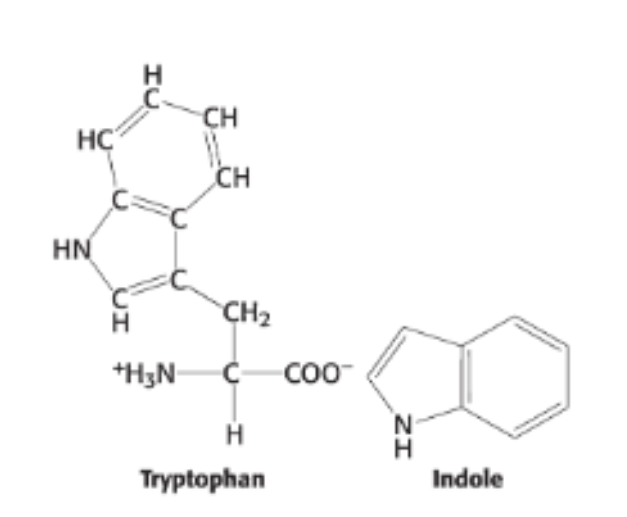
Why is indole more soluble than tryptophan in membranes?
Indole is uncharged, while tryptophan contains ions that cannot cross membranes.
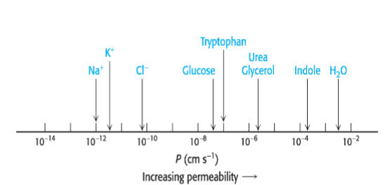
What is this graph showing?
Permeability coefficients of ions and molecules in a lipid bilayer
What must a cell exchange with its surroundings?
molecules and ions
What is the lipid bilayer highly permeable to?
small nonpolar molecules
What are some examples of small nonpolar molecules?
O2 (molecular mass 32 daltons) CO2 (44 daltons)
→ Rapidly diffuse across the membrane
What are some examples of uncharged polar molecules?
H2O (18 daltons) ethanol (46 daltons)
→ small enough to diffuse across
Does glucose or glycerol diffuse across the cell membrane more rapidly?
glycerol (92 daltons)- more rapidly
glucose (180 daltons)- less rapidly
(polar vs. nonpolar)
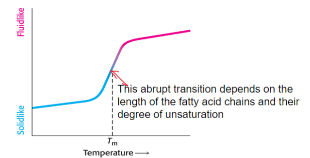
What do membrane processes depend on?
fluidity of the membrane
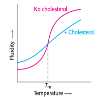
melting temperature
temperature at which a membrane transitions from being highly ordered to very fluid
What is the melting temperature dependent on (Tm)?
the length of the fatty acids in the membrane lipid
the degree of cis unsaturation
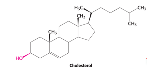
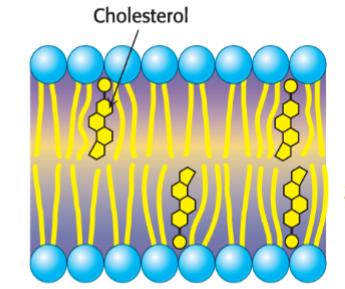
What substance helps maintain membrane fluidity?
cholesterol
What would happen if this structure had a lower melting point?
would be both saturated and unsaturated
What two things does cholesterol do to modulate the fluidity of the membrane?
Prevent solidifying at the temperature below Tm (prevent being too rigid)
Sterically blocks large motions of the fatty acid at the temperature above Tm (prevent being too fluid)
The _________ the fatty acid tail, the less tendency for interaction between the tails, which __________ the fluidity of the membrane.
longer, reduces
In addition, the more closely packed the fatty acid tails, the ____ fluid the membrane; saturated fatty acid tails have no double bonds and can pack _____ closely together.
less, more
What role do membrane lipids and membrane proteins play in the cell membrane?
membrane lipids establish a permeability barrier
membrane proteins allow transport of molecules and information across the membrane.
What is the range of protein content found in membranes?
as little as 18% to as much as 75%
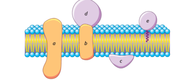
In what ways do proteins associate with the lipid bilayer?
Integral membrane proteins are embedded in the hydrocarbon core of the membrane.
Peripheral membrane proteins are bound to the polar head groups of membrane lipids or to the exposed surfaces of integral membrane proteins.
Some proteins are associated with membranes by
attachment to a hydrophobic moiety that is inserted into
the membrane.
What is a common structural feature of integral membrane proteins?
Membrane-spanning α helices.
can also use β strands to form a pore in the membrane or embed part of the protein into the membrane
What type of membrane proteins are very tightly associated with the membrane through hydrophobic interactions with hydrocarbon tails of membrane lipids?
Integral membrane proteins
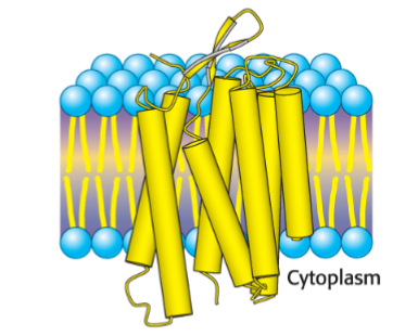
What is this structure?
Bacteriorhodopsin- integral protein (transmembrane protein) made of alpha helix
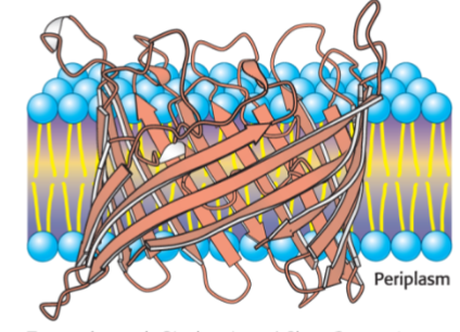
What is this structure?
Bacterial Porin- forms a pore, or channel. Mediates the free diffusion of ions and metabolites
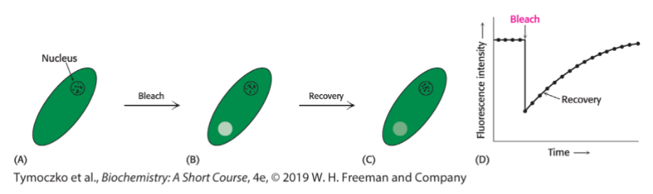
What does FRAP stand for and what does it measure?
Fluorescence Recovery After Photobleaching, and it measures the lateral mobility of membrane components.
How does FRAP work before fluorescence recovery?
A membrane component is attached to a fluorescent molecule. On a very small portion of the membrane, the dye is subsequently destroyed by high-intensity light, thereby bleaching a portion of the membrane
What is the relationship between the mobility of a fluorescently labeled membrane component and the recovery of fluorescence in a bleached area?
The mobility of the fluorescently labeled component is determined by how quickly the bleached area regains fluorescence.
What does the lateral diffusion of proteins depend on?
whether they are attached to other cellular or extracellular components.
What does this diagrammed technique show?
Fluorescence Recovery After Photobleaching. Measuring the rate of lateral diffusion of a membrane protein as molecules migrate back into the bleached area.
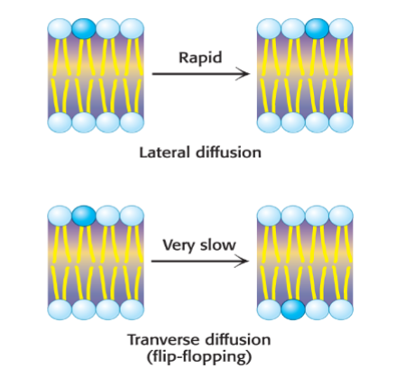
How do lipids primarily move within membranes?
Lipids rapidly diffuse laterally in membranes, while transverse diffusion or flip-flopping is rare without enzyme assistance
What accounts for the stability of membrane asymmetry?
The prohibition of transverse diffusion.
The individual lipid molecule are able to move in their own ______________.
monolayer
Why do lipids rarely flip-flop between the different faces of the bilayer?
the polar heads would have to contact the hydrophobic interior of the membrane.
A ____________ moves membrane lipids from the outer leaflet to the inner leaflet.
flippase
Under what two conditions will a small molecule spontaneously cross a membrane?
The concentration of the molecule is higher on one side of the membrane than the other
The molecule is lipophilic or soluble in nonpolar solutions
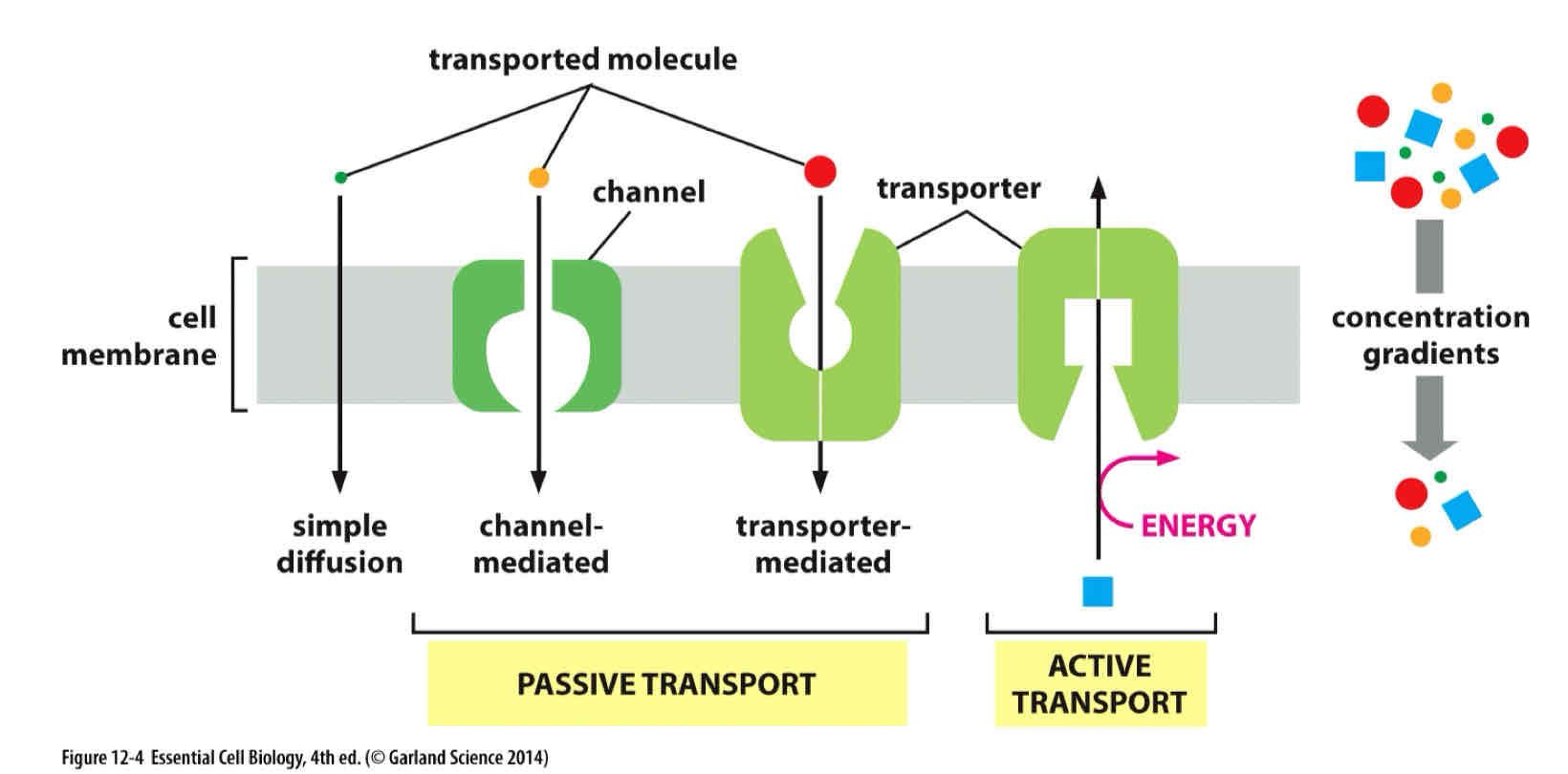
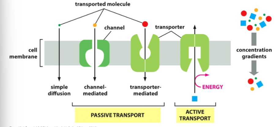
What are the two classes of membrane transport?
Transporters and channels.
Transporters
Transfer only those molecules or ions that fit into specific binding sites
Great specificity- often just one type of molecule
Conformational change
Channels
Mainly on the basis of size and electric charge
Molecule or ion that is small enough and carries the appropriate charge can pass through without conformational change
Selectivity and the rapid transport of ions
The _____________________________ of a solute determines if its movement across the membrane requires active or passive transport.
concentration gradient
What is the function of transport proteins in the cell membrane?
They function as pumps or channels to facilitate the flow of small molecules across the cell membrane.
What type of transport occurs when a molecule moves down its concentration gradient through a transport protein?
Passive transport or facilitated diffusion.
How do protein pumps move a molecule against its concentration gradient in active transport?
Protein pumps use energy with active transport.
Distinguish between passive and active transport.
Passive transport- allows molecules to move down their concentration gradients (downhill)
— No energy required
— Both transporters and channels can mediate passive transport
Active transport- allows molecules to move against their concentration gradients (uphill)
— requires an input of energy
— Only transporters can carry out active transport
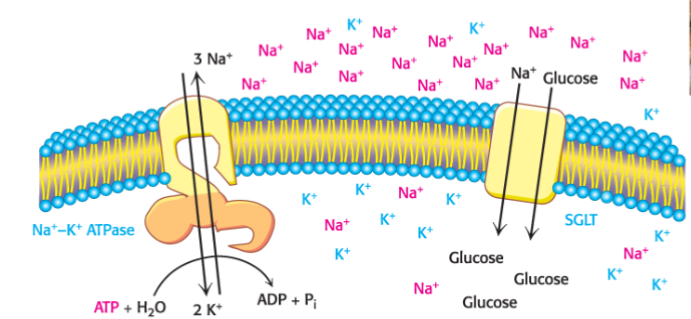
What is the primary function of the Na+–K+ ATPase (Na+–K+ pump)?
uses ATP hydrolysis to pump three Na+ ions out of the cell and two K+ ions into the cell against their concentration gradients.
Why are some pumps P-type ATPases?
Because the reaction includes an intermediate in which the enzyme is phosphorylated.
Name five facts about the Na+-K+ pump?
3 Na+ out, 2 K+ in
Keep the high concentration of Na+ outside
Uses the energy derived from ATP hydrolysis
Controls cell volume, renders neurons and muscle cells electrically excitable, and drives the active transport of sugars...
Na+ concentration gradient of across the membrane: stored Energy
Ion concentrations _______ inside and outside of a cell. Charges need to ________ inside and outside of the cell.
differ, balance
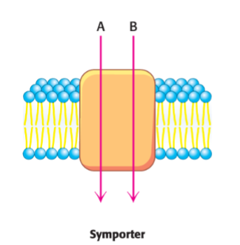
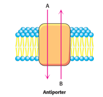
Secondary transporters use ___ concentration gradient to _____ the formation of another. They are also known as?
one, power… a.k.a coupled pump or cotransporter
symporters
power the transport of a molecule against its concentration gradient by coupling the movement to the movement of another molecule down its concentration gradient, with both molecules moving in the same direction
antiporters
use one concentration gradient to power the formation of another, but the molecules move in opposite directions
How is glucose moved into some animal cells against its concentration gradient?
By a symporter powered by Na+ ions moving down a concentration gradient.
Describe this diagram.
Diagram of Secondary Transport.
The electrochemical sodium gradient outside the cell powers the symport that co-transports glucose, along with sodium, into the cell. When Na+ moves into the cell down its electrochemical gradient, glucose is dragged into the cell along with it. Glucose is moving against its concentration gradient
ion channels
passive transport systems that allow specific and rapid transport of ions down their concentration gradients
What are the two ways in which ion channels can be activated?
voltage across a membrane (voltage-activated channels)
binding of specific molecules to the channels (ligand-activated channels)
In active transport by pumps, are the concentrations of most ions close to equilibrium across a cell membrane?
No, the concentrations of most ions are far from equilibrium across a cell membrane.
What typically happens when an ion channel opens?
Ions usually flow through it, moving rapidly down their electrochemical gradients.
The rapid shift of ions changes the ______________________________.
membrane potential
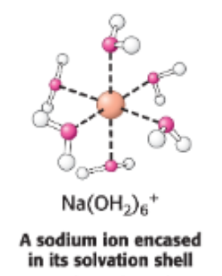
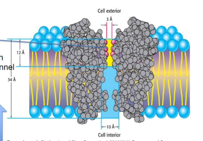
What does the structure of the potassium ion channel reveal?
The basis of ion specificity.
The ______ channel selectively and rapidly transports K+ across the cell membrane.
Larger ions are not transported because they are too ___ to enter the channel. Smaller ions are excluded because they cannot interact with the ____________________.
Such ions are small enough that the energy of ______________ cannot be compensated for by interactions with the ____________________
potassium, big, selectivity filter, desolvation, selectivity filter
selectivity filter
a region of ion-channel proteins that determines the specificity of a particular channel
Interpret this diagram.
Diagram of a Path Through a Channel.
K+ ions move toward exterior of cell. Only “naked” (no associated water molecules) K+ ions can move through the channel and interact with carbonyl group.
What is the function of ion channels?
Ion channels allow the entry of ions through a pore that contains the selectivity filter.
What determines the size of ions that can pass through the selectivity filter in ion channels?
The selectivity filter's width
What role do charged amino acid residues play in the selectivity filter of ion channels?
they repel ions of the wrong charge, allowing only ions of the correct charge to pass through.
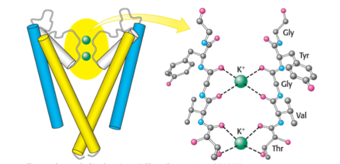
Interpret this diagram.
The selectivity filter of the potassium ion channel.
Potassium ions interact with the carbonyl groups of the selectivity filter, located at the 3-Å-diameter pore of the K+ channel. Only two of the four channel subunits are shown.
Interpret this diagram.
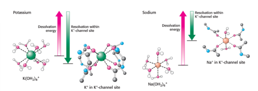
The energetic basis of ion selectivity.
The energy cost of dehydrating a potassium ion is compensated by favorable interactions with the selective filter. Because a sodium ion is too small to interact favorably with the selective filter, the free energy of desolvation cannot be compensated, and the sodium ion does not pass through the channel.
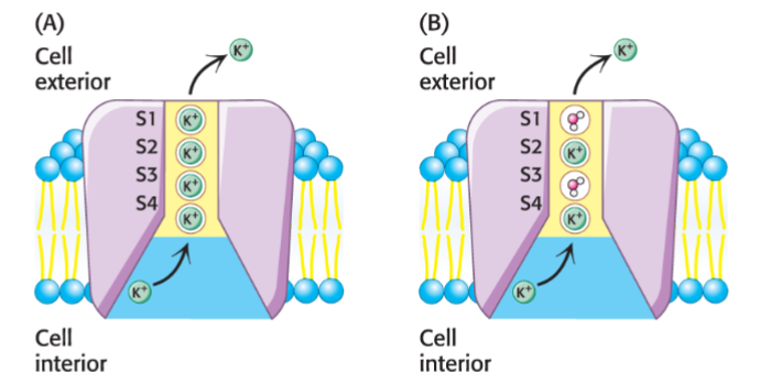
Interpret this diagram.
Model for K+-channel Transport
In the Hard-Knock model of ion transport (A), ions occupy adjacent sites. In the Knock-On model (B), the ions are separated by water molecules in alternate sites. In both models, electrostatic repulsion forces the ions apart.
Explain why phospholipids are capable of spontaneously assembling into the bilayer structure found in biological membranes but triacylglycerols are not.
Phospholipids, unlike triacylglycerols, are amphipathic and thus are driven by the hydrophobic effect to form a lipid bilayer in cells. The polar portions of fatty acid tails faces the aqueous internal and external environments and the nonpolar portion of the tails face inwards towards each other.
What are the forces that drive bilayer formation?
The primary force that drives bilayer formation is the hydrophobic effect. Nonpolar portions of phospholipids clump together and thus face inwards (away from the internal and external aqueous environments). Subsequently, polar portions of phospholipids face outwards. These interactions form a lipid bilayer
Explain the differences between integral and peripheral membrane proteins.
Integral proteins extend through the phospholipid bilayer. Peripheral membrane proteins are localized to one face of the bilayer.
What are the forces or bonds anchor an integral membrane protein in a biological membrane?
Hydrophobic interactions between the nonpolar amino acids located within the transmembrane portions of integral membrane proteins and the hydrophobic interior of the phospholipid bilayer anchor these proteins within biological membranes
What forces hold a peripheral proteins to the membrane?
A variety of non-covalent interactions (i.e. Van der Waals interactions, hydrophobic interactions, electrostatic attractions, and/ or hydrogen bonding) holds peripheral proteins to the cell membrane. Some peripheral proteins (I.e. those that are lipid- linked), covalently bind to lipids within the membrane to interact with the membrane.
How you can purify/isolate integral and peripheral proteins?
Integral membrane proteins can be isolated from the cell membrane via strong ionic detergents such as SDS. Peripheral membrane proteins can be isolated via weaker detergents such as Triton-X 100
When you say that biological membranes are asymmetric structures you mean that?
There is an uneven distribution of phospholipids across the two layers of the phospholipid bilayer (one face will usually have more phospholipids than the other).
The porin proteins utilize the _________ structural motif.
β sheet