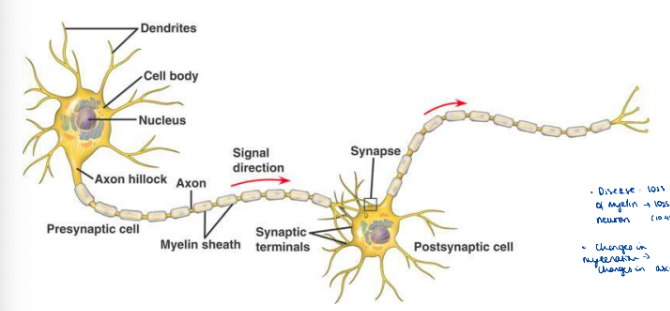
Intro to neuroscience

studied byStudied by 6 people
5.0(1)
get a hint
hint

Role of dendrite

1 / 49

Tags & Description

- Neurones - Myelination - myelination disorders - Glial cells - astrocytes

Studying Progress

0%
New cards
50
Still learning
0
Almost done
0
Mastered
0
50 Terms
1
New cards

Role of dendrite

Recieve electrical + chemical messages from other neurons

New cards
2
New cards


Role of cell body

Process incoming + outgoing signals

New cards
3
New cards

Axons

Send signals to axon terminals

New cards
4
New cards

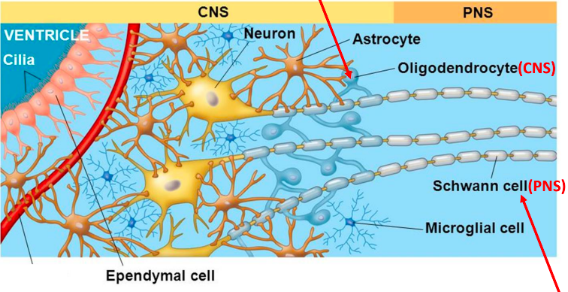
What is glia/glial cells? What are 2 types of myelinating glia?

  • Contribute to brain function mainly by insulating, supporting, nourishing neighbouring neurons

  • Oligodendroglial + Schwann cells

New cards
5
New cards

Axon terminals

Transmits signals to nearby cells

New cards
6
New cards

What is the resting membrane potential?

Approx -65mV

New cards
7
New cards


How is a resting membrane potential maintained? (5)

  • More K+ inside neurone, less Na+ outside CM.

  • Na+/K+ATPase - 3Na+ out, 2K+ in

  • Inside of neurone slightly negative compared to outside

  • Leak channels - more permeable to K+ so more K+ leaves

  • Energy-dependent process

New cards
8
New cards

Order of an action potential (graph) and what each one is caused by in terms of ion flow (4)

  1. Resting state - no ions move through VGC

  2. Depolarisation - by Na+ into cell

  3. Repolarisation - by K+ out of cell

  4. Hyperpolarisation - by K+ continuing to leave cell

New cards
9
New cards

What 2 types of potentials can be caused by a membrane potental?

  • Graded potentials (local changes in membrane potential that degrade with distance)

  • Action potentials (brief spike-change in membrane potential of a set amplitude and does not degrade, allows comm betwen neurons)

New cards
10
New cards

What do graded potentials determine?

  • Excitated neuron - generate AP

  • Inhibited neuron - less likely generate AP (at axon initial segment)

New cards
11
New cards

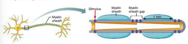
Roles of myelination + myelin sheath gaps

<ul><li><p><strong>Saltatory conduction</strong> - fast transmission of APs</p></li><li><p><strong>Myelin sheath</strong> - fast conduction, keeps current in axons. (voltage doesn’t decay much). APs are only generated in myelin sheath gaps, jump from gap to gap </p></li><li><p>Coordinated movement</p></li></ul>
  • Saltatory conduction - fast transmission of APs

  • Myelin sheath - fast conduction, keeps current in axons. (voltage doesn’t decay much). APs are only generated in myelin sheath gaps, jump from gap to gap

  • Coordinated movement

<ul><li><p><strong>Saltatory conduction</strong> - fast transmission of APs</p></li><li><p><strong>Myelin sheath</strong> - fast conduction, keeps current in axons. (voltage doesn’t decay much). APs are only generated in myelin sheath gaps, jump from gap to gap </p></li><li><p>Coordinated movement</p></li></ul>
New cards
12
New cards

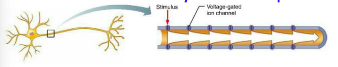
What is conduction like in nonmyelinated axons (compared to myelinated)?

<ul><li><p><strong>Continuous conduction</strong> (slow) - takes time for ions + gates to move, must occur before voltage can be regenerated </p></li><li><p>Voltage-gated Na+ + K+ channels regenerate APs at each point along axon so <mark data-color="yellow">voltage does not decay</mark> </p></li></ul>
  • Continuous conduction (slow) - takes time for ions + gates to move, must occur before voltage can be regenerated

  • Voltage-gated Na+ + K+ channels regenerate APs at each point along axon so voltage does not decay

<ul><li><p><strong>Continuous conduction</strong> (slow) - takes time for ions + gates to move, must occur before voltage can be regenerated </p></li><li><p>Voltage-gated Na+ + K+ channels regenerate APs at each point along axon so <mark data-color="yellow">voltage does not decay</mark> </p></li></ul>
New cards
13
New cards

When are axons myelinated

<ul><li><p><strong>Perinatal period</strong> (before birth)</p></li><li><p>Continues into adulthood, same axons, continued proliferation, differentiation + re-modelled</p></li></ul>
  • Perinatal period (before birth)

  • Continues into adulthood, same axons, continued proliferation, differentiation + re-modelled

<ul><li><p><strong>Perinatal period</strong> (before birth)</p></li><li><p>Continues into adulthood, same axons, continued proliferation, differentiation + re-modelled</p></li></ul>
New cards
14
New cards

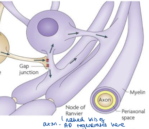
What are nodes of ranvier (3)

<ul><li><p>Unmyelinated gaps, exposes neurone membrane to external environment </p></li><li><p>Gaps have <mark data-color="yellow">lots</mark> of <mark data-color="yellow">Na+ &amp; K+ VGCs</mark></p></li><li><p>APs generated by one NOR, jumps to and is regenerated to the next one - <mark data-color="yellow">rapid</mark> transmission</p></li></ul>
  • Unmyelinated gaps, exposes neurone membrane to external environment

  • Gaps have lots of Na+ & K+ VGCs

  • APs generated by one NOR, jumps to and is regenerated to the next one - rapid transmission

<ul><li><p>Unmyelinated gaps, exposes neurone membrane to external environment </p></li><li><p>Gaps have <mark data-color="yellow">lots</mark> of <mark data-color="yellow">Na+ &amp; K+ VGCs</mark></p></li><li><p>APs generated by one NOR, jumps to and is regenerated to the next one - <mark data-color="yellow">rapid</mark> transmission</p></li></ul>
New cards
15
New cards

Approx how wide are nodes of ranvier?

1μm wide

New cards
16
New cards

What is coordinated movement in childhod?

  • Babies develop skills to move, continue to learn new skills as grow older

  • Between CNS + peripheral system (listen)

New cards
17
New cards


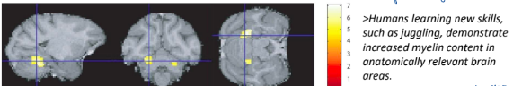
How does our brain learn new skills?

<ul><li><p>Changes in <strong>number + quality of contact</strong> between neurons (synapses)</p></li><li><p>Change in <strong>myelination</strong> (ability to make <mark data-color="yellow">new oligodendrocytes</mark> + <mark data-color="yellow">myelin</mark> important for learning) </p></li></ul>
  • Changes in number + quality of contact between neurons (synapses)

  • Change in myelination (ability to make new oligodendrocytes + myelin important for learning)

<ul><li><p>Changes in <strong>number + quality of contact</strong> between neurons (synapses)</p></li><li><p>Change in <strong>myelination</strong> (ability to make <mark data-color="yellow">new oligodendrocytes</mark> + <mark data-color="yellow">myelin</mark> important for learning) </p></li></ul>
New cards
18
New cards

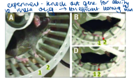
What gene is important for making new oligodendrocytes? What did an experiment prove about this gene in the ability to learn new skills?

<ul><li><p><strong>Myrf gene</strong> (myelin regulatory factor), affects ability to perform <mark data-color="yellow">complex tasks</mark> </p><p></p><p><u>Experiment:</u></p></li><li><p>Knockdown of Myrf gene in mice - less efficient ability to run</p></li></ul>
  • Myrf gene (myelin regulatory factor), affects ability to perform complex tasks

    Experiment:

  • Knockdown of Myrf gene in mice - less efficient ability to run

<ul><li><p><strong>Myrf gene</strong> (myelin regulatory factor), affects ability to perform <mark data-color="yellow">complex tasks</mark> </p><p></p><p><u>Experiment:</u></p></li><li><p>Knockdown of Myrf gene in mice - less efficient ability to run</p></li></ul>
New cards
19
New cards

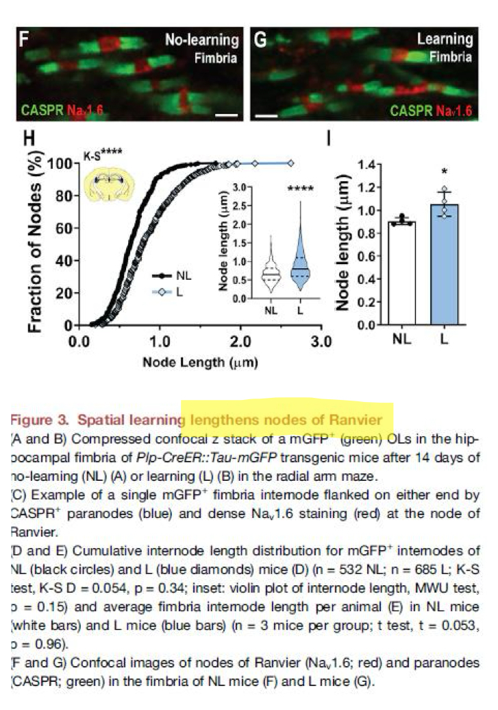
Alongside new myelin, what does extensive re-modelling of existing myelin affect? (3)

<ul><li><p><mark data-color="yellow">Compaction</mark> of the myelin sheath + <mark data-color="yellow">length</mark> of node of Ranvier </p></li><li><p><mark data-color="yellow">Spatial learning</mark> lengthens node of Ranvier</p></li><li><p>Modifying the <mark data-color="yellow">axon-glial configuration</mark> may be a mechanism that facilitates learning (in adult mouse brain) </p></li></ul><p></p>
  • Compaction of the myelin sheath + length of node of Ranvier

  • Spatial learning lengthens node of Ranvier

  • Modifying the axon-glial configuration may be a mechanism that facilitates learning (in adult mouse brain)

<ul><li><p><mark data-color="yellow">Compaction</mark> of the myelin sheath + <mark data-color="yellow">length</mark> of node of Ranvier </p></li><li><p><mark data-color="yellow">Spatial learning</mark> lengthens node of Ranvier</p></li><li><p>Modifying the <mark data-color="yellow">axon-glial configuration</mark> may be a mechanism that facilitates learning (in adult mouse brain) </p></li></ul><p></p>
New cards
20
New cards

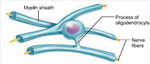
Schwann cell vs oligodendrocyte cell in making myelin

<p></p><ul><li><p>1 schwann cell makes <mark data-color="yellow">1 segment</mark> of myelin in <mark data-color="yellow">PNS</mark></p></li><li><p>1 (cell body of) <strong>oligodendrocyte</strong> can <mark data-color="yellow">myelinate many</mark> tens of axons at the same time, in <mark data-color="yellow">CNS</mark></p></li></ul>

  • 1 schwann cell makes 1 segment of myelin in PNS

  • 1 (cell body of) oligodendrocyte can myelinate many tens of axons at the same time, in CNS

<p></p><ul><li><p>1 schwann cell makes <mark data-color="yellow">1 segment</mark> of myelin in <mark data-color="yellow">PNS</mark></p></li><li><p>1 (cell body of) <strong>oligodendrocyte</strong> can <mark data-color="yellow">myelinate many</mark> tens of axons at the same time, in <mark data-color="yellow">CNS</mark></p></li></ul>
New cards
21
New cards

Why is more efficient for an oligodendrocyte cell body to myelinate multiple cells at the same time in the CNS?

listen to vid (18.20)

New cards
22
New cards

What is the node of Ranvier? What is found in the axonal membrane of NoR?

<ul><li><p>Naked (unmyelinated) bits of axon where APs are regenerated from 1 NoR to another</p></li><li><p><mark data-color="yellow">High conc. of VGC Na+ channels</mark> in axonal membrane</p></li></ul>
  • Naked (unmyelinated) bits of axon where APs are regenerated from 1 NoR to another

  • High conc. of VGC Na+ channels in axonal membrane

<ul><li><p>Naked (unmyelinated) bits of axon where APs are regenerated from 1 NoR to another</p></li><li><p><mark data-color="yellow">High conc. of VGC Na+ channels</mark> in axonal membrane</p></li></ul>
New cards
23
New cards

What is formed at the paranodal junction to limit the current leak from the node of Ranvier? (2)

  • Ends of each successive wrap of myelin form a specialised tight junction with the axonal membrane at the paranodal junction to limit the curent leak

  • Tight seal stops the flow of K+ in that direction (lateral diffusion/leakage away from nodal region)

New cards
24
New cards

How does the tight seal help with learning?

It seems to change as we learn

New cards
25
New cards

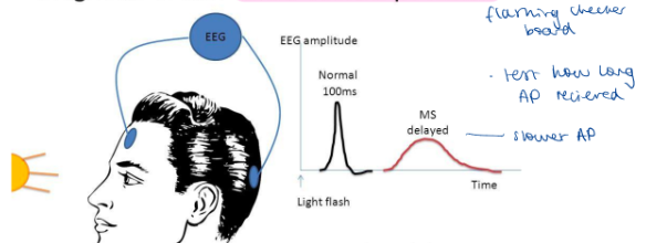
How have potential tests been used to detect inability to conduct APs caused by damage eg demyelination?

Measure electrical activity of the brain in response to stimulation

New cards
26
New cards

What is multiple sclerosis and how can it be diagnosed using electrical brain activity?

<p><u>Multiple sclerosis:</u></p><ul><li><p>Neurological disorder - <mark data-color="yellow">lack of coordination</mark>, impaired vision + speech. <mark data-color="yellow">Attacks myelin sheath</mark> of <mark data-color="yellow">bundles of axons</mark> in brain, spinal cord + optic nerves.</p></li></ul><p></p><p><u>Diagnosis (other than MRI scans)</u></p><ul><li><p><strong>Visual evoked potential</strong></p></li><li><p>Eg stimulate eye with checkerboard pattern and measure time of electrical response</p></li><li><p>Profound <mark data-color="yellow">slowing of conduction velocity</mark> and <mark data-color="yellow">bloc</mark>k in some fibres</p><p></p></li></ul>

Multiple sclerosis:

  • Neurological disorder - lack of coordination, impaired vision + speech. Attacks myelin sheath of bundles of axons in brain, spinal cord + optic nerves.

Diagnosis (other than MRI scans)

  • Visual evoked potential

  • Eg stimulate eye with checkerboard pattern and measure time of electrical response

  • Profound slowing of conduction velocity and block in some fibres

<p><u>Multiple sclerosis:</u></p><ul><li><p>Neurological disorder - <mark data-color="yellow">lack of coordination</mark>, impaired vision + speech. <mark data-color="yellow">Attacks myelin sheath</mark> of <mark data-color="yellow">bundles of axons</mark> in brain, spinal cord + optic nerves.</p></li></ul><p></p><p><u>Diagnosis (other than MRI scans)</u></p><ul><li><p><strong>Visual evoked potential</strong></p></li><li><p>Eg stimulate eye with checkerboard pattern and measure time of electrical response</p></li><li><p>Profound <mark data-color="yellow">slowing of conduction velocity</mark> and <mark data-color="yellow">bloc</mark>k in some fibres</p><p></p></li></ul>
New cards
27
New cards

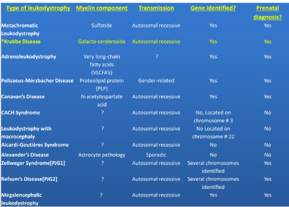
What are leukodystrophies and their characteristics? (5)

Dysmyelinating disorders with:

  • Primary lesion of myelin/oligodendrocyte (diseases of OD + myelin)

  • Genetic causes

  • Progressive clinical course

  • Predominant + confluent involvement of CNS white matter

New cards
28
New cards

What is a dysmyelinating disease, according to Poser 1957?

Heredofamilial disorders (genetic) where myelin not formed properly, or myelin formation is delayed/arrested, or disturbed maintenance of already formed myelin

New cards
29
New cards

Example of a leukodystrophy disease

<p>Krabbe disease </p>

Krabbe disease

<p>Krabbe disease </p>
New cards
30
New cards

What is Krabbe disease (aka globoid cell leukodystrophy)?

<ul><li><p><mark data-color="yellow">Autosomal-recessive</mark> condition, metabolic disorder, from <strong>deficiency</strong> of <strong>enzyme GALC (galactocerebrosidase)</strong></p></li></ul>
  • Autosomal-recessive condition, metabolic disorder, from deficiency of enzyme GALC (galactocerebrosidase)

<ul><li><p><mark data-color="yellow">Autosomal-recessive</mark> condition, metabolic disorder, from <strong>deficiency</strong> of <strong>enzyme GALC (galactocerebrosidase)</strong></p></li></ul>
New cards
31
New cards

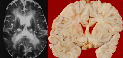
What is Krabbe disease characterised by?

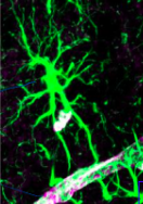
  • Rapid + nearly complete disappearance of myelin + myelin-forming cells from the CNS & PNS

  • Reactive astrocytic gliosis (hypertrophy, glial scar formation, signalling molecules released)

  • Infiltration of the unique and often multinucleated macrophages ("globoid cells“ – green arrow)

New cards
32
New cards

When do sympyoms of Krabbe disease develop normally and when does it usually result in death?

Before 6 months of age, results in death by 2yrs age

New cards
33
New cards

What is GALC (galactocerebrosidase) responsible for? How can it be toxic to OD and other cells?

  • Liposomal hydrolysis of galactolipids formed during white matter myelination

  • It catabolises galactosylsphingosine sphingosine and galactose

  • Accumulation of galactosylsphingosine is hugely toxic to oligodendrocytes and other cells.

New cards
34
New cards

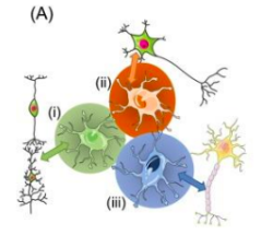
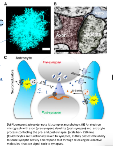
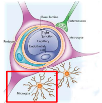
What is an astrocyte?

<ul><li><p>Type of <strong>glial cell</strong> that <strong>regulates BBB</strong>, coupling neuronal activity with <strong>metabolic support</strong> from blood, and interacts with other types of cells:</p><ul><li><p>Oligodendrocytes, microglia, blood vessels</p></li><li><p>Wraps around every blood vessel + capillary</p><p></p></li></ul></li></ul>
  • Type of glial cell that regulates BBB, coupling neuronal activity with metabolic support from blood, and interacts with other types of cells:

    • Oligodendrocytes, microglia, blood vessels

    • Wraps around every blood vessel + capillary

<ul><li><p>Type of <strong>glial cell</strong> that <strong>regulates BBB</strong>, coupling neuronal activity with <strong>metabolic support</strong> from blood, and interacts with other types of cells:</p><ul><li><p>Oligodendrocytes, microglia, blood vessels</p></li><li><p>Wraps around every blood vessel + capillary</p><p></p></li></ul></li></ul>
New cards
35
<p>Specialised astrocytes express<strong> distinct molecular signatures </strong>(the diff colours) to do what 3 roles? </p>
New cards
<p>Specialised astrocytes express<strong> distinct molecular signatures </strong>(the diff colours) to do what 3 roles? </p>

Specialised astrocytes express distinct molecular signatures (the diff colours) to do what 3 roles?

  • Modulate synaptic activity to clear neutrotransmitters (eg glutamate)

  • Release gliotransmitters

  • Provide metabolic support

New cards
36
New cards

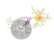
Why + how do astrocytes interact with oligodendrocytes?

<ul><li><p>To support axon and regulate myelination process, provide <strong>atrophic supporting factors</strong> - eg molecular signals, GFs, signalling molecules)</p></li><li><p>To regulate <strong>homeostasis</strong> of <strong>brain tissues</strong></p></li></ul><p></p>
  • To support axon and regulate myelination process, provide atrophic supporting factors - eg molecular signals, GFs, signalling molecules)

  • To regulate homeostasis of brain tissues

<ul><li><p>To support axon and regulate myelination process, provide <strong>atrophic supporting factors</strong> - eg molecular signals, GFs, signalling molecules)</p></li><li><p>To regulate <strong>homeostasis</strong> of <strong>brain tissues</strong></p></li></ul><p></p>
New cards
37
New cards

Why + how do astrocytes interact with microglia?

<p>To critically <strong>modulate</strong> their ability to <strong>support neurons </strong>during <strong>inflammation</strong></p>

To critically modulate their ability to support neurons during inflammation

<p>To critically <strong>modulate</strong> their ability to <strong>support neurons </strong>during <strong>inflammation</strong></p>
New cards
38
New cards

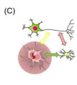
How do astrocytes help synaptic transmission?

<ul><li><p>Regulates chemical content of extracellular space. Has <strong>proteins</strong> in<strong> membrane</strong>s to <strong>remove + recycle excess </strong>released <mark data-color="yellow">neurotransmitter</mark>, hands it back to cell.</p><ul><li><p>Eg tightly controls conc. of K+ in extracellular fluid</p></li></ul></li><li><p>Can <strong>sense synaptic activity</strong> and respond to it by <mark data-color="yellow">releasing neuroactive molecules</mark> that can signal back to synapses</p></li></ul>
  • Regulates chemical content of extracellular space. Has proteins in membranes to remove + recycle excess released neurotransmitter, hands it back to cell.

    • Eg tightly controls conc. of K+ in extracellular fluid

  • Can sense synaptic activity and respond to it by releasing neuroactive molecules that can signal back to synapses

<ul><li><p>Regulates chemical content of extracellular space. Has <strong>proteins</strong> in<strong> membrane</strong>s to <strong>remove + recycle excess </strong>released <mark data-color="yellow">neurotransmitter</mark>, hands it back to cell.</p><ul><li><p>Eg tightly controls conc. of K+ in extracellular fluid</p></li></ul></li><li><p>Can <strong>sense synaptic activity</strong> and respond to it by <mark data-color="yellow">releasing neuroactive molecules</mark> that can signal back to synapses</p></li></ul>
New cards
39
New cards

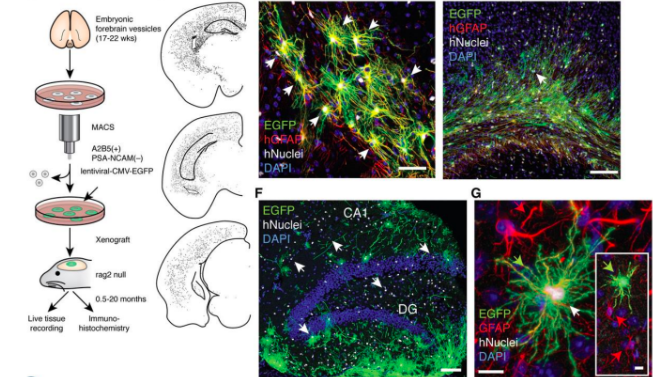
How has a study suggested that astrocytes affect learning and memory?

<ul><li><p>Human astrocytes - larger + more morphologically complex than rodent astrocytes</p></li><li><p>Transplant (hu)Astrocytes into the mouse brain and compared to controls</p></li><li><p><mark data-color="yellow">Improved synaptic function</mark> and mice are <mark data-color="yellow">‘smarter’</mark></p></li></ul>
  • Human astrocytes - larger + more morphologically complex than rodent astrocytes

  • Transplant (hu)Astrocytes into the mouse brain and compared to controls

  • Improved synaptic function and mice are ‘smarter’

<ul><li><p>Human astrocytes - larger + more morphologically complex than rodent astrocytes</p></li><li><p>Transplant (hu)Astrocytes into the mouse brain and compared to controls</p></li><li><p><mark data-color="yellow">Improved synaptic function</mark> and mice are <mark data-color="yellow">‘smarter’</mark></p></li></ul>
New cards
40
New cards

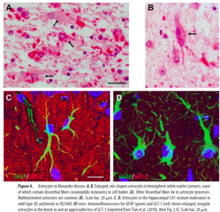
What is Alexander disease + what is it caused by?

  • Disease of astrocytes

  • A leukodystrophy characterised by abnormal protein deposits - Rosenthal fibres

  • Caused by dominant gain-of-function mutations in the glial fibrillary acidic protein (GFAP, expressed only in astrocytes).

New cards
41
New cards

Symptoms of Alexander disease

<ul><li><p>Enlarged brain and head</p></li><li><p>Seizures</p></li><li><p>Stiffness in the arms and/or legs</p></li><li><p>Learning difficulties</p></li><li><p>Delayed physical development.</p></li></ul><p></p><p>Most cases begin before 2 years old (infantile form) </p><p>Most children with the infantile form do not survive beyond 6yrs </p>
  • Enlarged brain and head

  • Seizures

  • Stiffness in the arms and/or legs

  • Learning difficulties

  • Delayed physical development.

Most cases begin before 2 years old (infantile form)

Most children with the infantile form do not survive beyond 6yrs

<ul><li><p>Enlarged brain and head</p></li><li><p>Seizures</p></li><li><p>Stiffness in the arms and/or legs</p></li><li><p>Learning difficulties</p></li><li><p>Delayed physical development.</p></li></ul><p></p><p>Most cases begin before 2 years old (infantile form) </p><p>Most children with the infantile form do not survive beyond 6yrs </p>
New cards
42
New cards

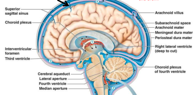
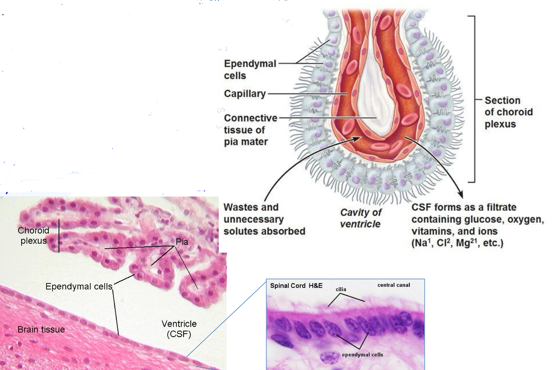
Cerebospinal fluid role

<ul><li><p>Bathes brain + spinal chord</p></li><li><p>Rich in nutrients, carries metabolites, helps <mark data-color="yellow">clear waste products</mark></p><p></p></li></ul>
  • Bathes brain + spinal chord

  • Rich in nutrients, carries metabolites, helps clear waste products

<ul><li><p>Bathes brain + spinal chord</p></li><li><p>Rich in nutrients, carries metabolites, helps <mark data-color="yellow">clear waste products</mark></p><p></p></li></ul>
New cards
43
New cards

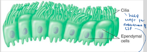
What are ependymal cells? What do they include and what are they important for?

<ul><li><p>Line the <strong>CSF-filled ventricles</strong> of <strong>brain</strong> + <strong>spinal chord</strong> to provide <mark data-color="yellow">physical barrier</mark> between CSF and brain</p><ul><li><p>Important for CNS development</p></li></ul></li><li><p>Like <mark data-color="yellow">epithelial cells</mark> and have a<strong> basement membrane</strong>, <strong>cell-cell junctions</strong> and <strong>motile cilia </strong>on the ventricles </p></li></ul>
  • Line the CSF-filled ventricles of brain + spinal chord to provide physical barrier between CSF and brain

    • Important for CNS development

  • Like epithelial cells and have a basement membrane, cell-cell junctions and motile cilia on the ventricles

<ul><li><p>Line the <strong>CSF-filled ventricles</strong> of <strong>brain</strong> + <strong>spinal chord</strong> to provide <mark data-color="yellow">physical barrier</mark> between CSF and brain</p><ul><li><p>Important for CNS development</p></li></ul></li><li><p>Like <mark data-color="yellow">epithelial cells</mark> and have a<strong> basement membrane</strong>, <strong>cell-cell junctions</strong> and <strong>motile cilia </strong>on the ventricles </p></li></ul>
New cards
44
New cards

Role of cilia

  • Wafts to help circulate CSF around ventricles + over brain

  • Otherwise CSF will build up in brain → disorders

New cards
45
New cards

What is the choroid plexus?

<ul><li><p>In <mark data-color="yellow">ventricles</mark> of brain, <strong>produce CSF</strong></p></li><li><p>Formed from<strong> specialised ependymal cells </strong></p></li><li><p>Have lots of <mark data-color="yellow">blood vessels </mark>to filter substances (eg nutrients, electrolytes) from blood into CSF</p></li></ul>
  • In ventricles of brain, produce CSF

  • Formed from specialised ependymal cells

  • Have lots of blood vessels to filter substances (eg nutrients, electrolytes) from blood into CSF

<ul><li><p>In <mark data-color="yellow">ventricles</mark> of brain, <strong>produce CSF</strong></p></li><li><p>Formed from<strong> specialised ependymal cells </strong></p></li><li><p>Have lots of <mark data-color="yellow">blood vessels </mark>to filter substances (eg nutrients, electrolytes) from blood into CSF</p></li></ul>
New cards
46
New cards

What can a choroid plexus tumour or dysfunction in cilia motility (rare) lead to?

  • Hydrocephalus (water on the brain)

  • Hyperproduction or buildup of CSF fluidventricles swell

  • Brain tissue squeezes and loses consciousness

New cards
47
New cards

What are microglia (2) and their roles (2) in defending the CNS?

<ul><li><p><strong>Resident tissue macrophage</strong> of <strong>CNS</strong>, 15% of brain cells </p></li><li><p><strong>Primary defense</strong> of CNS - first cells to respond to injury/infection </p></li></ul><p><u>Roles:</u></p><ul><li><p>Remove cellular debris (general maintenance + surveillance)</p></li><li><p><strong>Neuroprotection</strong> - trophic support for neurones </p></li></ul>
  • Resident tissue macrophage of CNS, 15% of brain cells

  • Primary defense of CNS - first cells to respond to injury/infection

Roles:

  • Remove cellular debris (general maintenance + surveillance)

  • Neuroprotection - trophic support for neurones

<ul><li><p><strong>Resident tissue macrophage</strong> of <strong>CNS</strong>, 15% of brain cells </p></li><li><p><strong>Primary defense</strong> of CNS - first cells to respond to injury/infection </p></li></ul><p><u>Roles:</u></p><ul><li><p>Remove cellular debris (general maintenance + surveillance)</p></li><li><p><strong>Neuroprotection</strong> - trophic support for neurones </p></li></ul>
New cards
48
New cards

What does dysfunctional microglia lead to?

  • Disurpts normal brain homeostasis

  • Neurotoxic - neurodegeneration

  • Common variants in microglial-experssed genes linked to increased risk of Alzheimer’s

New cards
49
New cards

As microglia are reactive to changes in the brain (they proliferate and found in high numbers), what can they be used for?

Useful indicator of brain health, disease + response to treatment

New cards
50
New cards

What does PET (radioactive) imaging reveal about microglial density?

<ul><li><p>Indicates their activation</p></li><li><p>Expression of TSPO (translocator protein) on mitochondrial membranes of</p><p>microglia (and other cells)</p></li><li><p>Radioligands, such as [11C]PK11195, report areas of increased microglial density</p></li></ul>
  • Indicates their activation

  • Expression of TSPO (translocator protein) on mitochondrial membranes of

    microglia (and other cells)

  • Radioligands, such as [11C]PK11195, report areas of increased microglial density

<ul><li><p>Indicates their activation</p></li><li><p>Expression of TSPO (translocator protein) on mitochondrial membranes of</p><p>microglia (and other cells)</p></li><li><p>Radioligands, such as [11C]PK11195, report areas of increased microglial density</p></li></ul>
New cards

Explore top notes

note Note
studied byStudied by 7 people
Updated ... ago
5.0 Stars(1)
note Note
studied byStudied by 12 people
Updated ... ago
5.0 Stars(1)
note Note
studied byStudied by 24 people
Updated ... ago
5.0 Stars(1)
note Note
studied byStudied by 3 people
Updated ... ago
5.0 Stars(1)
note Note
studied byStudied by 11 people
Updated ... ago
5.0 Stars(1)
note Note
studied byStudied by 53 people
Updated ... ago
5.0 Stars(1)
note Note
studied byStudied by 14 people
Updated ... ago
4.7 Stars(3)
note Note
studied byStudied by 7275 people
Updated ... ago
4.9 Stars(88)

Explore top flashcards

flashcards Flashcard75 terms
studied byStudied by 2 people
Updated ... ago
5.0 Stars(1)
flashcards Flashcard96 terms
studied byStudied by 5 people
Updated ... ago
5.0 Stars(1)
flashcards Flashcard55 terms
studied byStudied by 16 people
Updated ... ago
5.0 Stars(2)
flashcards Flashcard179 terms
studied byStudied by 4 people
Updated ... ago
5.0 Stars(2)
flashcards Flashcard207 terms
studied byStudied by 11 people
Updated ... ago
5.0 Stars(1)
flashcards Flashcard69 terms
studied byStudied by 1 person
Updated ... ago
5.0 Stars(1)
flashcards Flashcard46 terms
studied byStudied by 3 people
Updated ... ago
5.0 Stars(1)
flashcards Flashcard464 terms
studied byStudied by 33458 people
Updated ... ago
4.1 Stars(392)