Looks like no one added any tags here yet for you.
What are carbohydrates rich in?
they are carbon-based molecules that are rich in hydroxyl (C-OH) - water soluble
________________________ are complex carbohydrates - polymers of covalently linked monosaccharides
polysaccharides
What are the four types of polysaccharides?
glycogen- energy storage in animal
starch- energy storage in plant
chitin- structural component of insect
cellulose- structural component of plant cell wall
oligosaccharides
a few sugars (2 to 12) that are covalently linked
What are some functions of carbohydrates?
major energy sources: carbohydrate
essential structural/protective components
cell-cell recognition signals
What are the simplest carbohydrates?
monosaccharides
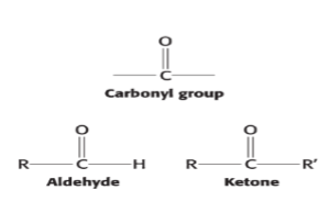
contain a single carbonyl group (aldehydes or ketones) and two or more alcohol groups
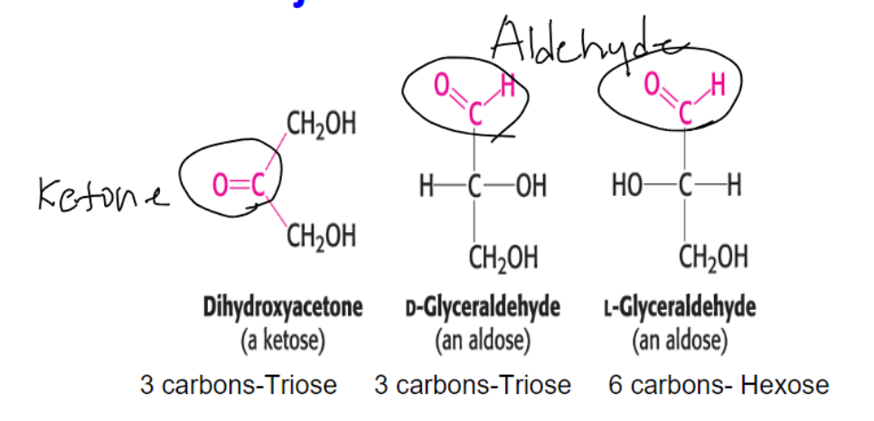
have the general formula Cn(H2O)n Lowest n=3
The smallest monosaccharides are composed of how many carbons?
three.
dihydroxyacetone
D- and L-glyceraldehyde
What are the three components that contribute to monosaccharide classification?
based on carbon number: Cn(H2O)n
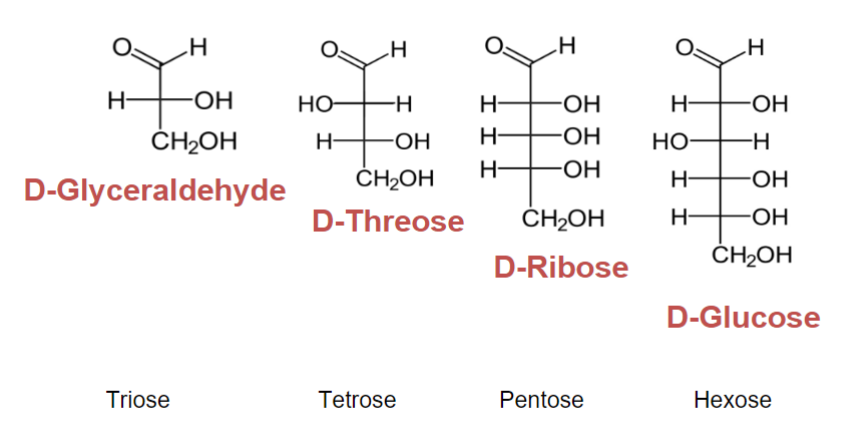
n= 3 Tri- n=4 Tetr- n=5 Pent- n=6 Hex- n=7 Hept- n=8 Oct-
based on position of carbonyl group
aldehyde = aldose
ketone = ketose
based on the isomeric forms
enantiomers
epimers
anomers
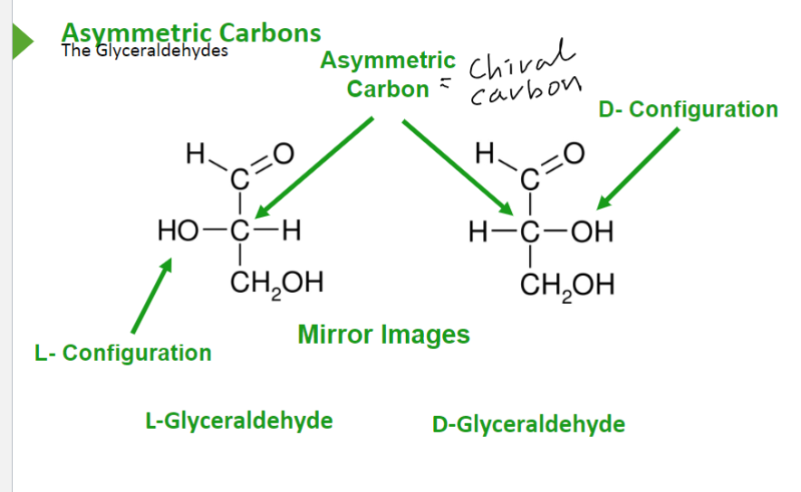
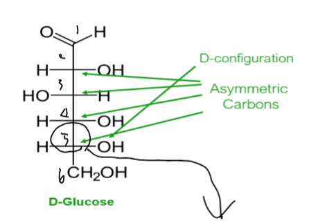
Explain how Fischer projections work.
In a Fischer projection of a molecule, atoms joined to an asymmetric tetrahedral carbon atom by horizontal bonds are in front of the plane of the page and those joined by vertical bonds are behind the plane.
(should say three carbons on right)
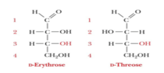
List some examples of monosaccharide Fischer projections.
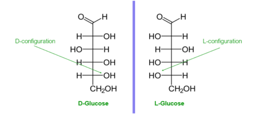
Compare the structures of L-glyceraldehyde and D-glyceraldehyde
What is special about the carbon that is written at the top and designated as C-1?
it is the most highly oxidized
D configuration —OH is on the right of the highest-numbered chiral carbon
L configuration —OH is on the left of the highest-numbered chiral carbon
What are the four common monosaccharides that we should memorize?
Glyceraldehyde (C3H6O3)
Glucose (C6H12O6)
Fructose (C6H12O6)
Ribose (C5H10O5)
Describe this diagram.
This shows the isomeric forms of carbohydrates.
enantiomers
stereoisomers that are mirror images
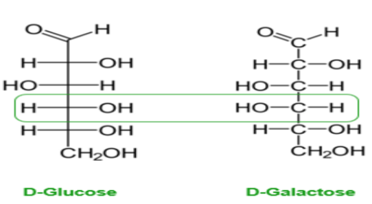
diastereomers
stereoisomers that are not mirror images
epimers
diastereomers differing in configuration of one carbon only
What are the formulaic equivalents of number of stereoisomers and number of chiral centers?
2^n, n
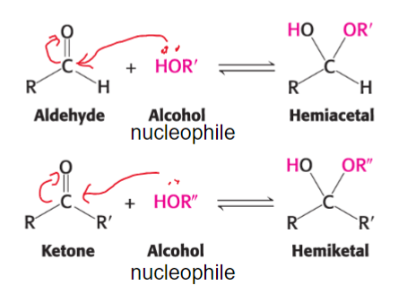
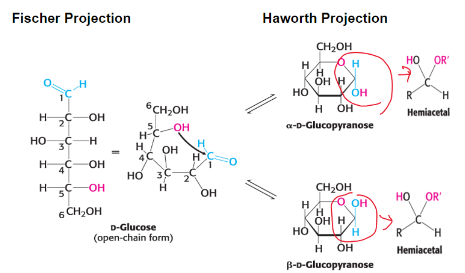
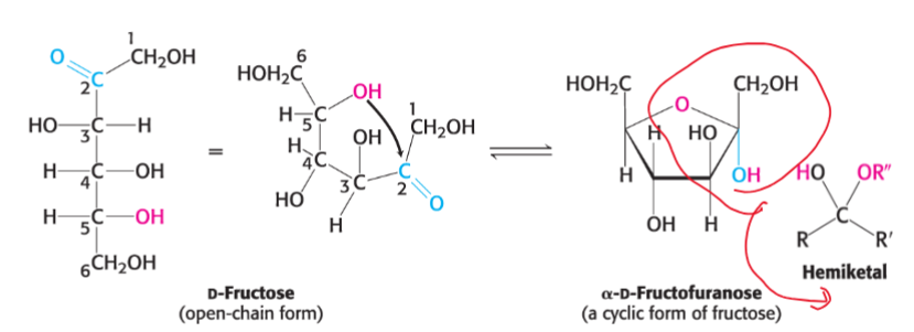
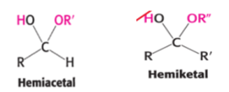
What is the chemical basis for ring formation?
an aldehyde can react with an alcohol to form a hemiacetal, whereas a ketone can react with an alcohol to form a hemiketal
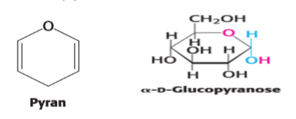
What does pyranose result from and what is its general structure?
it results from glucose as an intramolecular hemiacetal that is a six-membered ring (name is due to resemblance to pyran)
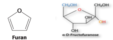
What does furanose result from and what is its general structure?
it results from ketohexose fructose as an intramolecular hemiketal that is a five-membered ring (name is due to resemblance to furan)
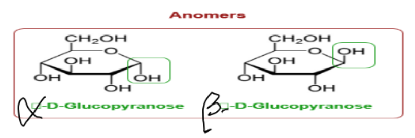
What does formation of a cyclic hemiacetal create?
another diastereoisomeric form called an anomer
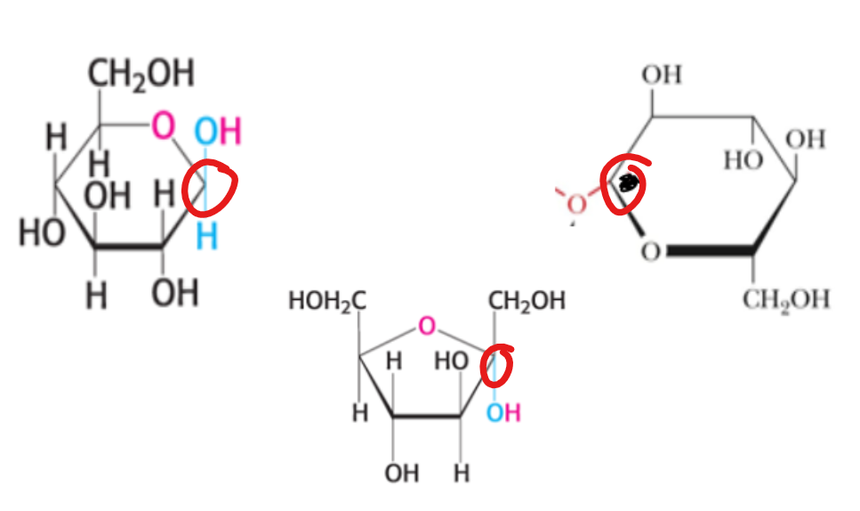
What does the α and β form mean for the hydroxyl at C-1 (anomeric carbon)?
α means below the plane of the ring
β means above the plane of the ring
What is special about the anomeric carbon?
it is the only carbon in a carbohydrate ring that is covalent bonded to 2 oxygen atoms
Describe this diagram.
This shows pyranose formation. For cyclization, an OH group must attack C=O group within the same sugar.
What form of fructose can also exist in anomeric forms in which the α and β forms refer to the orientation of the hydroxyl at C-2?
the furanose form
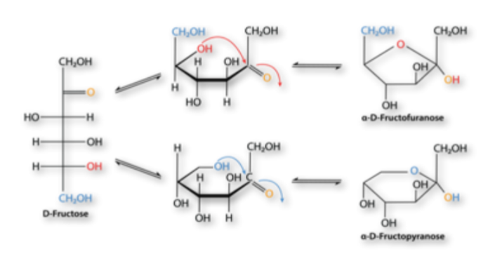
Which two forms of fructose exist?
pyranose form: predominates when fructose is free in solution
furanose form: commonly seem in fructose derivatives
Describe this diagram.
This shows furanose formation. The open-chain form of fructose cyclizes to a five-membered ring when the C-5 hydroxyl group attacks carbon C-2 of the ketone to form an intramolecular hemiketal. Two anomers are possible, but only the α anomer is shown.
Describe this diagram.
This shows ring structures of fructose. Fructose can form both five-membered furanose and six-membered pyranose rings. In each case, both α and β anomers are possible.
Remember that the anomeric carbon is the carbonyl carbon atom of a sugar involved in ring formation.
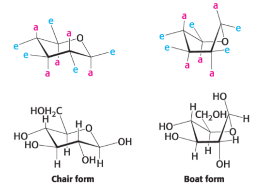
What are the two types of conformation that pyranose can form?
boat and chair
In the chair form of pyranose, what two orientations do substituents on the carbon ring atoms have?
axial and equatorial
Why does β-D-glucopyranose adopt the chair conformation?
the axial positions are occupied by hydrogen atoms (reduces steric hindrance)
Describe this diagram.
This shows the chair and boat forms of β-D-glucopyranose. The chair form is more stable owing to less steric hindrance because the axial positions are occupied by hydrogen atoms.
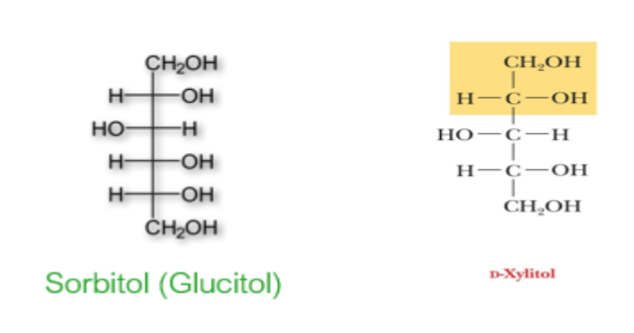
sugar alcohols (alditols)
the carbonyl group of sugar is reduced to an alcohol (-OH) group
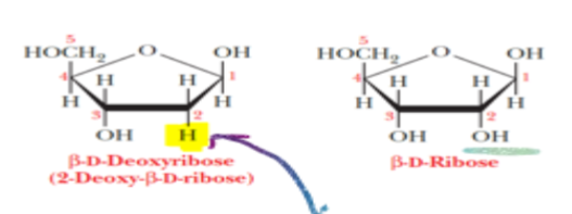
deoxy sugars
reduced sugars in which a hydrogen atom is substituted for one of the hydroxyl groups of the sugar
What fractions of glucose is α anomer, β anomer, and open-chain form, respectively?
1/3 α anomer
2/3 β anomer
<1% open-chain form
Why is the open-chain form of glucose able to freely react with oxidizing agent?
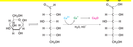
What is the difference between reducing and nonreducing sugars?
reducing- sugars that react with oxidizing agents
nonreducing- sugars that don’t react with oxidizing agents
reducing sugars
a sugar that converts into a form with a free aldehyde group that is readily oxidized and can thus reduce another compound
have a free aldehyde group (either hemiacetal or hemiketal)
What test is used to detect the presence of reducing agents?
Tollens reagent test.
Ag(NH3)2+ is oxidizing agent
if anomeric carbon is free, the result is positive
What does glucose, a reducing sugar, form when it reacts with hemoglobin?
glycosylated hemoglobin (hemoglobin A1c) - fully functional
What does determining the amount of HbA1c in the blood allow?
monitoring of the long-term control of blood glucose levels in diabetics
Reactions between ______________________ and ______________ often impair protein function. Such modifications, called _____________________________________________, have been implicated in a number of pathological conditions.
carbohydrates, proteins, advanced glycation end products
O-glycosidic bond
bond formed between the anomeric carbon atom of sugar and hydroxyl group of another (product is called glycoside)
N-glycosidic bond
bond formed between the anomeric carbon atom of sugar and an amine
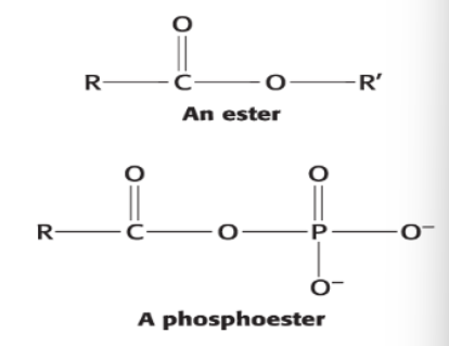
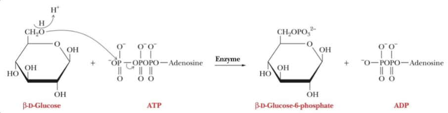
Carbohydrates form ester linkages to __________________.
phosphates
phosphate esters
intermediates in the breakdown of carbohydrates provide energy
formed by transfer of a phosphate group from ATP to give the phosphorylated sugar and ADP
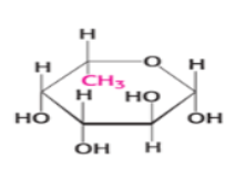
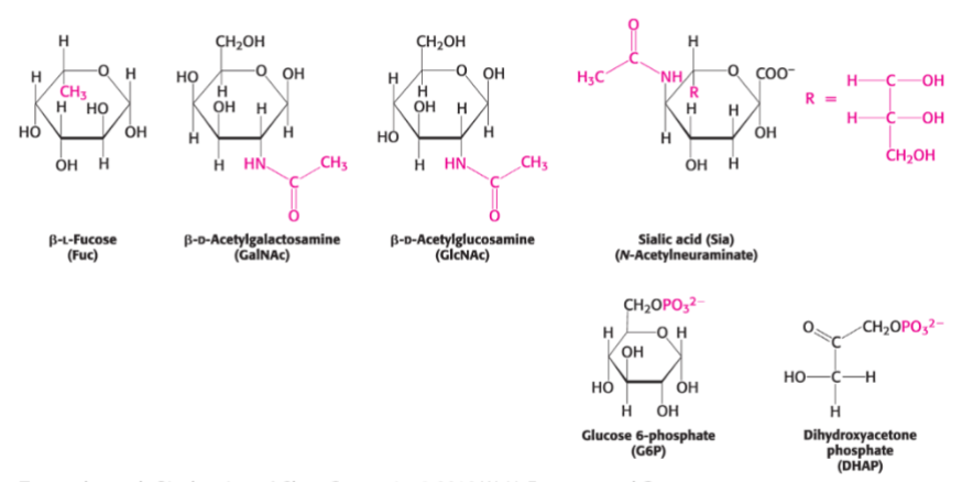
Describe this diagram.
This shows modified monosaccharides. Carbohydrates can be modified by the addition of substituents (shown in red) other than hydroxyl groups. Such modified carbohydrates are often expressed on cell surfaces.
oligosaccharides
contain two or more monosaccharides linked by O-glycosidic bonds
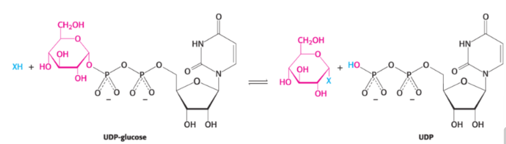
glycosyltransferases
large class of enzymes that catalyze formation of glycosidic bonds
monosaccharide substrates for glycosyltransferases are activated by attachment to uridine diphosphate (UDP)
Enzymes on the outer surface of __________________________ cleave common disaccharides.
intestinal epithelium
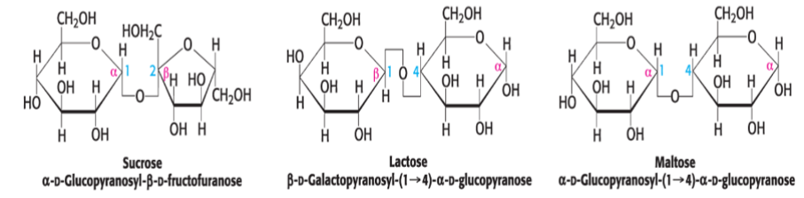
What do sucrase, lactase, and maltase cleave, respectively?
sucrase- sucrose (table sugar)
lactase- lactose (milk sugar)
maltase- maltose
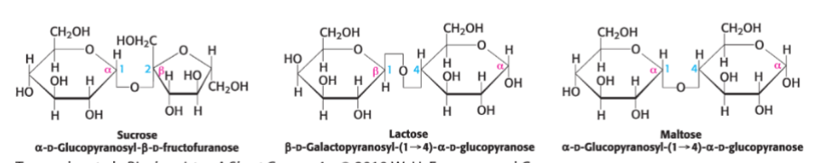
What are the three most common disaccharides made of?
sucrose (glucose + fructose)
lactose (glucose + galactose)
maltose (glucose + glucose)
What are large polymeric oligosaccharides called?
polysaccharides
homopolymer
the name for a polysaccharide when all of the monosaccharides in the polysaccharide are the same
What is the storage form of glucose in animals?
the homopolymer and polysaccharide glycogen
Most __________ units in glycogen are linked by _____________________, with branches formed by _________________________ every __ glucose units.
glucose
α-1, 4-glycosidic bonds
α-1,6-glycosidic bonds
12
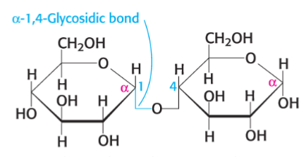
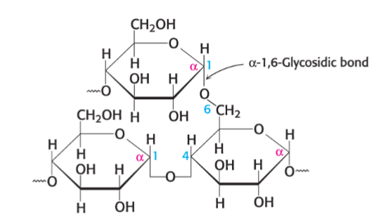
Describe this diagram.
This shows the branch point in glycogen. Two chains of glucose molecules
joined by α-1,4-glycosidic bonds are linked by an α-1,6-glycosidic bond to
create a branch point. Such an α-1,6-glycosidic bond forms at approximately
every 10 glucose units, making glycogen a highly branched molecule.
What is glucose stored as in plants?
starch
What are the two forms of starch?
amylose- linear polymer of glucose units linked by α- 1,4-glycosidic bonds
amylopectin- branched polymer with an α-1,6- glycosidic bond for every 30 α-1,4-glycosidic bonds
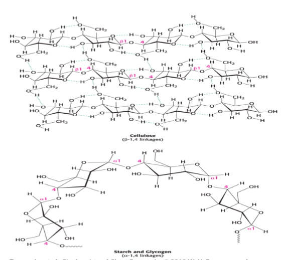
What is cellulose linked by?
β-1,4-glycocidic bonds
Compare the α and β linkages in cellulose?
The β linkage yields a straight chain capable of interacting with other cellulose molecules to from strong fibrils.
The α linkages of starch and glycogen form compact hollow cylinders suitable for accessible storage.
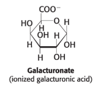
What is an example of a soluble fiber in animals that aids in digestion?
polygalacturonic acid (even though mammals can’t digest cellulose + other plant fibers)
Describe this diagram.
This shows that glycosidic bonds determine polysaccharide structure. The β-1,4 linkages favor straight chains, which are optimal for structural purposes. The α-1,4 linkages favor bent structures, which are more suitable for storage.
What are proteins with oligosaccharides attached called?
glycoproteins
What are the three main classes of glycoproteins?
Glycoproteins: The protein is the largest component by weight. Glycoproteins play a variety of roles, including as membrane proteins.
Proteoglycans: The protein is attached to a particular type of polysaccharide called a glycosaminoglycan. By weight, proteoglycans are mainly carbohydrate. Proteoglycans play structural roles or act as lubricants.
Mucins or mucoproteins: Like proteoglycans, mucins are predominantly carbohydrate. The protein is characteristically attached to the carbohydrate by N-acetylgalactosamine. Mucins are often lubricants.
In all cases of glycoproteins, carbohydrates (generally oligosaccharides) are attached to which two atoms?
the nitrogen atom in the side chain of asparagine (N-linkage)
or
the oxygen atom of the side chain of serine or threonine (O-linkage)
What do a N-linked polysaccharide consist of?
a common pentasaccharide core made of
three mannoses
six carbon sugar
two N-acetylgalactosamine units
additional monosaccharide may be attached to the core
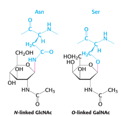
Describe this diagram.
This shows glycosidic bonds between proteins and carbohydrates.
Glycoproteins - proteins linked to oligosaccharides
N-linked - joined to asparagine in protein - E.R. and Golgi apparatus
O-Linked - joined to serine/threonine in protein - Golgi apparatus
What are proteoglycans attached to?
glycosaminoglycans (make up 95% of proteoglycan by weight)
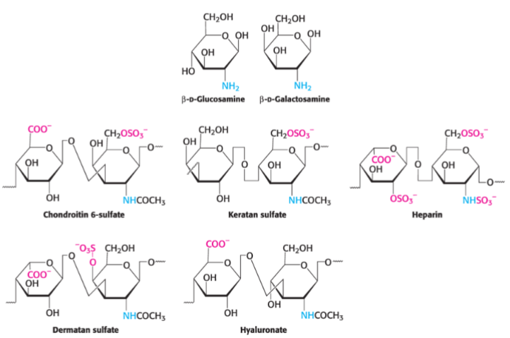
glycosaminoglycans
composed of repeating units of a disaccharide, one of which is a derivative of an amino sugar and one of which carries a negative charge, either as a carboxylate or sulfate
proteoglycans
key components of the extracellular matrix and serve as lubricants
mucopolysaccharidoses
pathological conditions that result from the inability to degrade proteoglycans (ex. Huler disease)
Describe this diagram.
This shows repeating units of glycosaminoglycans.
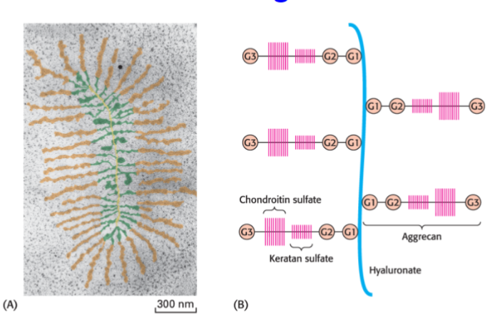
What is cartilage composed of?
the proteoglycan aggrecan and collagen
What does the glycosaminoglycan component of aggrecan do?
it cushions joints by releasing water on impact and then rebinding water
chitin
a glycosaminoglycan found in the exoskeleton of insects… one of the most abundant carbohydrates in the world
Describe this diagram.
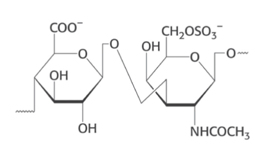
This shows the structure of chondroitin sulfate
Describe this diagram,
This shows the structure of proteoglycan from cartilage
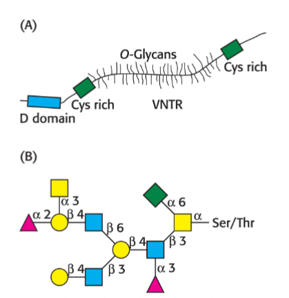
Describe the structure of the protein component in mucins.
it is extensively glycosylated to serine and threonine residues beginning with N-acetylgalactosamine
site of glycosylation
a region of the protein backbone rich in serines and threonines, called variable number of tandem repeats (VNTR)
Describe this diagram.
This shows the structure of mucin structure.
The human ABO blood groups reflect the specificity of _______________________.
glycosyltransferases
What oligosaccharide foundation do all blood groups share?
O