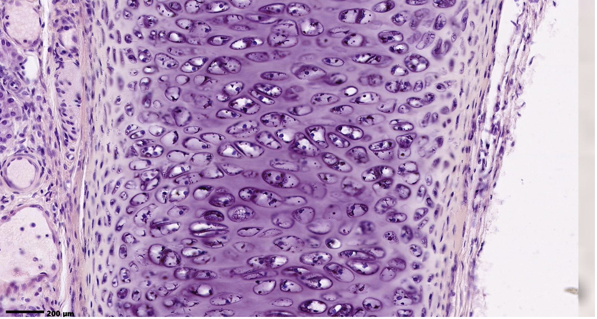
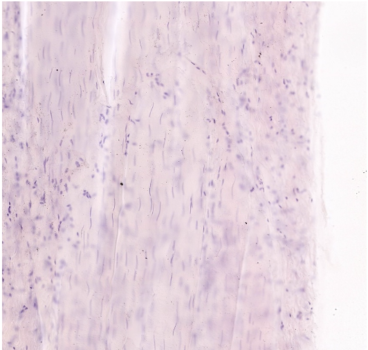
ID the predominant tissue in the field of view. Where can it be found?
hyaline cartilage; fetal skeletal tissue epiphyseal plates articular surface of synovial joints costal cartilages of rib cage cartilages of nasal cavity, larynx, rings of trachea plates in bronchi
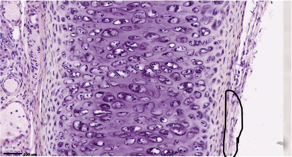
ID the layer within the circle.
Perichondrium (dense irregular tissue)
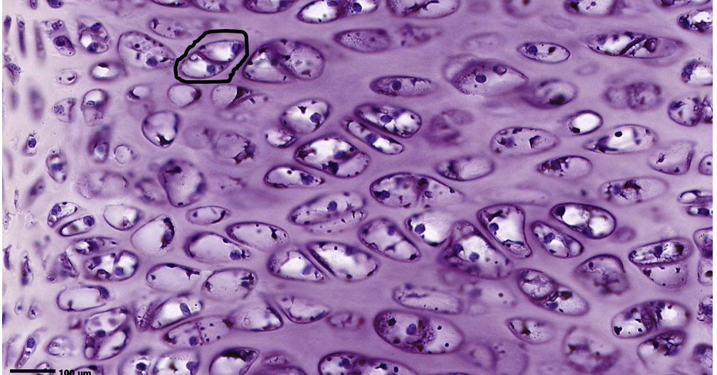
ID the entire structure within the circle.
Isogenous group
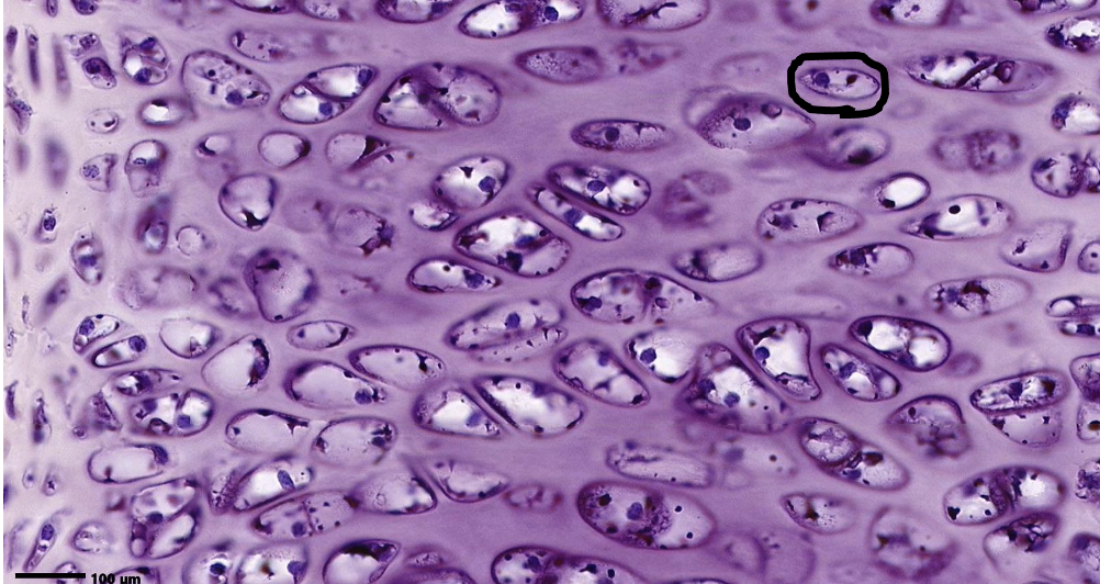
ID the cell within the circle
Chondrocyte
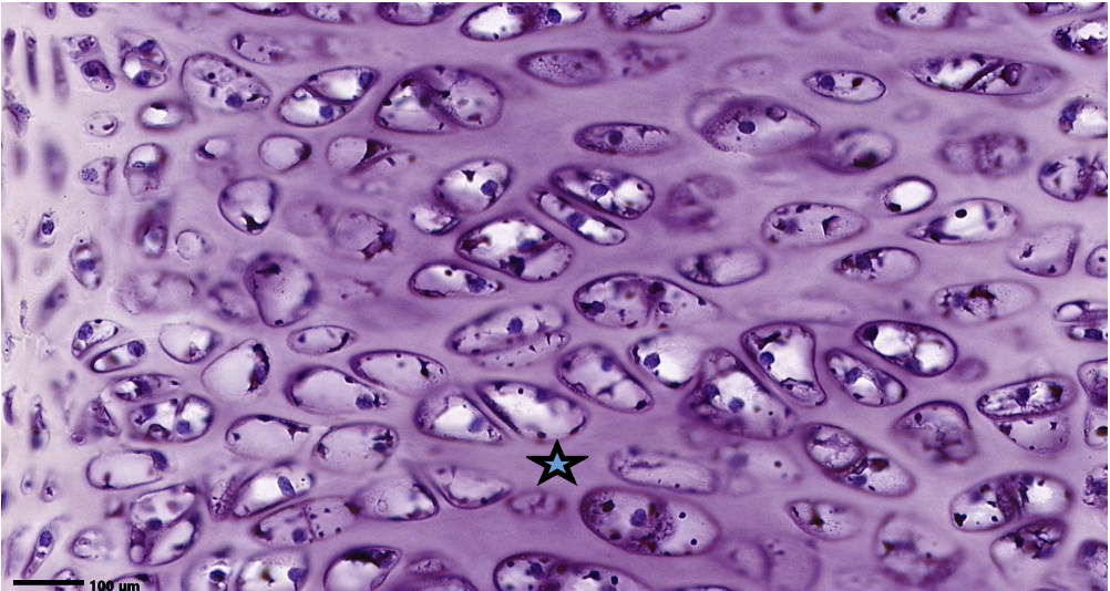
What is the predominant protein at the star?
type II collagen
ID the tissue in the field of view. What type of collagen is found here? Where can it be found?
Elastic cartilage Type II collagen (hyaline + elastin) external ear, the walls of the external acoustic meatus, the auditory (Eustachian) tube, the epiglottis and some of the larynx.
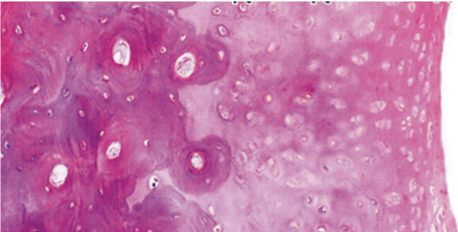
ID the tissue.
Auricular cartilage (left side is bone)
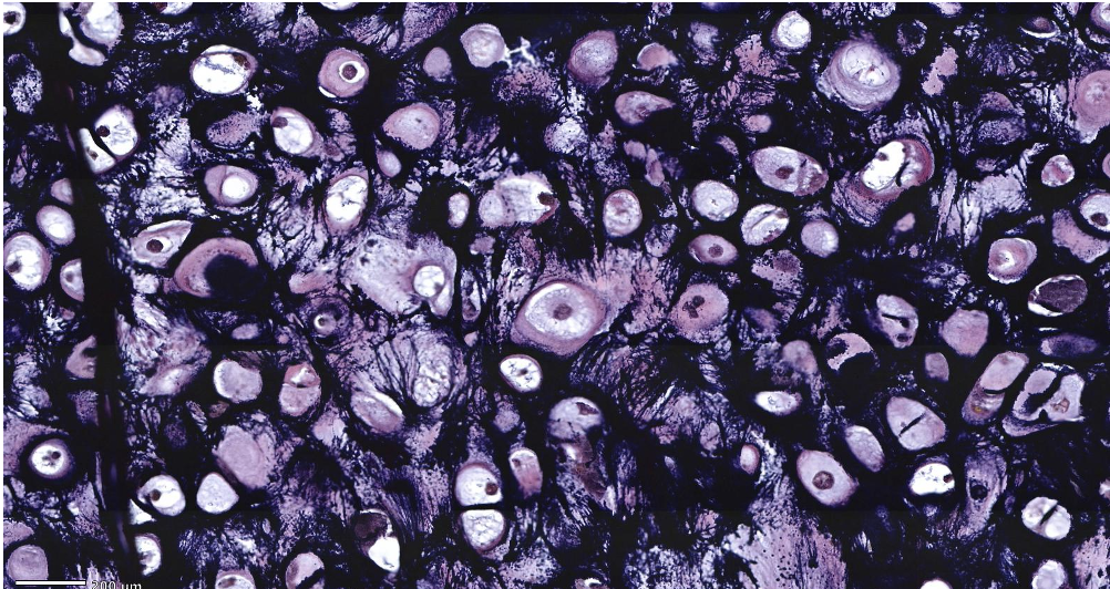
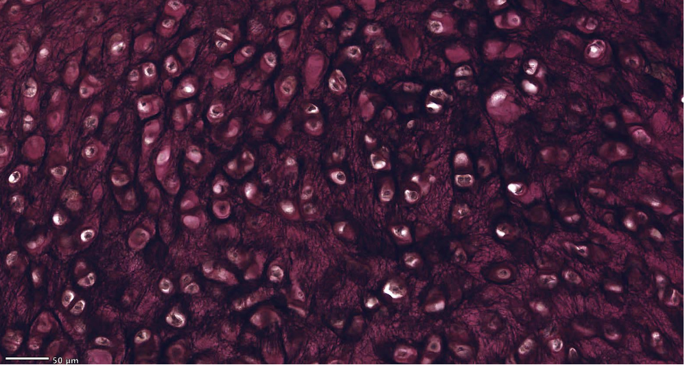
ID the tissue.
Elastic cartilage
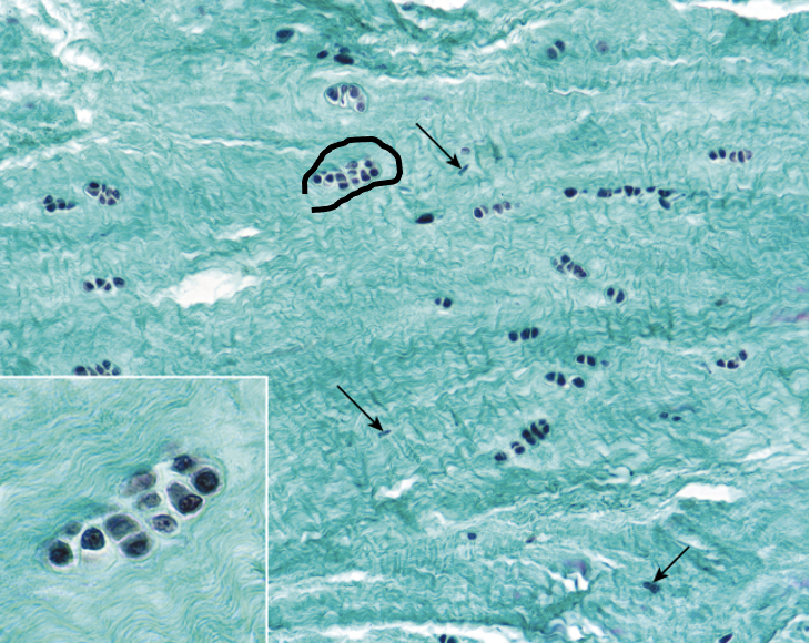
ID the cells within the circle. ID the tissue. Where is this found?
Chondrocytes Fibrocartilage intervertebral discs (IVD), pubic symphysis, articular discs of sternoclavicular joint and temporomandibular joint, menisci of knee joint, triangular fibrocartilage complex of wrist, and sites of tendon attachment to bone
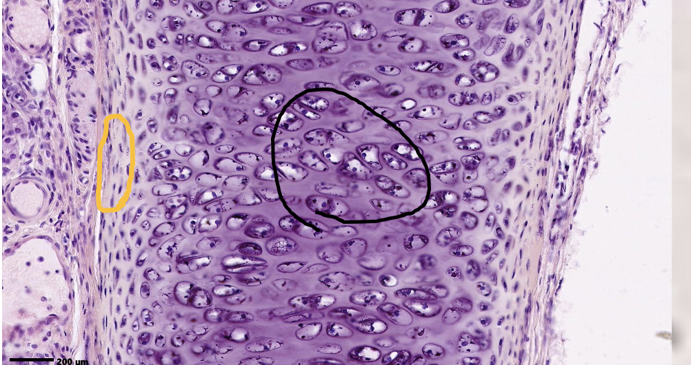
What is the mechanism of tissue growth within the black circle? What is the mechanism of tissue growth within the orange circle?
interstitial or mitosis of cells appositional
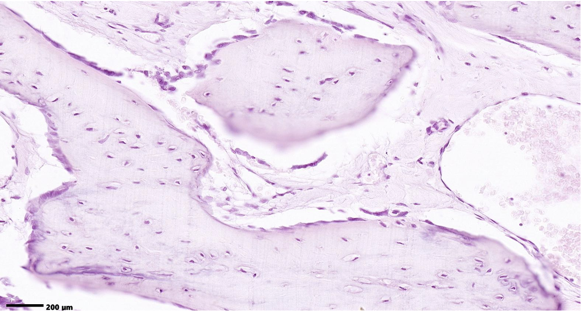
ID the tissue in the field of view.
Bone
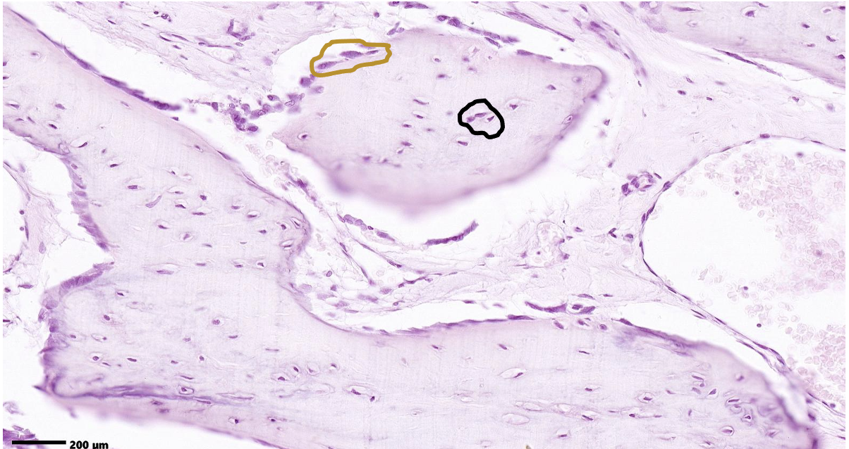
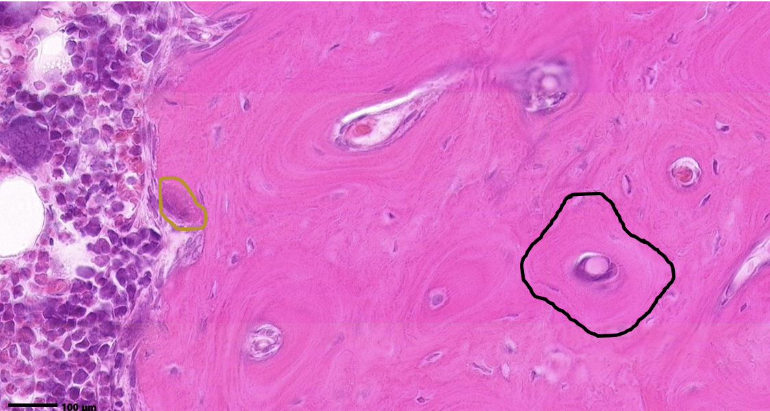
What method of bone formation is occurring here? ID the cells in the black circle. ID the cells in the brown circle.
intermembraneous ossification osteocytes osteoblasts
ID the entire structure within the black circle. ID the cell within the brown circle. Does it have PTH receptors?
osteon osteoclast no, osteoblasts do
ID the cells within the brown circle & their marker enzyme. ID the cells within the black circle.
osteoblasts with alkaline phosphatase osteocytes
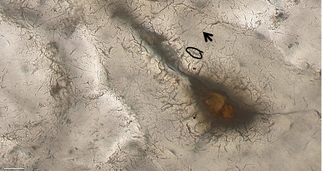
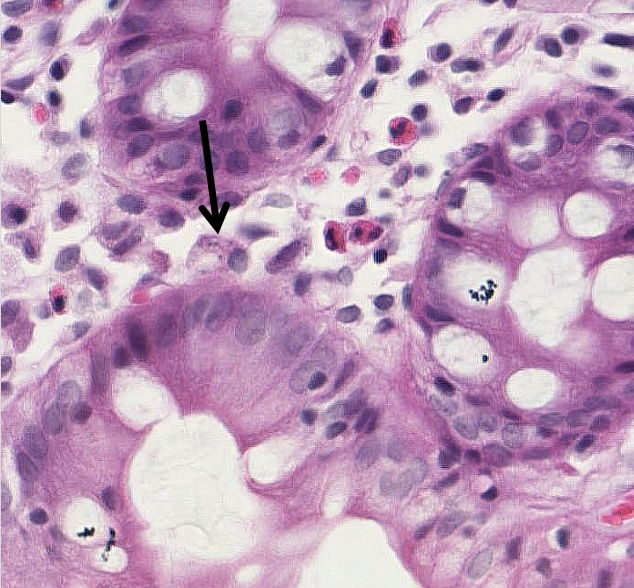
ID the cell within the black circle. ID the structure at the tip of the arrow. What is found inside this structure at the arrow?
osteocyte canaliculi that contain the cytoplasmic processes of osteocytes communicating with each other via gap junctions.
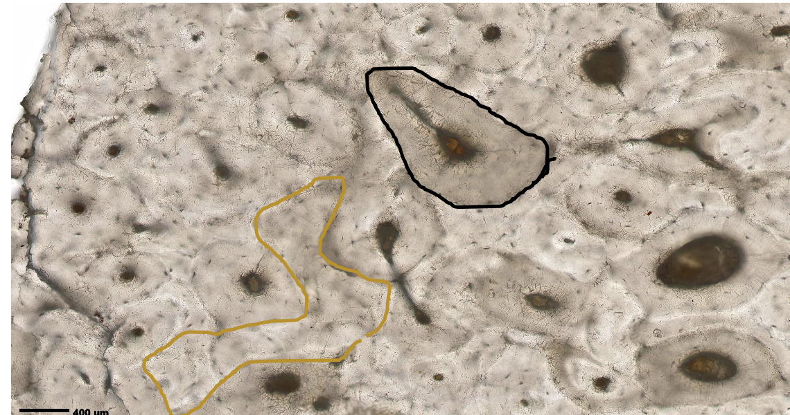
ID the entire structure within the black circle. ID the entire structure within the brown circle.
osteon with a Haversian canal in the middle & a Volkman canal coming off it interstitial lamellae (remodeled osteon)
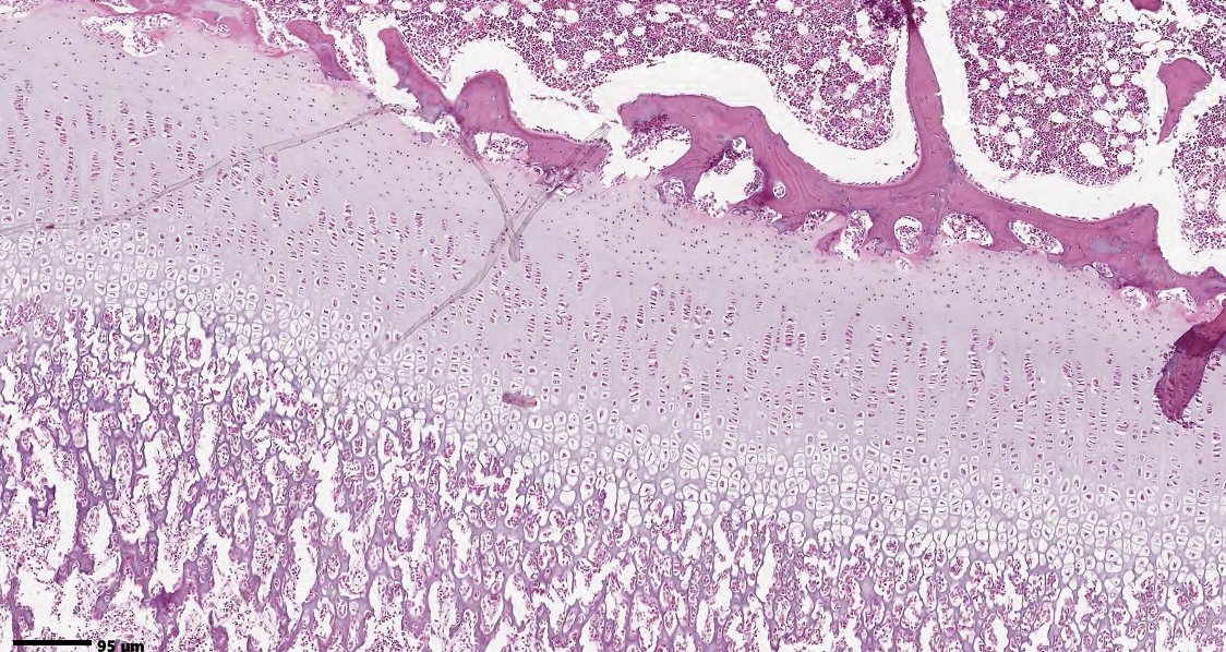
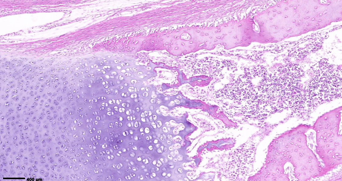
What is the mechanism of bone formation shown in this slide? Where is the first site of bone formation with this mechanism.
endochondrial ossification bony collar that forms at the diaphysis via appositional growth
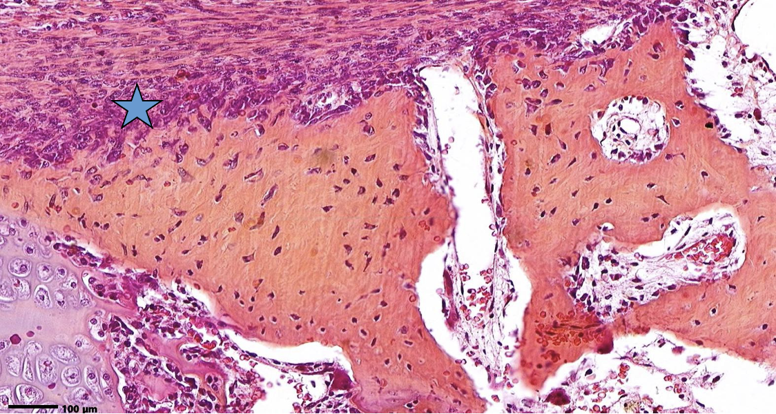
ID the layer at the star.
periosteum
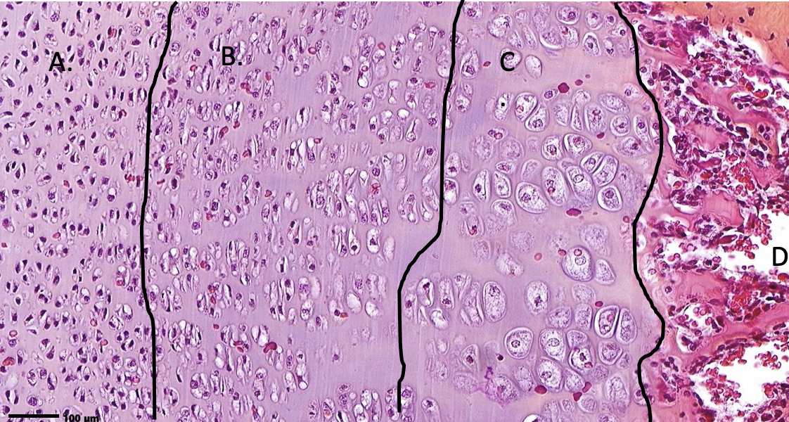
ID the zones. ID the 2 types of tissue.
A = zone of reserve cartilage B = zone of proliferationC = zone of hypertrophy D = zone of calcified cartilage, top part is bone hyaline cartilage (type II collagen) and bone (type I collagen)
ID the zones on your own. How do long bones grow in length and width?
Length by interstitial growth of hyaline cartilage (zone of proliferation) Width by appositional growth (precursors in periosteum become osteoblasts producing new bone matrix.)
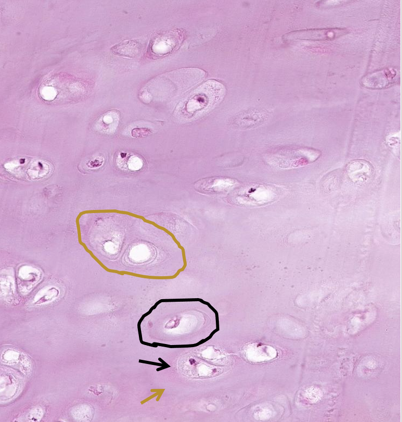
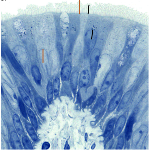
ID what is in the brown & black circle. What types of matrix are found at the brown & black arrows?
isogenous group chondrocyte territorial matrix (further out would be interterritorial) capsular matrix
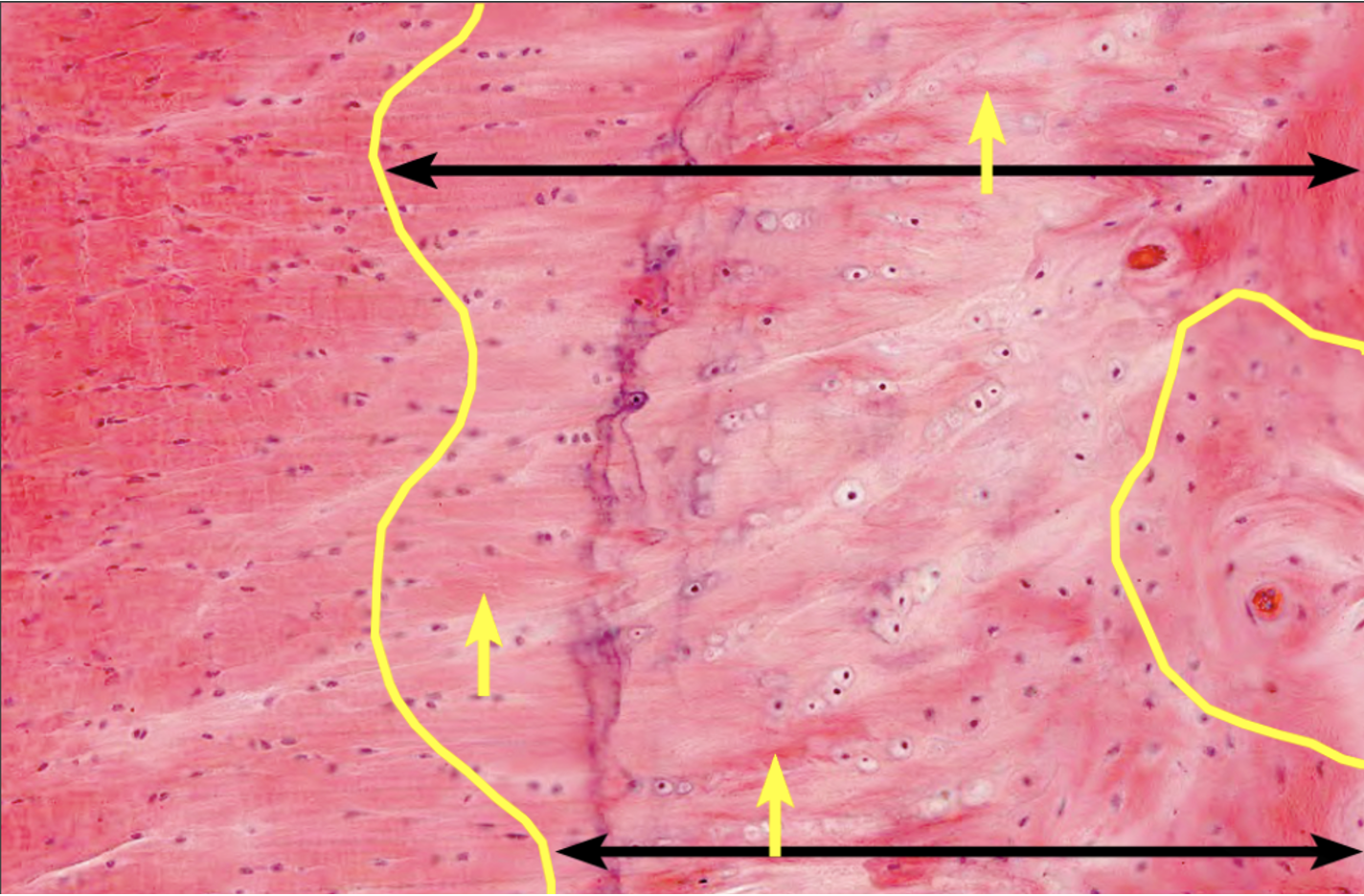
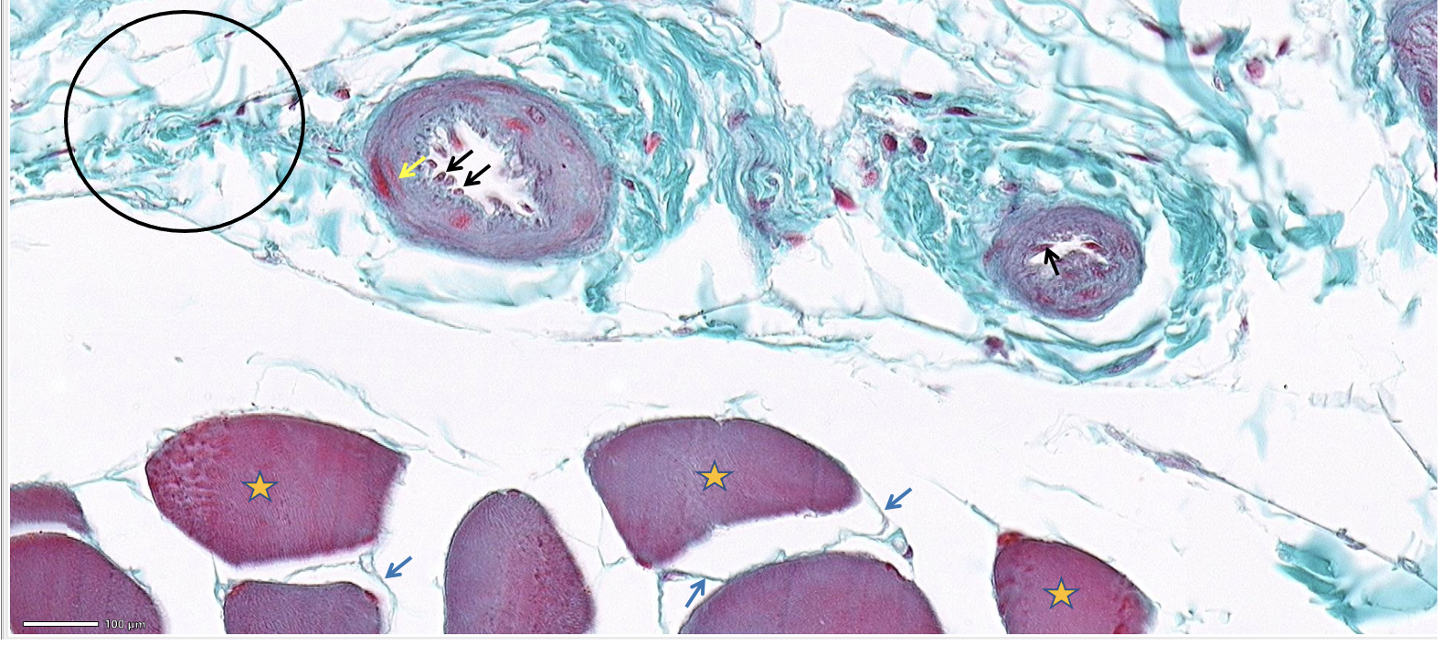
What tissue is found to the left of the yellow line, in between the black arrows, and in the yellow circle?
dense connective tissue fibrocartilage bone
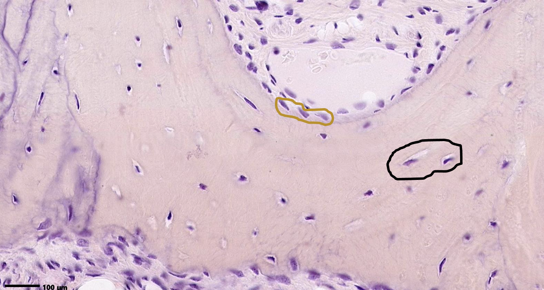
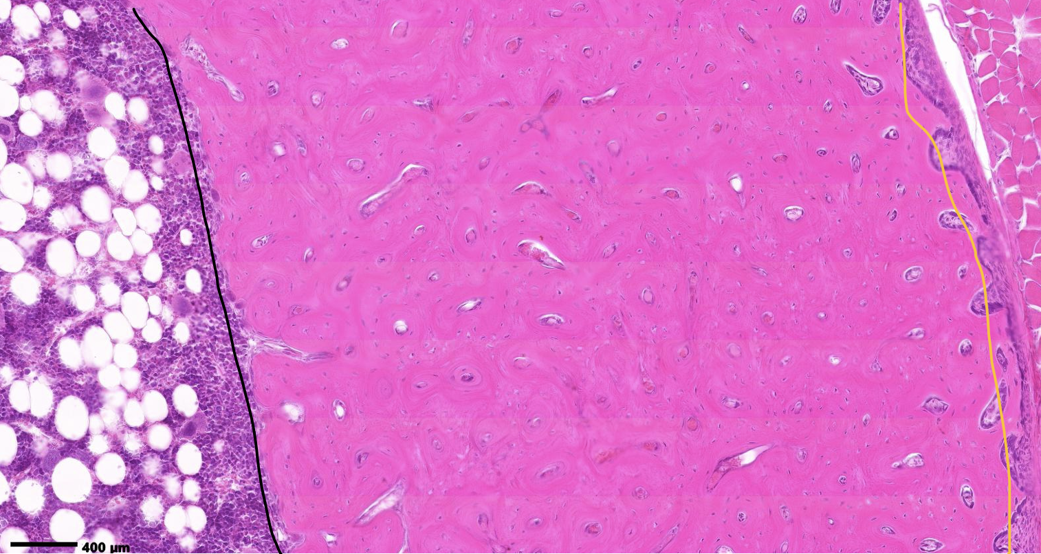
What is found at the black and gold lines?
Endosteum & periosteum
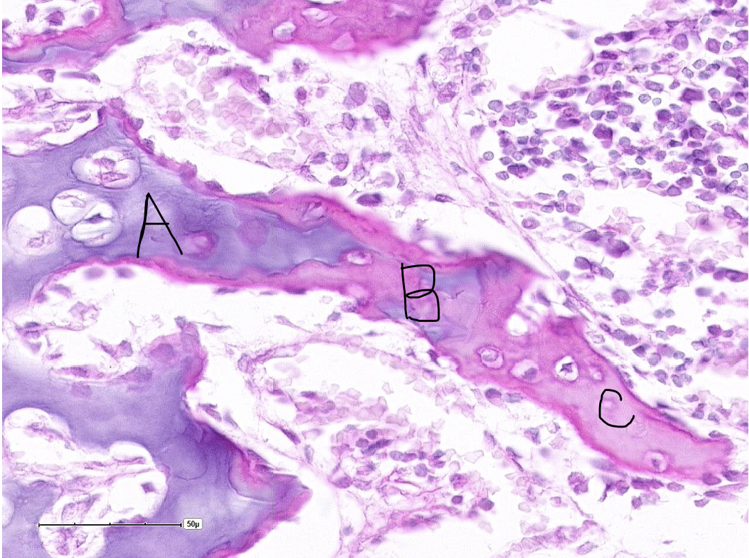
What is found at each of the letters?
A: calcified cartilage B: mixed spicule C: bone
ID the cells at the tips of the black arrows
ID the cell at the tip of the yellow arrow AND what did Dr. Harp call these cells?
ID the layer and classify the tissue within the black circle.
ID the layer at the tip of the blue arrow.
ID the cells with the orange stars.
endothelial cells
smooth muscle cells AKA leimyocyte
perimyosium & dense irregular CT
endomyosium
skeletal muscle cells
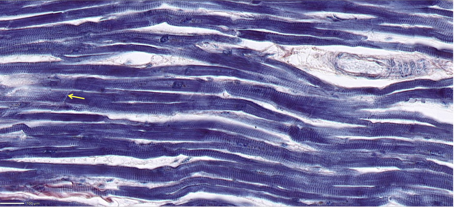
Classify the predominant tissue in the field of view.
ID the structure at the tip of the arrow and ID 3 of its components.
cardiac muscle
intercalated disc which contains gap junctions, desmosomes, and fascia adherens.
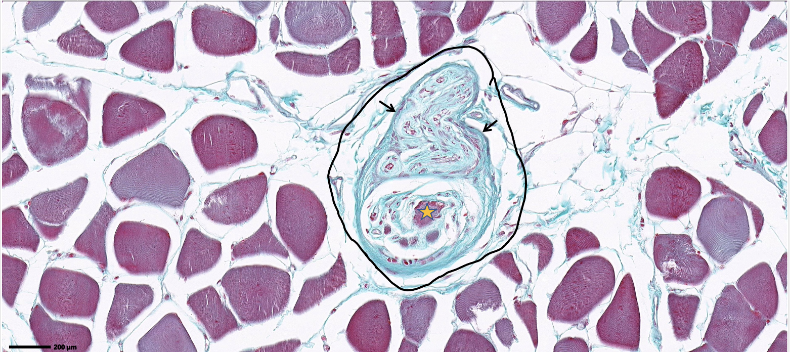
ID the entire structure within the circle and where is this structure found?
ID the cell with the orange star
ID the structure at the tip of the black arrow
muscle spindle organ, found in the perimyosium layer of CT
intrafusal fiber.
CT capsule that surrounds the muscle spindle organ.
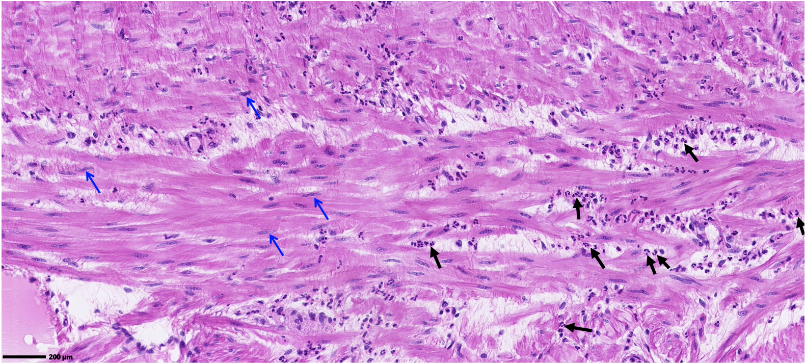
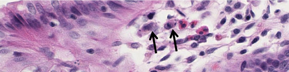
ID the cells at the black arrows.
ID the cells at the blue arrows
neutrophils
smooth muscle cells
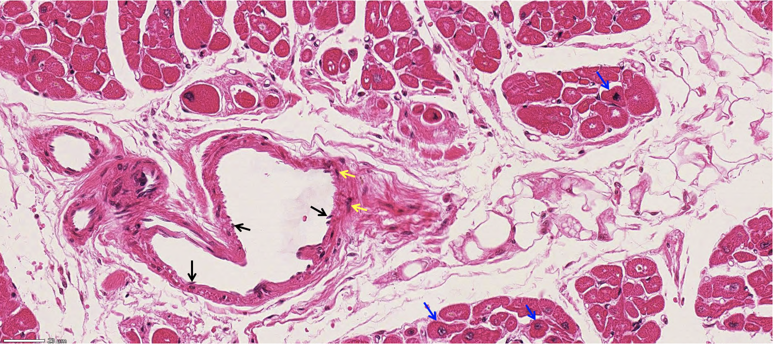
ID the cells at the tip of the blue arrows.
ID the cells at the tips of the black arrows.
ID the cells at the tip of the yellow arrows.
cardiac muscle cells
endothelial cells
smooth muscle cells
Classify the predominant tissue. What cells are at the tip of the blue arrows & what layer are they found in?
cardiac muscle purkinje fibers found in the subendocardial layer which is below the endocardium
Classify the epithelium
ID the predominant connective tissue
ID the cells at the arrows
transitional
dense irregular CT (made up of type I collagen)
smooth muscle cells
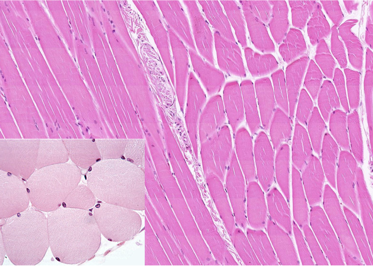
What type of tissue is this? What are the cells in the bottom left corner?
skeletal muscle & skeletal muscle cells
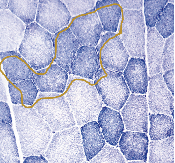
What type of fiber is in the gold circle?
type I fibers (slow-twitch)
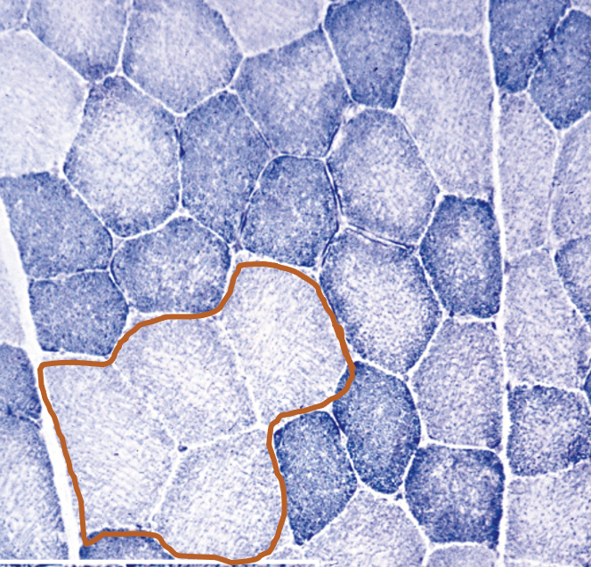
What type of fiber is in the brown circle?
type IIb fibers (fast-twitch)
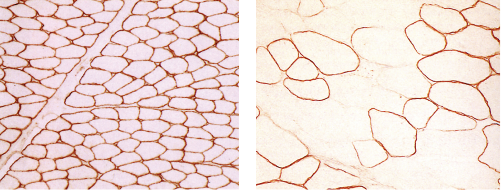
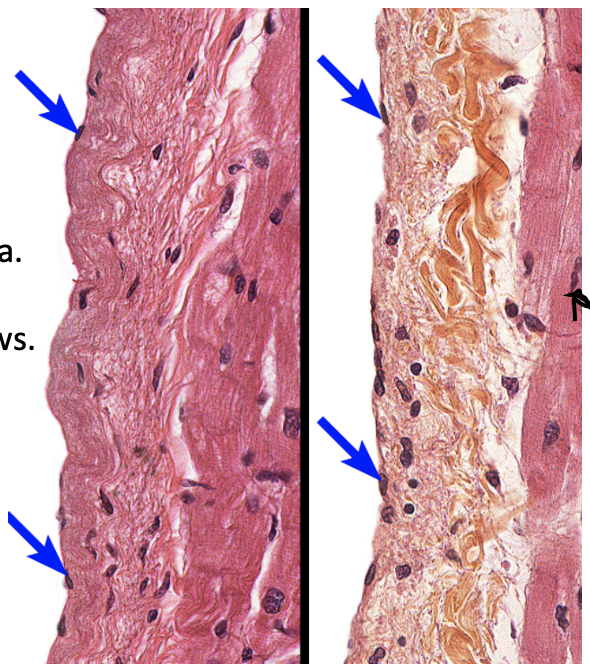
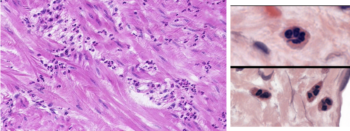
What do these 2 images represent?
Left: healthy dystrophin Right: dystrophin in duchenne muscular dystrophy
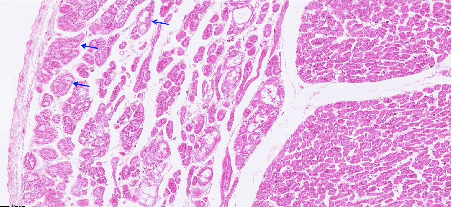
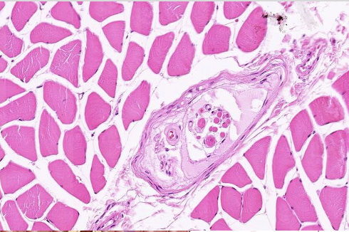
What is found in the middle of this skeletal muscle? What nerve fibers does it contain?
muscle spindle organ with sensory afferent & motor efferent nerve fibers
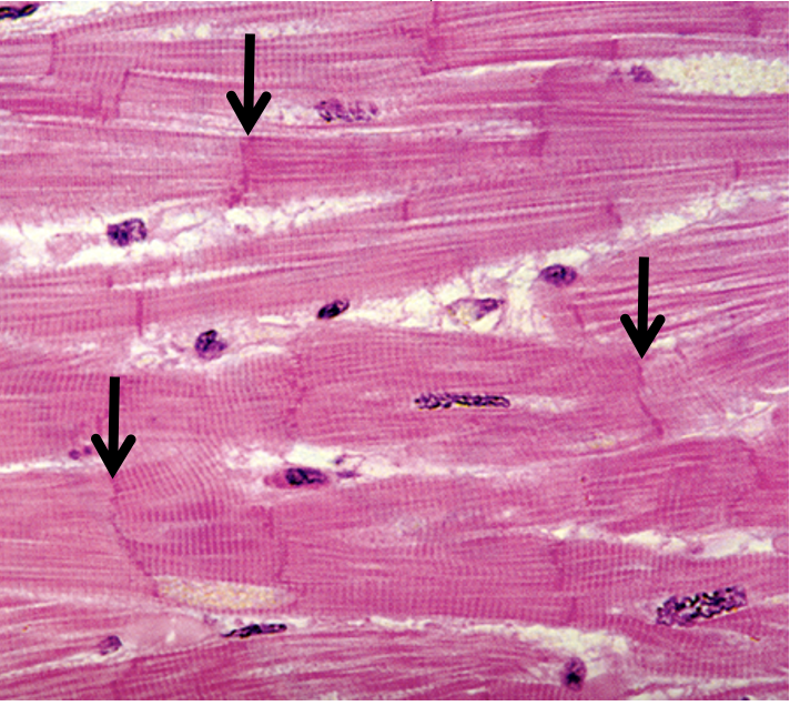
What type of tissue is this?
What is at the tip of the black arrows?
cardiac muscle
intercalated discs
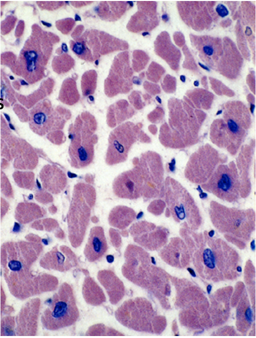
What tissue is shown in this cross-section? What cells are found here?
cardiac muscle & cardiac muscle cells
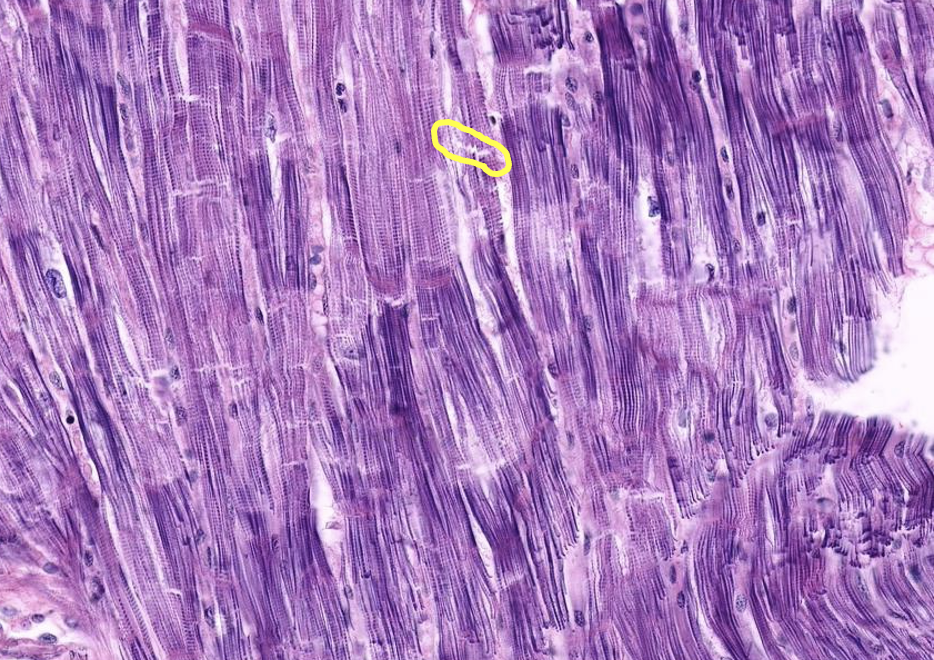
What tissue is shown?
cardiac muscle
What is in the yellow circle?
intercalated disc of cardiac muscle
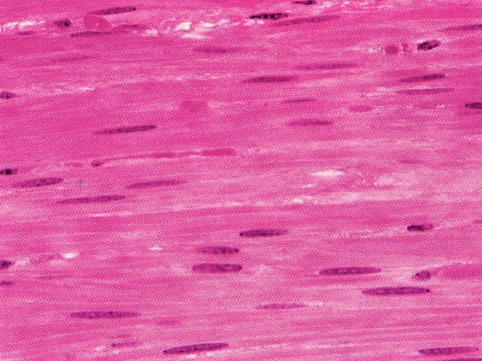
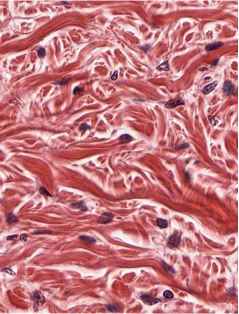
What tissue is shown?
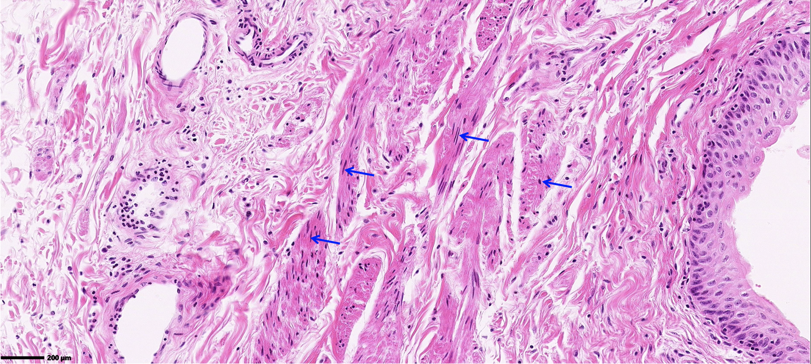
smooth muscle
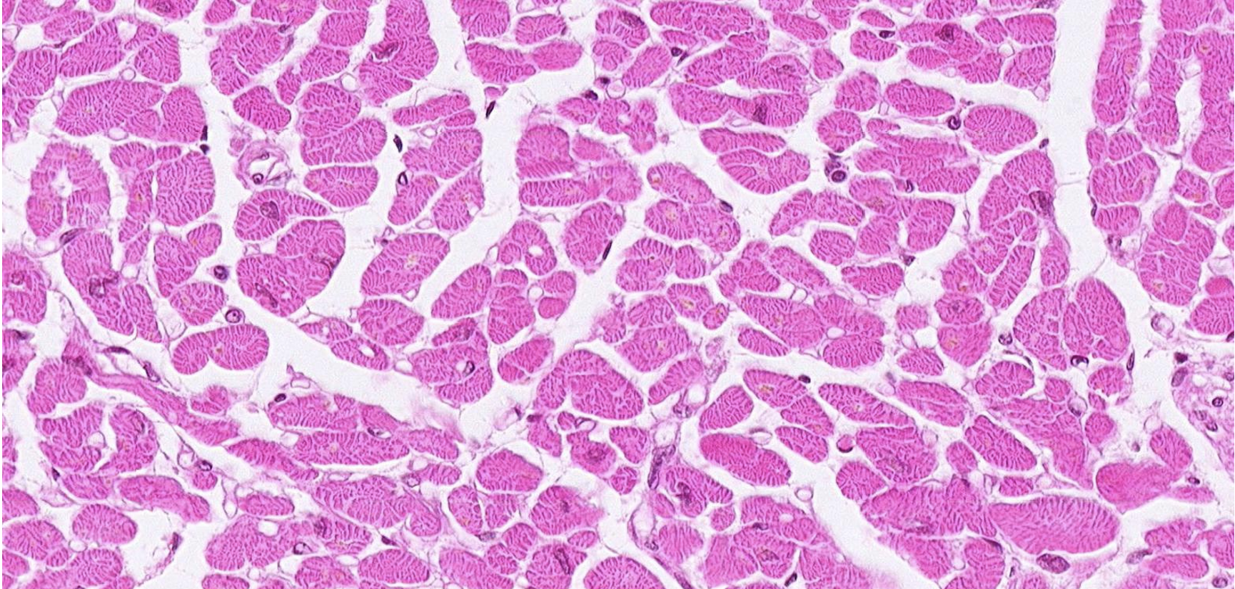
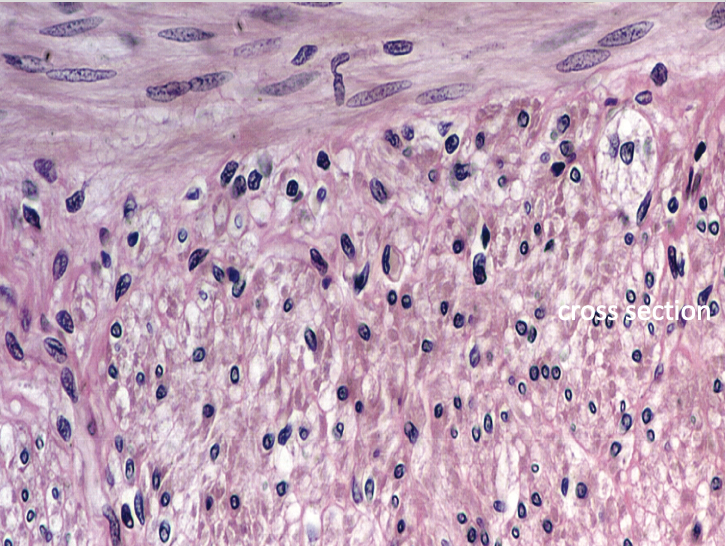
What tissue does this cross-section show?
smooth muscle
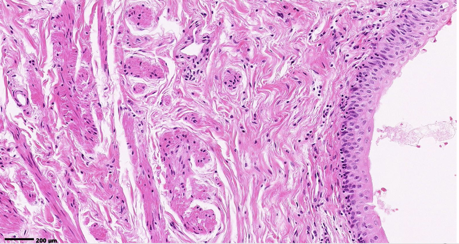
What tissue is shown? bonus point if you know where it is
smooth muscle, ureter
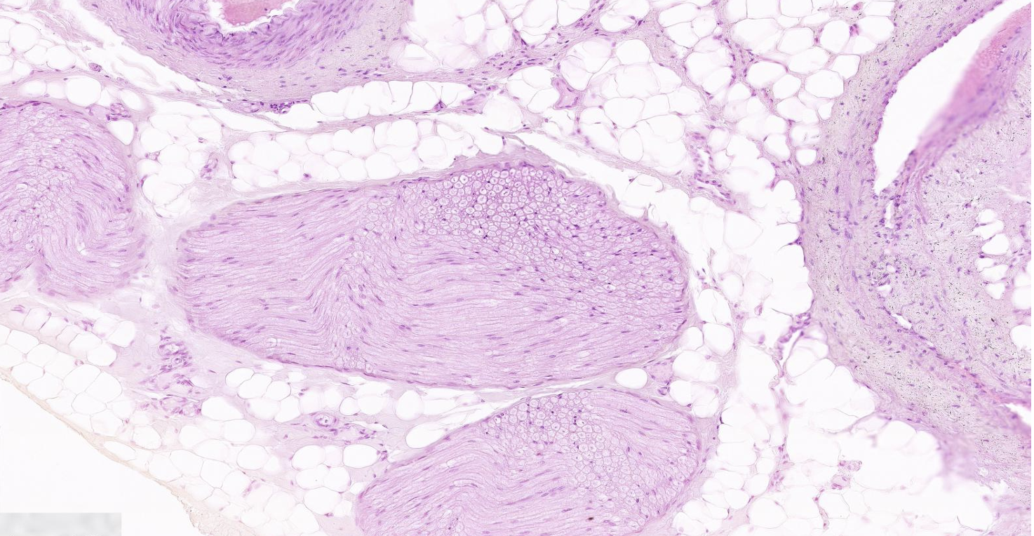
What does this image show?
peripheral nerve
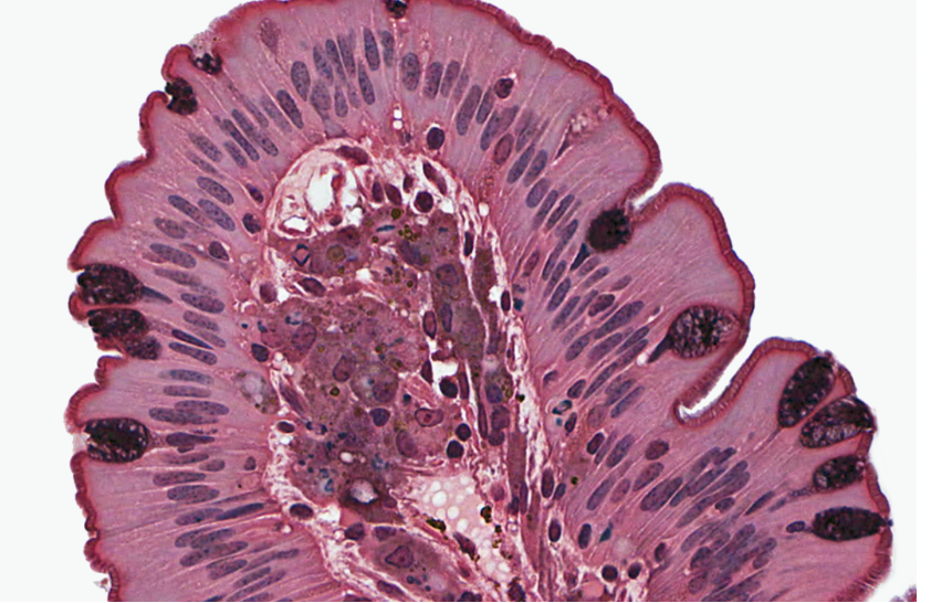
Classify the epithelium. What is the function of this epithelium?
simple columnar epithelium with goblet cells and microvilli. epithelium is absorption & goblet cells is lubrication.
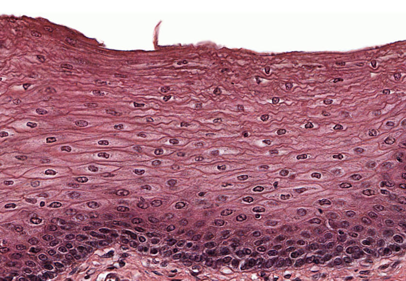
Classify the epithelium. What is the function of this epithelium?
stratified squamous wet (regardless of whether it is wet or dry) is protection.
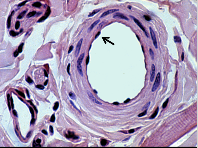
ID the cell at the arrow then classify the epithelium at the arrow.
endothelial cells & squamous epithelium
ID the cell at the arrow.ID the product secreted.
goblet cell (unicellular) that secretes mucous
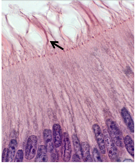
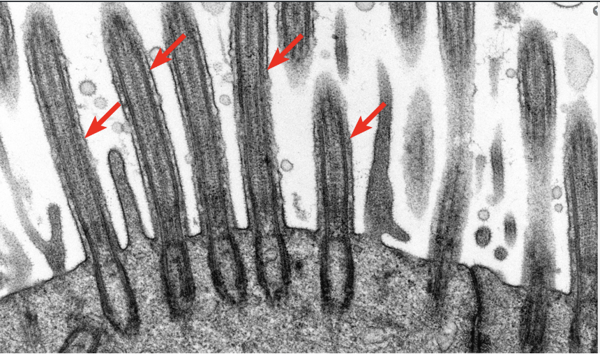
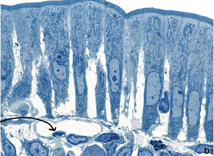
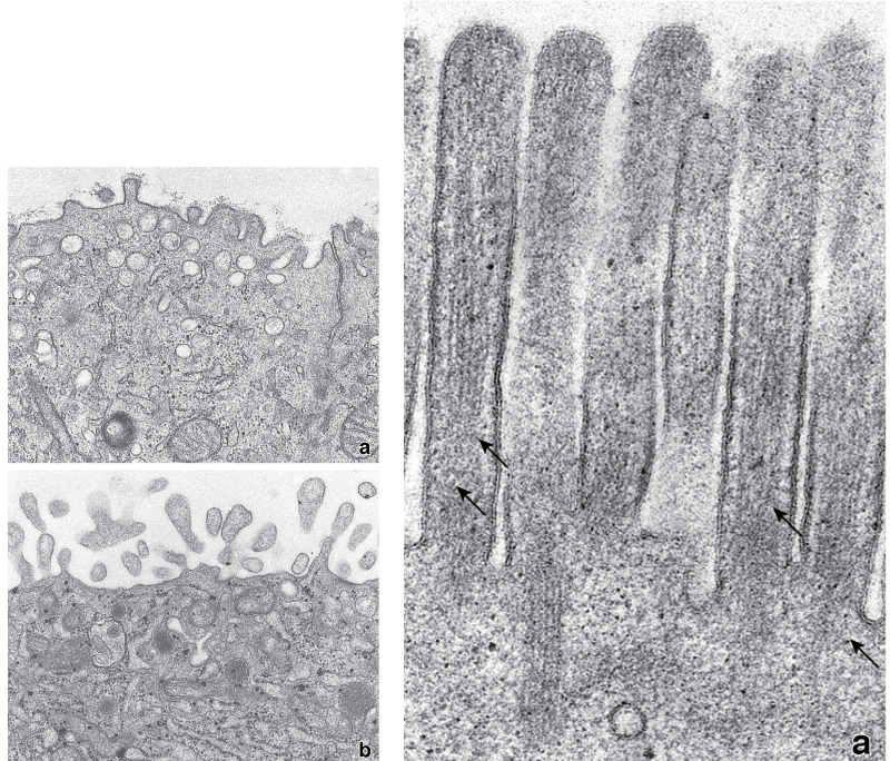
ID the border modification at the arrow and then state what forms its core.
stereocilia, with a core of microfilaments or actin filaments.
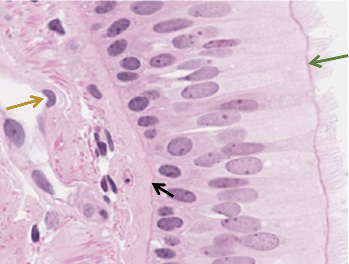
Classify the epithelium ID the structure at the black arrow ID the cell at the gold arrow ID the organelle at the green arrow
pseudostratified columnar with cilia basement membrane endothelial cell lining a blood vessel the basal body
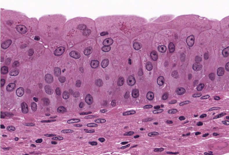
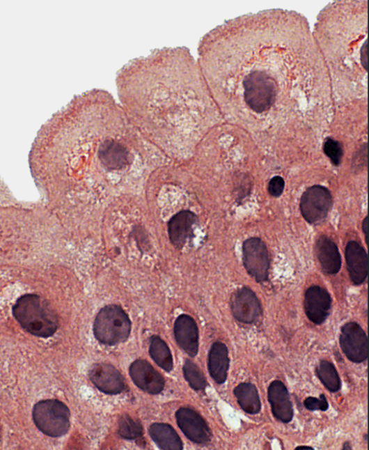
Classify the epithelium.
transitional epithelium
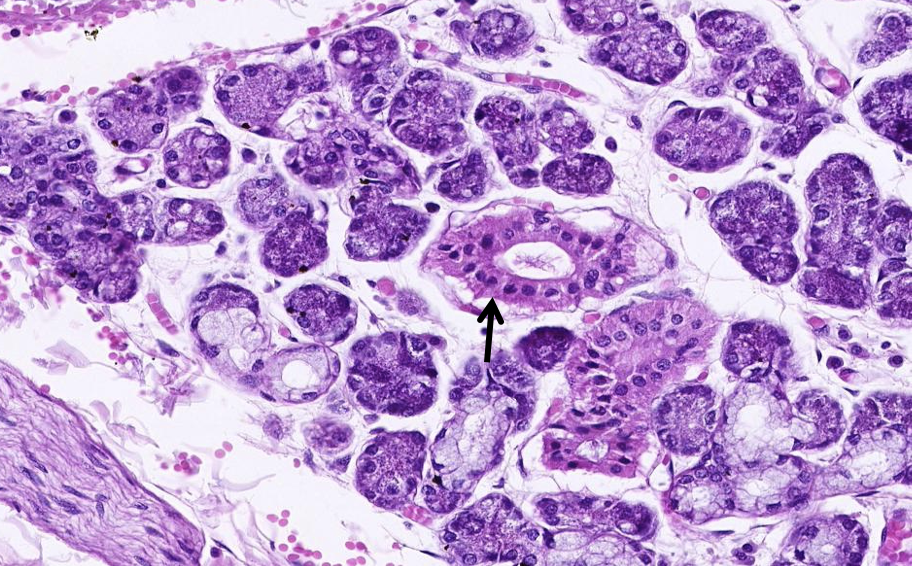
ID the structure at the arrow then classify its epithelium.ID the gland by product secreted.
a duct with simple cuboidal epitheliumThe gland is secreting both serous and mucous products and would be classified as a mixed gland. Note the washed out cytoplasm and flat nuclei of the mucous secreting cells compared to the basophilic (purple) cytoplasm and round nuclei of the serous secreting cells.
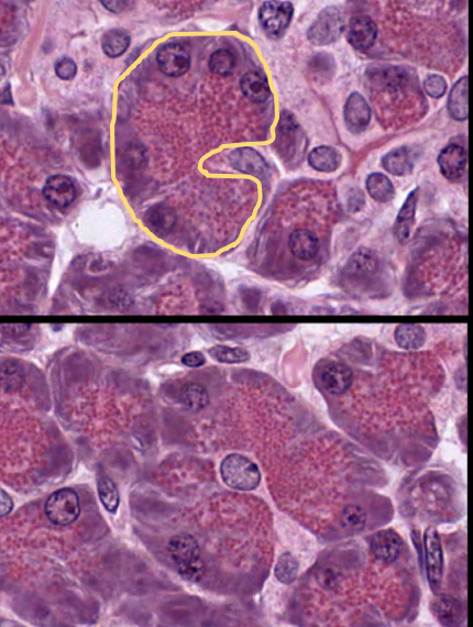
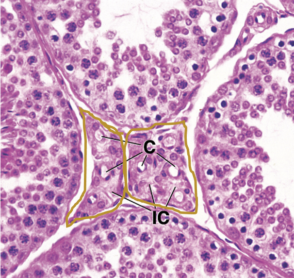
This is a section of the pancreas. ID the gland by the product secreted. Glandular cells are outlined.
The cells outlined have round nuclei with the nuclei located at the base of the cell while the secretory granules are apically located. These cells will secrete apically releasing their granular contents into a duct (exocrine secretion). The round nuclei is consistent with serous glands. No lipid secreting cells in the field of view.
This is a section of the kidney. A. ID the epithelium at the star. B. ID the epithelium at the arrow.
A. simple cuboidal (star is within the lumen of a collecting duct BUT JUST AN FYI) B. simple squamous. (The cell at the tip of the arrow is an endothelial cell)
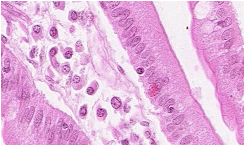
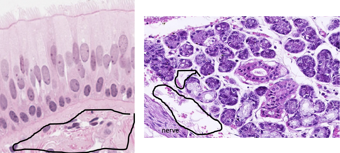
ID the epithelium. What function is it involved in?
simple columnar epithelium with microvilli (notice it looks like a crew cut) This type of epithelium is involved in absorption.
On this slide you should also be able to identify plasma cells underneath the epithelium and two round nuclei within the epithelium that are lymphocytes.
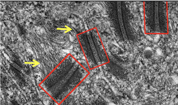
ID the structures in the boxes and at the arrows.
red boxes are desmosomes that are joining adjacent cells together. yellow arrows are keratin intermediate filaments.
ID the border modification.
The red arrows are indicating motile cilia. (Primary cilia are found only one per cell.)
What cells are fund within the gold square?
epitheloid cells
What type of cell is the arrow pointing to?
endothelial cells = simple squamous
What type of tissue are the blue arrows pointing to?
endocardium = simple squamous epithelium
What type of tissue is the black arrows pointing to?
mesothelium = simple squamous epithelium
What type of epithelium is shown?
transitional
What border modification is shown?
microvilli
What border modification is shown?
cilia
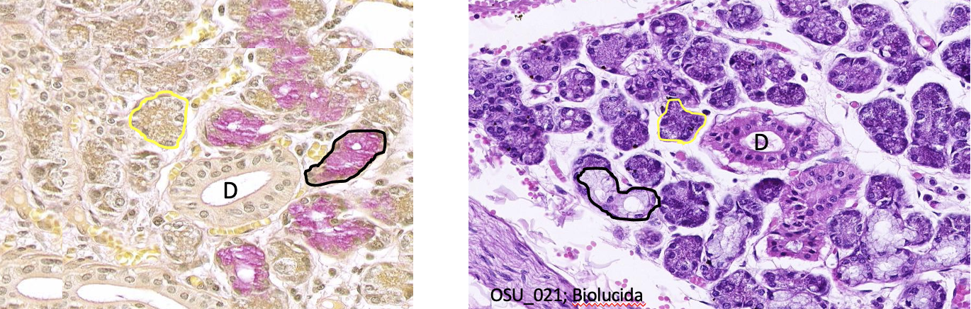
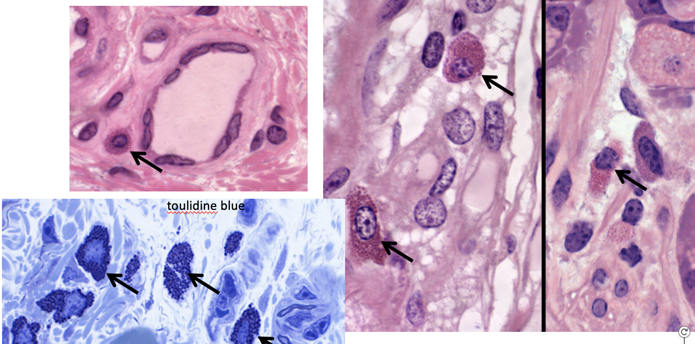
What is found in the yellow & black circles in these images? What is represented by "D"?
serous secreting cells & mucous secreting cells ducts for secretions [Together the serous and mucous secreting cells as well as their ducts make up the parenchyma of the gland (functional tissue)]
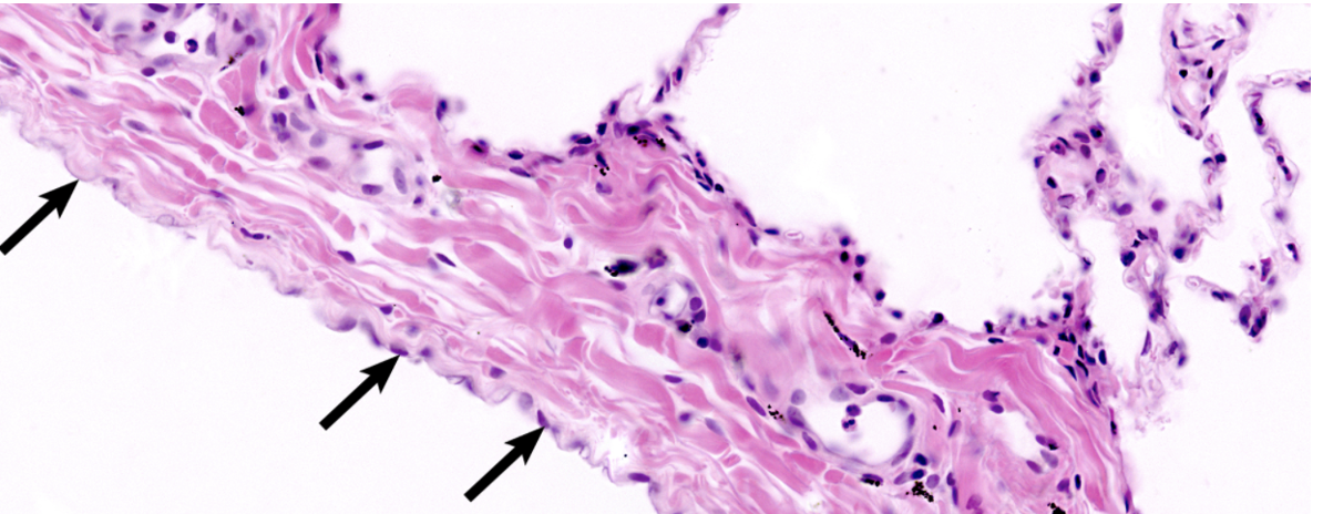
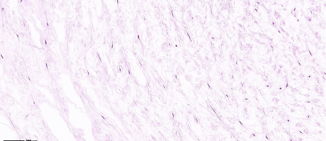
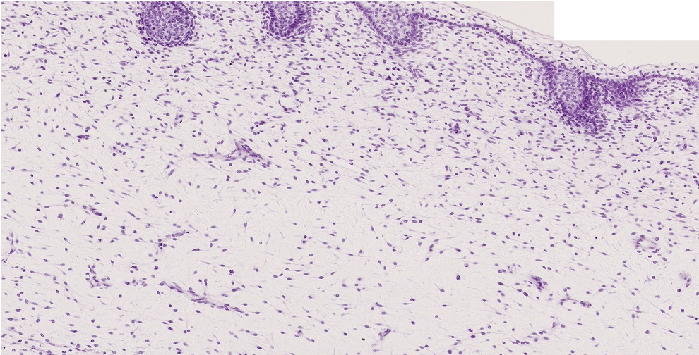
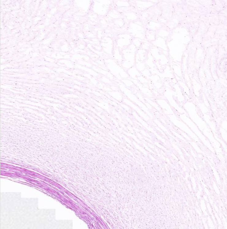
Classify the predominant CT in the field of view.
mucous CT, It is fiber rich and cell poor
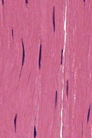
Classify the predominant CT in the field of view.
dense regular CT aka tendon
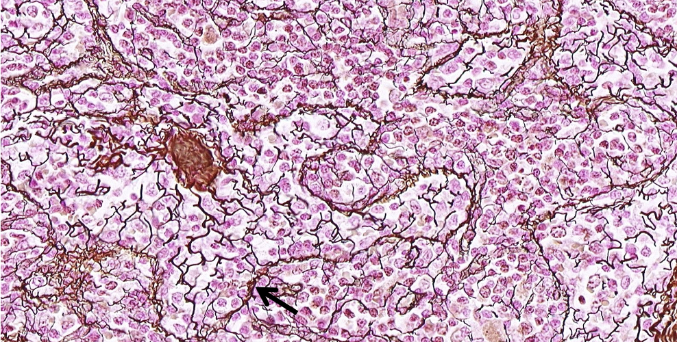
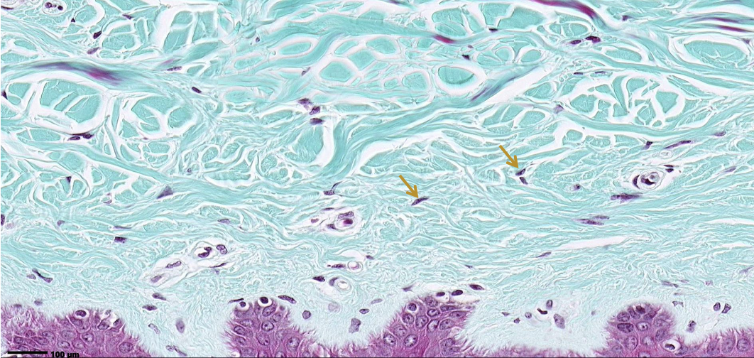
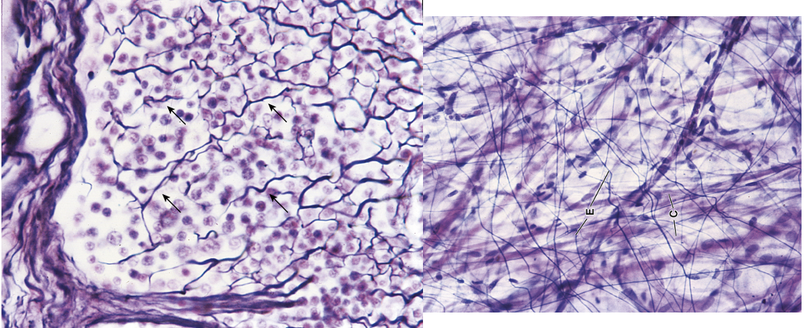
A. ID the structure at the arrow. B. ID the protein at the arrow
A. reticular fibers B. type III collagen
A. ID the cells at the arrows. B. List 5 products
A. mast cells B. histamine, heparin, eosinophilic chemotactic factor (ECF), neutrophilic chemotactic factor (NCF), and tryptase
ID the predominate CT in the field of view.
mesenchymal CT
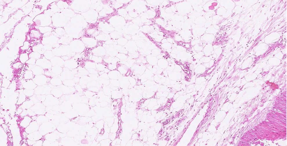
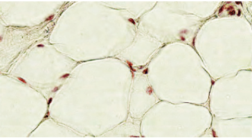
ID the predominant CT in the field of view.
adipose tissue
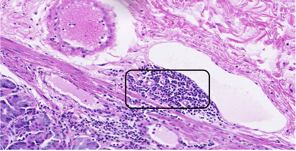
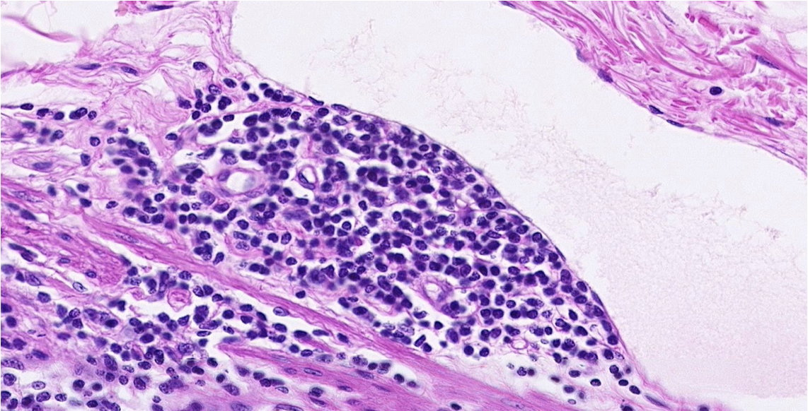
ID the cells in the circle. Classify the predominant CT.
lymphocytes & dense irregular CT
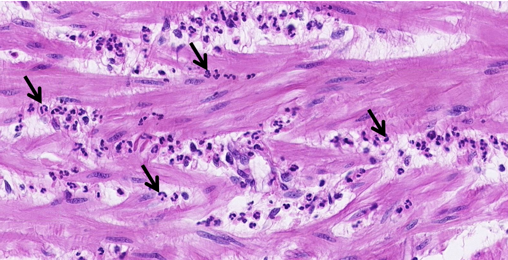
ID the cells at the arrows.
neutrophils
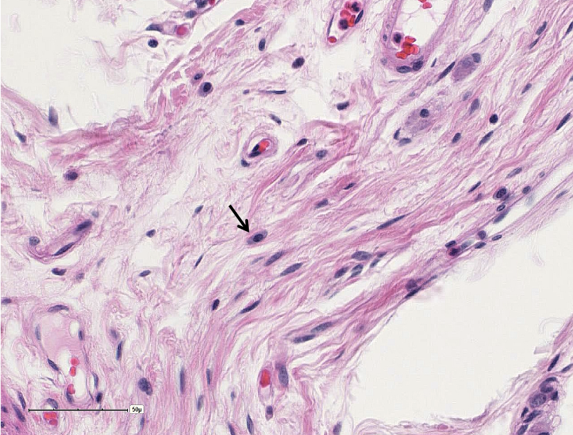
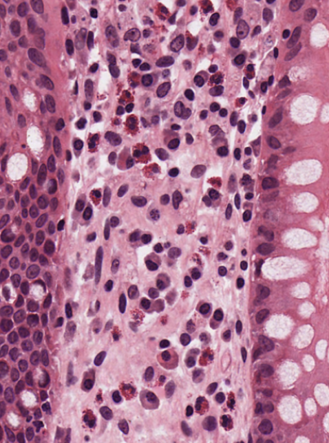
Classify the predominant CT in the field of view. ID the cell at the arrow.
dense irregular mast cell
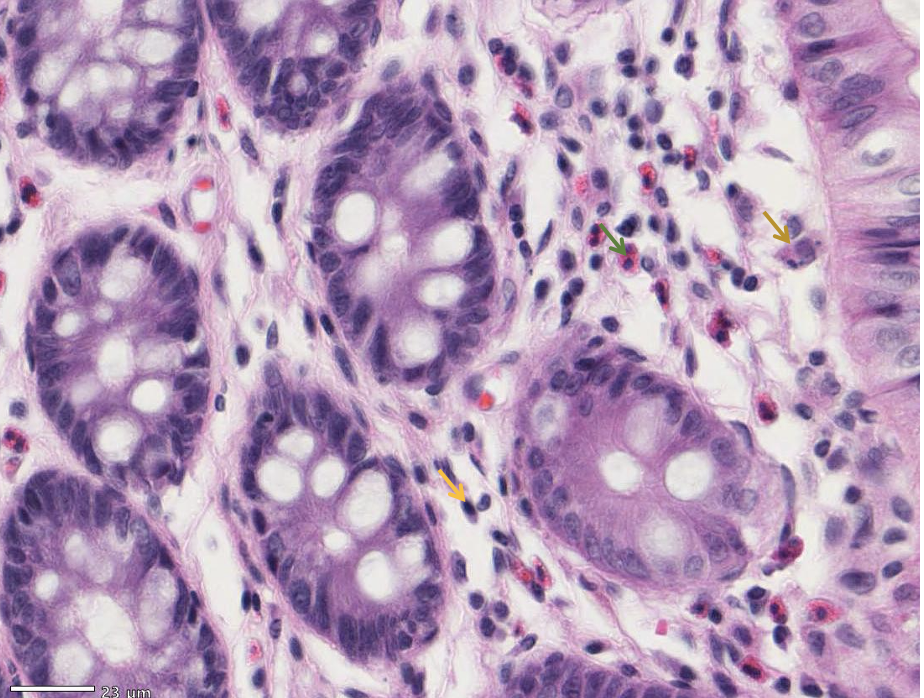
ID the cells at the arrows.
brownish-gold arrow – a macrophage. green arrow - an eosinophil. orange arrow - a plasma cell.
Classify the predominant CT. ID the cells at the arrows.
dense irregular & fibroblasts (make the type I collagen)
What tissue is found in the black circles? basic.
connective tissue
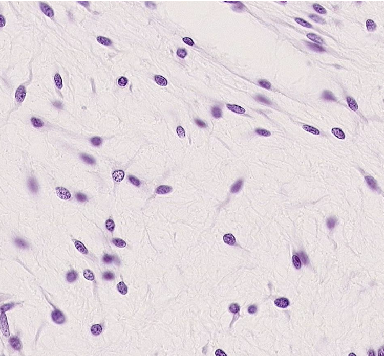
What type of tissue is shown here?
mucous CT
What type of tissue is shown here?
mesenchyme CT
What type of tissue is shown here?
loose CT
What type of tissue is shown here?
dense irregular CT
What type of tissue is shown here? What cell type is shown?
dense regular CT & fibroblasts
What type of tissue is shown here?
adipose
What type of fiber is shown here? What collagen is it made of?
reticular fibers - type III collagen
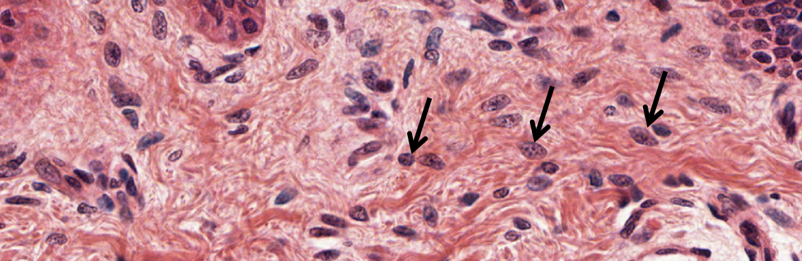
What cells are the black arrows pointing to?
fibroblasts
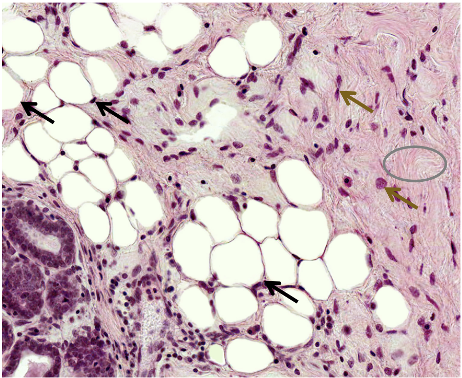
What is shown at the black arrow, gold arrow, and gray circle?
Black arrow = adipocytes; white adipose tissue (adult) Gold arrow = fibroblasts Gray circle = type I collagen
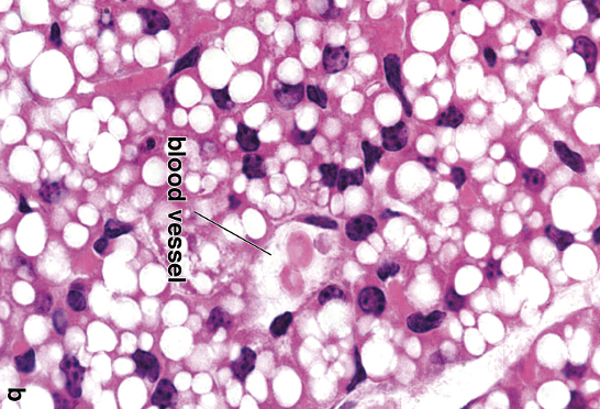
What type of tissue is shown?
brown adipose (fetal)
What is shown at the black arrow?
macrophage (aka histiocyte)
What is shown at the black arrow?
macrophage (aka histiocyte)
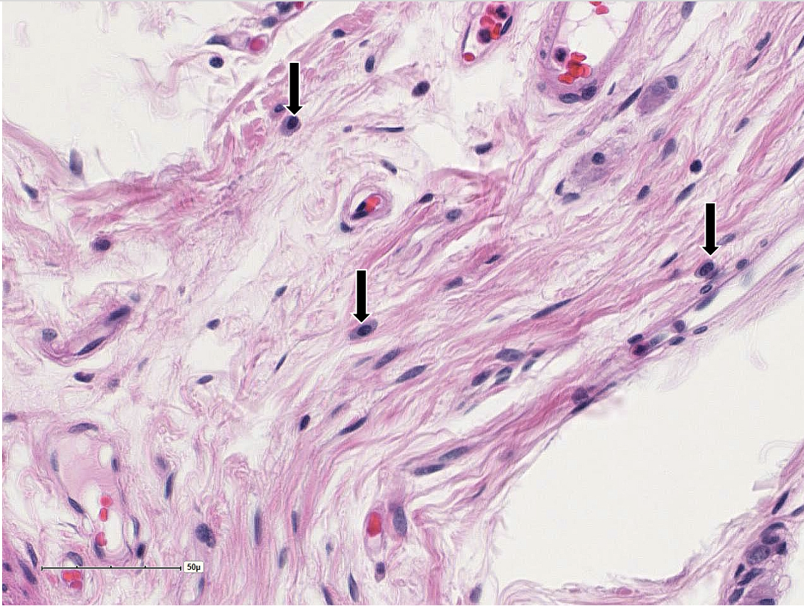
What is shown at all these black arrows?
mast cells
What is shown at the black arrows?
mast cells
What cells are shown in this image? (purple)
lymphocytes
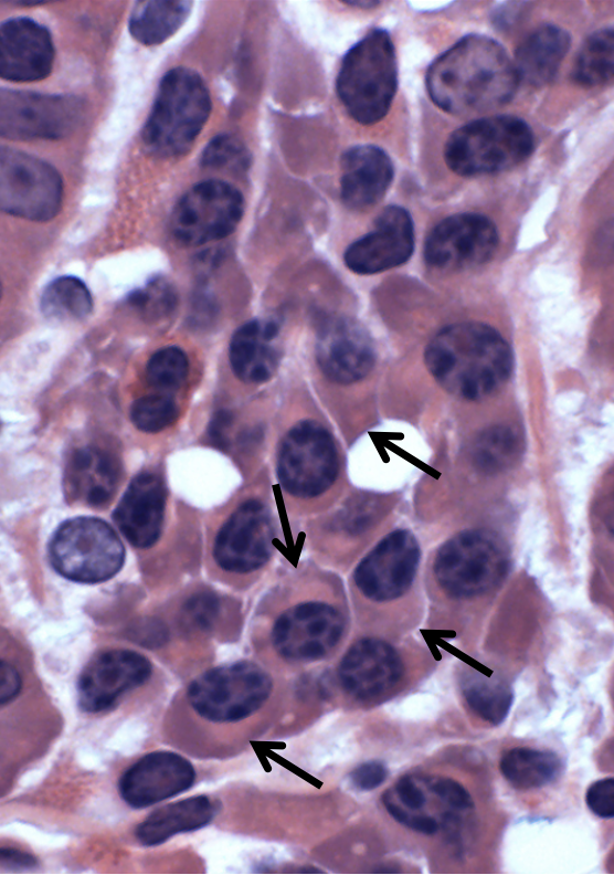
What cells are shown at the black arrows?
plasma cells
What cell is shown in these images?
neutrophils
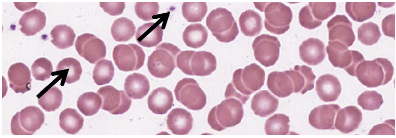
What is the left & right arrow pointing at?
erythrocyte & thrombocyte (platelet)
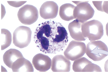
What is the cell shown?
neutrophil (made from granulocyte)
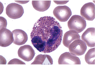
What is the cell shown?
eosinophil (made from granulocyte)
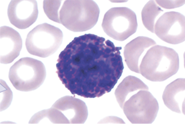
What is the cell shown?
basophil (made from granulocyte)
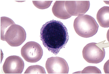
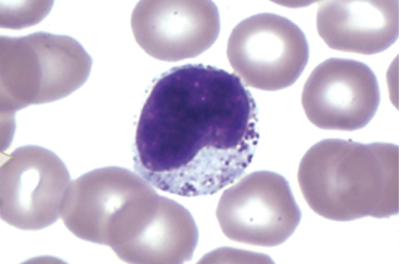
What is the cell shown?
lymphocyte (made from agranulocyte)
What is the cell shown?
monocyte (made from agranulocyte)