General characteristics of the heart:
Arteries
Carry blood high in oxygen (excluding pulmonary arteries)
Veins
Carry blood low in oxygen (except for pulmonary veins)
Great vessels→ arteries and veins entering and leaving the heart; relatively large size
Heart anatomy ensures that unidirectional flow of blood occurs; backflow is prevented by valves within the heart
The heart acts like two side-by-side pumps that work at the same rate and pump the same volume of blood; one side directs blood to the lungs for respiratory gas exchange; the other directs blood to body tissues for nutrient and respiratory gas delivery
The heart develops blood pressure by alternating cycles of the wall contraction and relaxation.
Blood pressure→ the force of blood pushing against the inside walls of the vessels
The Cardiovascular System:
Right side of heart:
Receives blood from body
Pumps out to lungs
Pulmonary circulation:
Conveys deoxygenated blood from the right side of the heart through blood vessels to the lungs; oxygen is picked up; carbon dioxide realesed; the blood returns to the left side of the heart
Pulmonary veins
Pulmonary arteries
Right atrium
Right ventricle
Left side of heart
Receives blood from lungs
Pumps out of body
Systemic circulation:
Moves oxygenated blood from the left side of the heart through blood vessels to the systematic cells such as those in the liver, skin, muscle, etc.
Left atrium
Left ventricle
Aorta to systemic arteries
Systemic veins
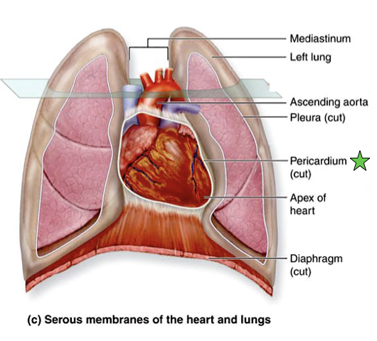
The Heart
Sits in middle of chest, between lungs
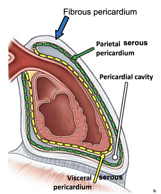
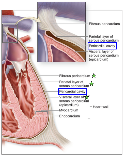
Surrounded by pericardial sac (pericardium)
Fibrous pericardium
Outer portion of the pericardium that is made of tough, dense connective tissue
Attached inferiorly to the diaphragm and superiorly to the base of the great vessels
Creates a space around the heart
Prohibits heart from moving within the thoracic cavity, and prevents the heart from overfilling with blood
Fluid filled, protective, anti-friction
Has multiple layers
Serous Membrane
Parietal serous pericardium
Shiny inner surface
Visceral serous pericardium (epicardium)
Light reflects on membrane
Pericardial cavity
Narrow space between the parietal and visceral layers of the serous membrane
Potential space for thin lining of serous fluid
Too much fluid is called inflammation
The Heart→ Superficial Anatomy
Base→ the posterosuperior surface of the heart, formed primarily by the left atrium
Superior border→ formed by the great arterial trunks (ascending aorta and pulmonary trunk), and the superior vena cava
Inferior border→ formed by the right ventricle
Apex
Chambers
The Heart→ Internal Anatomy
Epicardium
The outermost heart layer; aka the visceral layer
Composed of serous membrane and areolar connective tissue
Myocardium
Middle layer of the heart wall; thickest of the layers
Composed of cardiac muscle tissue
Contracts cardiac muscles to pump blood
Endocardium
The internal surface of the heart chamber and external surface of the heart valves
Composed of simple squamous epithelium and an underlying layer of areolar tissue
Chambers
Ventricles
Difference in appearance in 2 ventricles
Reflects the different loads places on them in their jobs
Left ventricle is thicker than the right ventricle; pushes blood through arteries to systematic system; right valve only pushes to lungs
Separated by interventricular septum
Trabeculae carneae
Atria (atriums)
Left atrium
Receives blood from the pulmonary circulation
Right atrium
Receives deoxygenated blood from the systemic circulation
Interatrial septum
Fossa Ovalis (of septum)
Pectinate muscles
Valves
Pulmonary Semilunar Valve
Right Atrioventricular Valve
Left Atrioventricular valve
Aortic Semilunar valve
Chordae Tendineae
Papillary Muscles
Prevent atrioventricular valves from everting and flipping into the atria when ventricles contract
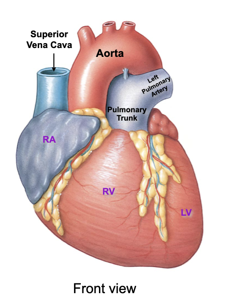
The Heart→ Attached Vessels
Superior Vena Cava
Drains blood from the head, neck, upper limbs, and superior regions of the trunk
Pulmonary Trunk
Transports blood from the right ventricle into the pulmonary circulation
Aorta
Conducts blood from the left ventricle into the systemic circulation
Left pulmonary veins
Left pulmonary artery
Right pulmonary veins
Inferior vena cava
Drains blood from the lower limbs and trunk
Coronary Sinus
Drains blood from the heart wall
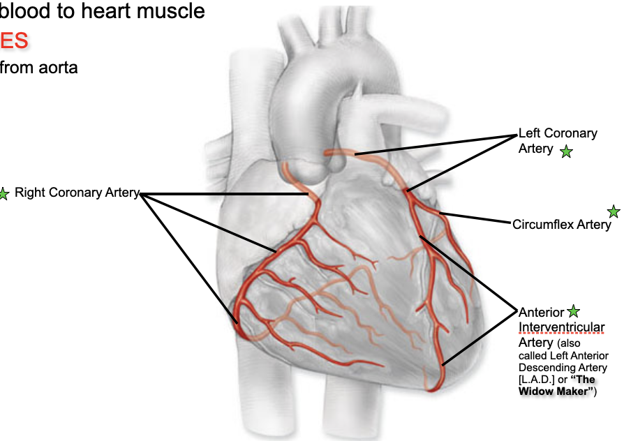
Coronary Circulation→ Arteries
Left and right coronary arteries travel with the coronary sulcus of the heart to supply blood to the cells of the heart walls.
Coronary arteries are considered functional end arteries; act like end arteries; easily blocked and can lead to dying arteries from lack of blood
These arteries are the only branches of the ascending aorta.
Coronary Vessels
Provide blood to heart muscles
Arteries → transports blood away from the heart; arise at aorta
Left Coronary Artery
Branches into the anterior interventricular artery; supplies the anterior surface of both ventricles and most of the interventricular septum
Branches into circumflex artery; supplies left atrium and ventricle
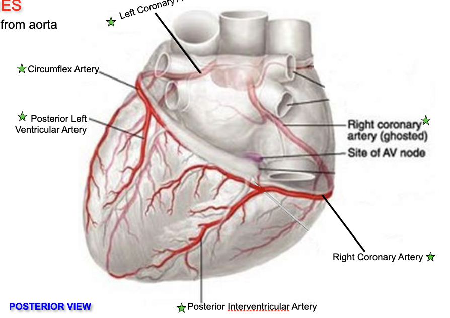
Right Coronary Artery
Branches in right marginal artery; supplies blood to the right border of the heart
Branches into posterior interventricular artery; supplies the posterior surface of both the left and right side of the heart
Anterior Interventricular Artery
Circumflex Artery
Posterior Left Ventricular Artery
Posterior Interventricular Artery
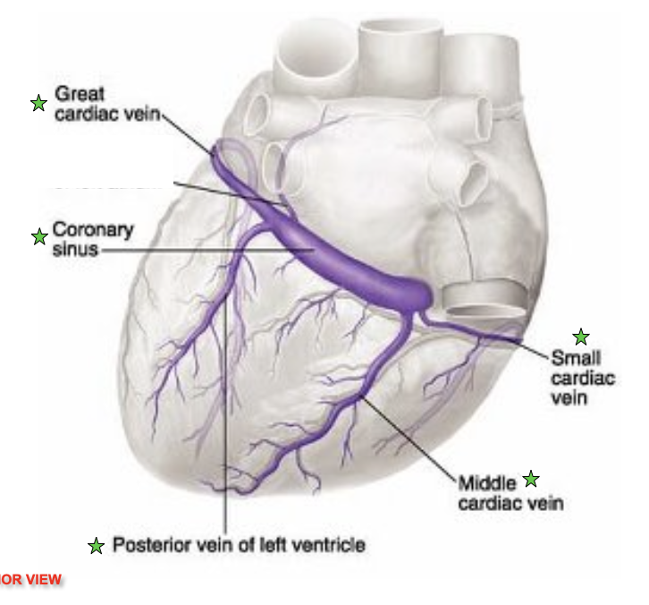
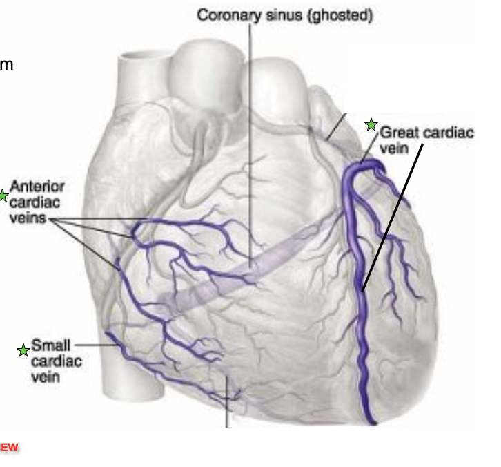
Coronary Circulation→ Veins
Veins→ transports blood to the heart
Dump into coronary sinus
Sinus dumps into right atrium
Great Cardiac vein
Coronary sinus
Drains directly into the right atrium
Posterior vein of left ventricle
Anterior cardiac veins
Middle cardiac vein
Drains into coronary sinus
Small cardiac vein
Drains into coronary sinus
Great cardiac vein
Drains into coronary sinus
Ventricular myocardium is compressed during contraction; most coronary flow occurs during ventricular relaxation
Normal flow is evenly distributed; however under certain circumstance coronary flow may be reduced
Tachycardia→ an increased heart rate that shortens diastole
Hypotension→ reduces the ability of blood flow through the ventricular myocardium
Contraction of Heart Muscles
Angiogram
Myocardial infarction (M.I) → Heart attack
blocked lumen in branch of left coronary artery
Anterior infarct
Area of dying tissues due to loss of blood/oxygen supply
Coronary Artery Bypass Graft (C.A.B.G)
Contraction of the Heart Muscles
Cardiac muscles cells contract as a single unit; an impulse distributes immediately and simultaneously thought the cell from the stria to the ventricles
Intercalated discs have numerous desmosomes and gap junction
Desmosomes prevent cardiac muscle cells from pulling apart
Gap junctions provide low-resistance pathway for ions to move between adjoining cardiac muscle cells
Allows muscle impulse to travel easily and instantaneously among cardiac muscle groups
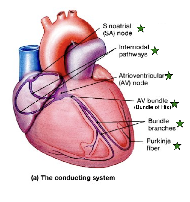
Intrinsic Conduction System
Sinoatrial (SA) node
Initiates the heartbeat; located in the posterior wall of the right atrium
Cells act as a the pacemaker
Initiates the heart impulse 70-80 times per minute
Internodal pathways
Atrioventricular (AV) node
Located in the floor of the right atrium between the right AV valve and the opening of the coronary sinus
Normally slows conduction of the impulse as it travels from the atria to the the ventricles; providing a delay between activation of the atria and ventricles
AV bundle
Receives muscle impulse from the AV node and extends into the interventricular septum before dividing into left and right bundles
Left/Right Bundles
Conduct impulse to conduction fibers called Purkinje Fibers that begin within the apex of the heart and extend through the walls of the ventricles
Purkinje fiber
Larger than other cardiac cells
Rapid conduction impulses, consistent with the large size of the cells; impulse spreads immediately throughout the ventricular myocardium
Fibrous
The fibrous skeleton of the heart is formed from dense regular connective tissue
Located between the atria and ventricles
Provide structural support at the boundary between the atria and the ventricles
Forms supportive fibrous rings to anchor the heart valves
Provides a rigid framework for the attachment of cardiac muscle tissue
Acts as an electrical insulator; prevents ventricles from from contracting at the same time as the atria
Blood Flow Through the Heart
A cardiac cycle includes all the events within the heart from the start of one heartbeat to the initiation of the next
Systole→ the contraction of a heart chamber
During this period, the contraction of the myocardium forces blood either into another chamber (from atrium to ventricle) or into a blood vessel (from ventricle into attached large artery)
Diastole→ the relaxation phase of a heart chamber
During this period between contraction phases, the myocardium of each chamber relaxes; chamber fills with blood
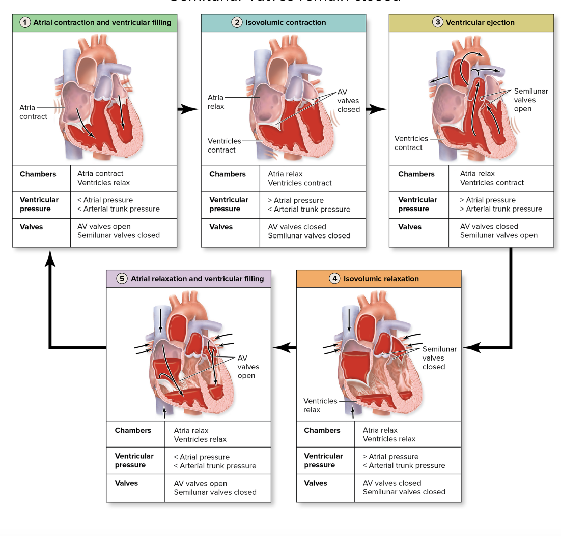
Steps in the Cardiac Cycle
Atrial contraction and ventricular filling
Occurs at the beginning of the cardiac cycle.
Brief contraction of the atrial myocardium initiated by the SA node occurs
Contraction of the atria finishes filling the ventricles through the open AV valve while the ventricles are in the diastole
Semilunar valves remain closed
Isovolumic contraction
Occurs at the beginning of the ventricular contraction
AV valves are forced cloths; produce “lubb” sound
Semilunar valve remain closed
Atria remain in diastole
Ventricular Ejection
Takes place later in the ventricular contraction; when pressure on blood in the ventricles forces the semilunar valves to open
Blood is ejected into the arterial trunks
Atria remain in diastole; Av valves remain closed
Isovolumic Relaxation
Occurs at the start of ventricular relaxation
Semilunar valves close to prevent blood backflow into the ventricles; produces “dupp” sound
The AV valve remain closed and the atria remain in diastole
Atrial relaxation and ventricular filling
Occur during the continuation of ventricular relaxation
Atria remain in diastole
The AV valve opens; passive filling of the ventricles from the atria begins and continues as most of the ventricular filling occurs
Semilunar valves remain closed
Heart Sounds
Caused by closing of valves
Changes in normal sound can be diagnostic
Heart murmur
First indication of heart valve problems
Result of turbulence of the blood as it passes through the heart
May cause valvular leakage, decreased valve flexibility, or misshapen valve
Valvular insufficiency
Occurs when one or more of the cardiac valves leaks because the valve cusps do no close tightly enough
May be caused by inflammation or disease
Valvular Stenosis
Scarring of the valve cusps so that they become rigid or partially fused and cannot completely open
Narrows and presents resistance to the flow of blood, decreasing chamber output
Often the affected chamber undergoes hypertrophy and dilates
Primary cause is rheumatic heart disease
Electrical Activity in the Heart
Stepwise progress of Electrical Impulse
Step 1: SA node activity and atrial activation begin (elapsed time=0)
Step 2: Atrial Depolarization; Stimulus spreads across the atrial surfaces and reaches the AV node (elapsed time= 50 msec)
Step 3: There is a 100-msec delay at the AV node. Atrial contraction begins (elapsed time= 150 msec)
Step 4: The impulse travels along the interventricular septum within the AV bundle and the bundle branches to the purkinje fibers and, via the moderator band, to the papillary muscles of the right ventricle (elapsed time= 175 msec)
Step 5: ventricular depolarization; The impulse is distributed by Purkinje fibers and relayed throughout the ventricular myocardium. Atrial contraction is completed, and ventricular contraction begins (elapsed time =225 msec)