


A&P UNIT 4 EXAM
Cardiovascular System
The cardiovascular system, also known as the circulatory system, is responsible for the transportation of blood, nutrients, and oxygen throughout the body. It consists of the heart, blood vessels, and blood.
Heart
The heart is a muscular organ that pumps blood throughout the body. It has four chambers:
Right atrium
Right ventricle
Left atrium
Left ventricle
Blood Vessels
Blood vessels are tubes that transport blood throughout the body. There are three types of blood vessels:
Arteries: carry oxygenated blood away from the heart
Veins: carry deoxygenated blood back to the heart
Capillaries: connect arteries and veins, allowing for the exchange of nutrients and waste products
Blood
Blood is a fluid that carries oxygen, nutrients, and waste products throughout the body. It consists of:
Red blood cells: carry oxygen
White blood cells: fight infections
Platelets: help with blood clotting
Plasma: a liquid that carries blood cells and nutrients
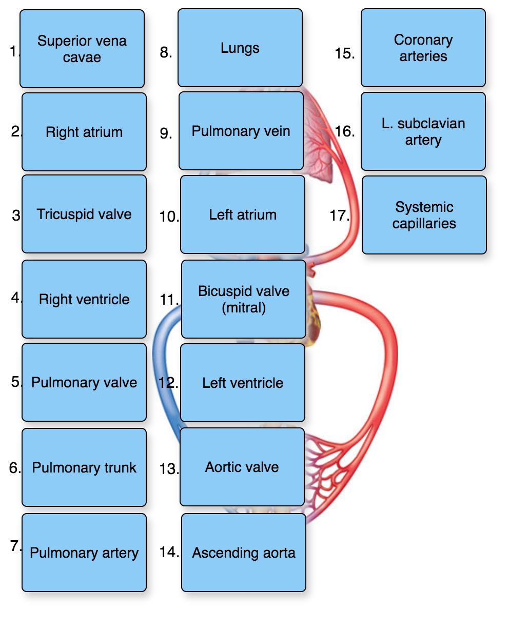
Blood vessels in order: 1. heart, 2. large arteries, 3. medium arteries, 4. arterioles, 5. capillaries, 6. venule, 7. medium vein, 8. large vein, 9. heart
BLOOD FLOW DIAGRAM*
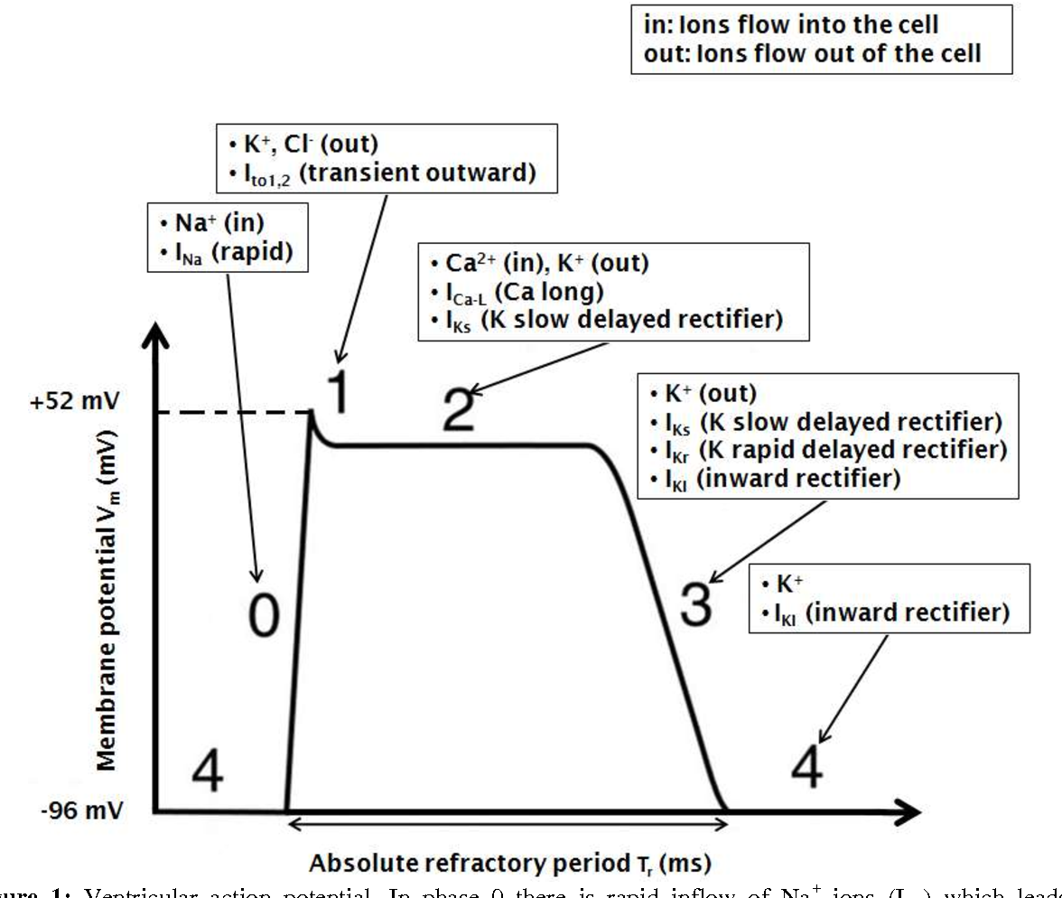
Cardiac Action Potential Graph*
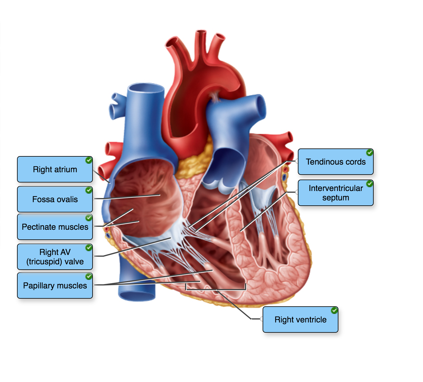
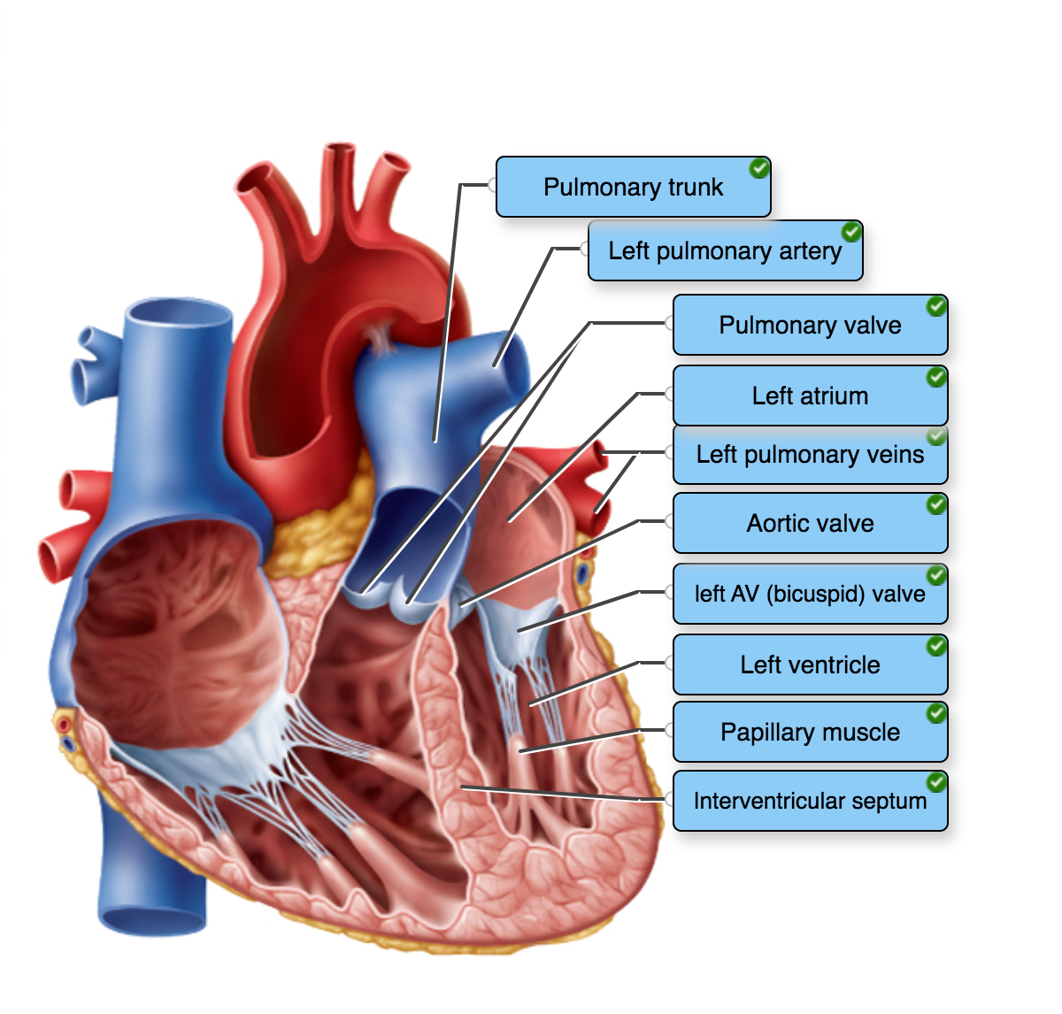
Anatomy of the Heart
The heart is a muscular organ that pumps blood throughout the body. It is located in the chest, between the lungs, and is about the size of a fist. The anatomy of the heart can be divided into four main parts:
Pericardium
A double-layered sac that surrounds the heart and protects it from friction.
Consists of an outer fibrous layer and an inner serous layer.
Heart Wall
Consists of three layers: epicardium, myocardium, and endocardium.
Epicardium: the outermost layer of the heart wall, also known as the visceral pericardium.
Myocardium: the middle layer of the heart wall, composed of cardiac muscle tissue.
Endocardium: the innermost layer of the heart wall, lines the chambers and valves of the heart.
Chambers
The heart has four chambers: two atria and two ventricles.
Atria: the upper chambers of the heart, receive blood from the veins.
Ventricles: the lower chambers of the heart, pump blood out of the heart.
Valves
The heart has four valves: two atrioventricular valves (AV) and two semilunar valves (SL).
AV valves: located between the atria and ventricles, prevent backflow of blood into the atria.
SL valves: located at the base of the pulmonary artery and aorta, prevent backflow of blood into the ventricles.
Cardiac Conduction
Definition: The process of electrical signal transmission through the heart that initiates and coordinates the contraction of the heart muscles.
Components of the cardiac conduction system:
Sinoatrial (SA) node: The natural pacemaker of the heart located in the right atrium.
Atrioventricular (AV) node: Located in the lower part of the right atrium, it receives the electrical signal from the SA node and delays it before transmitting it to the ventricles.
Bundle of His: A bundle of specialized fibers that transmit the electrical signal from the AV node to the ventricles.
Purkinje fibers: Specialized fibers that distribute the electrical signal throughout the ventricles, causing them to contract.
Steps in the cardiac conduction process:
The SA node generates an electrical signal that spreads through the atria, causing them to contract.
The electrical signal reaches the AV node, where it is delayed to allow the atria to fully contract before the ventricles begin to contract.
The electrical signal is transmitted through the bundle of His and Purkinje fibers, causing the ventricles to contract from the bottom up.
Factors that can affect cardiac conduction:
Heart disease or damage to the conduction system.
Electrolyte imbalances, such as low potassium or high calcium levels.
Medications that affect the heart's electrical activity, such as beta-blockers or calcium channel blockers.
Age-related changes in the conduction system.
HEART DIAGRAMS*
Circulation
Blood flows through the body in two circuits:
Pulmonary circulation: carries deoxygenated blood from the heart to the lungs and oxygenated blood back to the heart
Systemic circulation: carries oxygenated blood from the heart to the rest of the body and deoxygenated blood back to the heart
Functions
The cardiovascular system has several important functions:
Transporting oxygen and nutrients to the body's tissues
Removing waste products from the body's tissues
Regulating body temperature
Fighting infections and diseases
Maintaining fluid balance in the body
Endocrine System
The endocrine system is a complex network of glands and organs that produce and secrete hormones. These hormones regulate a wide range of bodily functions, including growth and development, metabolism, and reproductive processes.
Works more slowly than nervous system, however the effects last much longer.
Glands of the Endocrine System
Pituitary gland
Thyroid gland
Parathyroid glands
Adrenal glands
Pancreas
Ovaries (in females)
Testes (in males)
Hormones Produced by the Endocrine System
Growth hormone (GH)
Thyroid-stimulating hormone (TSH)
Adrenocorticotropic hormone (ACTH)
Follicle-stimulating hormone (FSH)
Luteinizing hormone (LH)
Prolactin
Oxytocin
Vasopressin
Insulin
Glucagon
Cortisol
Adrenaline (epinephrine)
Noradrenaline (norepinephrine)
Testosterone (in males)
Estrogen (in females)
Functions of the Endocrine System
Regulation of growth and development
Regulation of metabolism
Regulation of fluid and electrolyte balance
Regulation of reproductive processes
Response to stress
Maintenance of homeostasis
Regulation of blood sugar levels
Regulation of blood pressure
Regulation of heart rate
Regulation of body temperature
Disorders of the Endocrine System
Diabetes mellitus
Hypothyroidism
Hyperthyroidism
Cushing's syndrome
Addison's disease
Acromegaly
Gigantism
Dwarfism
Polycystic ovary syndrome (PCOS)
Infertility
Erectile dysfunction
Hormones Secreted by Gland
Pituitary Gland (Anterior & Posterior)
Growth Hormone (GH)
growth- most target cells
Prolactin (PRL)
milk making
Adrenocorticotropic Hormone (ACTH)
stims cortisol prod.
Thyroid-Stimulating Hormone (TSH)
stims thyroid
Follicle-Stimulating Hormone (FSH)
stims production of sperm in men
menstrual cycle and stims ovaries to make eggs
Luteinizing Hormone (LH)
sexual development
Antidiuretic Hormone (ADH)
water retention of kidneys
Oxytocin
sexual arousal
contractions/labor
mother/baby bonding
Thyroid Gland
Thyroxine (T4)
energy
Triiodothyronine (T3)
energy
Calcitonin
reduces blood calcium levels
Parathyroid Gland
Parathyroid Hormone (PTH)
inhibits osteoclast activity
calcium release
Adrenal Gland
Adrenaline (Epinephrine)
fight or flight- heart
Noradrenaline (Norepinephrine)
fight or flight- blood vessels
Cortisol (cortex)
steroid
metabolism
immune response
Aldosterone (cortex)
water & salt
Androgens
male sexual reproduction
Pancreas
Insulin
decreases glucose levels
Glucagon
raises glucose levels
Somatostatin
Ovaries
Estrogen
Progesterone
Testes
Testosterone
Pineal Gland
Melatonin
sleep/wake cycle
Thymus
Thymosin
t cells (immune support)
Hypothalamus
Gonadotropin-Releasing Hormone (GnRH)
Growth Hormone-Releasing Hormone (GHRH)
Somatostatin (SS)
Thyrotropin-Releasing Hormone (TRH)
Corticotropin-Releasing Hormone (CRH)
Prolactin-Releasing Hormone (PRH)
Prolactin-Inhibiting Hormone (PIH)
Respiratory System
The respiratory system is responsible for the exchange of gases between the body and the environment. It consists of the following parts:
Nose and Mouth
Air enters the body through the nose and mouth.
Hairs in the nose filter out large particles.
Mucus in the nose and mouth traps smaller particles.
Pharynx
The pharynx is a muscular tube that connects the nose and mouth to the larynx.
It serves as a passageway for air and food.
Larynx
The larynx is located at the top of the trachea.
It contains the vocal cords, which vibrate to produce sound.
The epiglottis, a flap of tissue, covers the larynx during swallowing to prevent food from entering the airway.
Trachea
The trachea, or windpipe, is a tube that connects the larynx to the bronchi.
It is lined with cilia and mucus-producing cells that help to trap and remove particles.
Bronchi
The bronchi are two tubes that branch off from the trachea and lead to the lungs.
They are also lined with cilia and mucus-producing cells.
Lungs
The lungs are the main organs of the respiratory system.
They are divided into lobes and contain millions of tiny air sacs called alveoli.
Oxygen from the air is exchanged for carbon dioxide from the blood in the alveoli.
Diaphragm
The diaphragm is a dome-shaped muscle that separates the chest cavity from the abdominal cavity.
It contracts and relaxes to help with breathing.
When it contracts, it flattens and increases the volume of the chest cavity, allowing air to enter the lungs.
When it relaxes, it returns to its dome shape and decreases the volume of the chest cavity, forcing air out of the lungs.
Pathway (airway passages) lower respiratory system- through the lungs; 1. trachea, 2. main primary bronchi, 3. secondary bronchi, 4. tertiary bronchi 5. bronchioles, 6. terminal bronchioles, 7. Respiratory bronchioles, 8. alveolar ducts, 9. alveoli
Anatomy of the Respiratory System
The respiratory system is responsible for the exchange of gases between the body and the environment. It consists of the following structures:
Nasal Cavity
Lined with mucous membranes and cilia
Warms, moistens, and filters air
Pharynx
Connects the nasal cavity and mouth to the larynx
Contains the tonsils
Larynx
Contains the vocal cords
Connects the pharynx to the trachea
epiglottis
Trachea
Lined with cilia and mucus-producing cells- pseudostratified ciliated columnar epithelium
Divides into the left and right bronchi
Bronchi
Divides into smaller bronchioles
Lined with smooth muscle and mucus-producing cells
Alveoli
Tiny air sacs where gas exchange occurs
Surrounded by capillaries
Lungs
Paired organs that contain bronchi, bronchioles, and alveoli
Surrounded by pleural membranes
Diaphragm
Dome-shaped muscle that separates the thoracic and abdominal cavities
Contracts during inhalation, allowing the lungs to expand
Blood Anatomy:
Introduction to blood anatomy
Composition of blood
Plasma
Formed elements
Red blood cells (erythrocytes)
White blood cells (leukocytes)
Platelets (thrombocytes)
Red blood cells (erythrocytes)
Structure
Function
Production
White blood cells (leukocytes)
Structure
Function
Types
Platelets (thrombocytes)
Structure
Function
Production
Blood groups and types
ABO blood group system
Rh blood group system
Blood circulation
Systemic circulation
Pulmonary circulation
Blood disorders
Anemia- iron deficienty
Leukemia- decrease in wbc production
Hemophilia
Thrombocytopenia
Types of Leukocytes
Leukocytes, also known as white blood cells, are a crucial component of the immune system. There are five main types of leukocytes, each with unique functions and characteristics:
Neutrophils
Most abundant type of leukocyte
First responders to bacterial infections
Phagocytize and destroy bacteria
Lymphocytes
Second most abundant type of leukocyte
Responsible for adaptive immunity
Two main types: B cells and T cells
Monocytes
Largest type of leukocyte
Precursors to macrophages and dendritic cells
Phagocytize and destroy pathogens
Eosinophils
Involved in allergic reactions and parasitic infections
Release enzymes that destroy parasites
Play a role in asthma and other allergic diseases
Basophils
Least abundant type of leukocyte
Involved in allergic reactions and inflammation
Release histamine and other chemicals that promote inflammation
Leukocytes work together to protect the body from infections and diseases. Understanding the different types of leukocytes and their functions is essential for maintaining a healthy immune system.