
Nervous System Study Guide - Biology 275
Nervous System
Protection and Support of the Brain:
Meninges and their Function

- Three connective tissue layers that separate and support the soft tissue of the brain from the bones of the cranium
- Enclose and protect some of the blood vessels that supply the brain
- Circulation of CSF
*Superficial to Deep*
Dura Mater:
- Outer, dense irregular connective tissue covering
2 fused layers
- Meningeal Layer: immediately superficial to the arachnoid
- Periosteal Layer: forms the periosteum on the internal surface of the cranial bones
- Dural Venous Sinuses: where the two layers are separated forming blood-filled spaces; drain blood from the brain
Two potential spaces
- Epidural Space: contains the arteries and veins that nourish the meninges and bones of the cranium
- Subdural Space: positioned between the arachnoid mater and dura mater
Arachnoid Mater:
- Arachnoid trabeculae: partially composed of a delicate web of collagen and elastic fibers. Lies external to pia mater
- Subarachnoid space: contains CSF. Lies deep to the arachnoid mater
- Both support cerebral arteries and veins within subarachnoid space
Pia Mater:
- Innermost of the cranial meninges
- Thin layer of areolar connective tissue
- Covers small blood vessels that enter the brain and help CSF formation in the ventricles
Cranial Dura Septa:
– the meningeal layer of the dura mater extends as flat partitions into the cranial cavity
Falx Cerebri:
- Large, sickle-shaped, vertical fold of dura mater
- Located in the midsagittal plane and projects into the longitudinal fissure between the left and right cerebral hemispheres
Tentorium Cerebelli:
- A horizontally oriented fold of dura mater that separates both the occipital and the temporal lobes of the cerebrum from the cerebellum
- Anterior surface has a gap called the tentorial notch, to allow for the the passage of the brainstem
- Falx cerebelli extends through this
Diaphragma Sellae:
- Smallest
- Forms a roof over the sella turcica of sphenoid bone
- A small opening within it allows for the passage of a thin stalk, called the infundibulum, that attaches the pituitary gland to the base of the hypothalamus
Ventricles in the Brain:
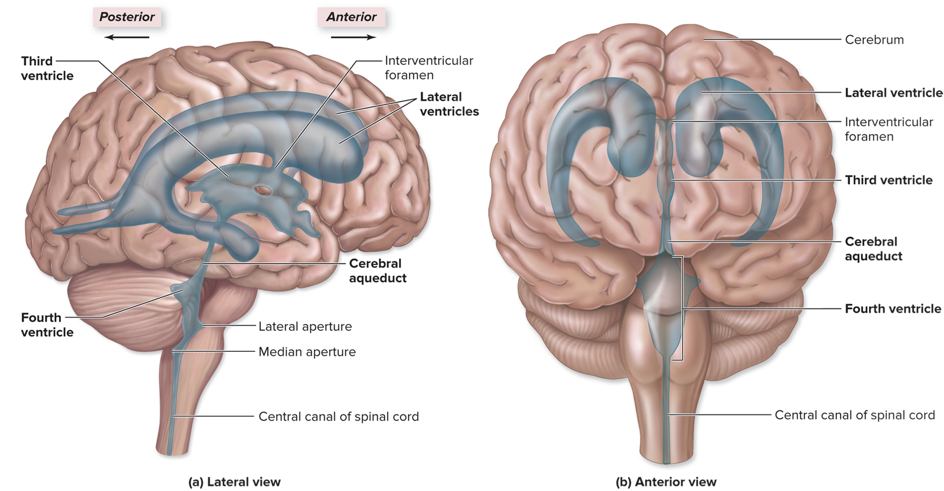
- Cavities or expansions within the brain that are derived from the neural canal
- Lined with ependymal cells and contain CSF
- Connected with one another as well as with the central canal of the spinal cord
Lateral Ventricles: separated by a thin medial partition called the septum pellucidum.
Third Ventricle:
- Located within the diencephalon
- Each lateral ventricle is connected through this by the opening called the interventricular foramen
Cerebral Aqueduct:
- Passes through the midbrain and connects the third ventricle with the tetrahedron shaped fourth ventricle
Fourth Ventricle:
- Located between the cerebellum and brainstem (pons and medulla oblongata)
- Opens to the subarachnoid space via paired lateral apertures and a single median aperture
- Narrows at its inferior end before it emerges with the slender central canal of the spinal cord
Location of Cerebrospinal fluid - what produces, flow, and functions

Production and Circulation:
- CSF is produced by the choroid plexus in the ventricles
- CSF flows from the lateral ventricles, through the interventricular foramen into the third ventricle, and then through the cerebral aqueduct into the fourth ventricle
- CSF in the fourth ventricle passes through the paired lateral apertures and the single median aperture, and into the subarachnoid space, as well as the central canal of the spinal cord
- As the CSF flows through the subarachnoid space, it provides buoyancy to support the brain
- Excess CSF flows into the arachnoid villi, then drains into the dural venous sinuses. The greater pressure on the CSf in the subarachnoid space ensures that CSF moves into the venous sinuses without permitting venous blood to enter the subarachnoid space
Function:
- Buoyancy:
- The brain floats within the CSF, to prevent the brain from being crushed by its own weight
- Without CSF, the portions of the brain would sink through the foramen magnum of the skull
- Protection:
- CSF provides a liquid cushion to protect delicate neural structures from sudden movements
- Pov: when someone hits the brakes too hard, your movements are slowed as the water acts as a “movement buffer”
- CSF helps slow movements of the brain in response to sudden movement
- Environmental stability:
- CSF transports nutrients and chemical messengers to the brain and removes waste products from the brain
- Protects nervous tissue from chemical fluctuations that would disrupt neuron function
- Excess CSF is eventually transported into the veins of the cardiovascular system
Blood-Brain Barrier (BBB):

- The blood-brain barrier strictly regulates which substances can and cannot be filtered from the blood to enter the interstitial fluid of the brain
- This helps prevent exposure of neurons in the brain to drugs, waste products in the blood, and variations in levels of normal substances (ions, hormones), that could alter brain function
- Lipid-soluble molecules such as nicotine, alcohol, and some anesthetics, can diffuse past the blood-brain barrier and the interstitial fluid of the CNS to reach neurons
- Note: there is no BBB in the choroid plexus, hypothalamus, and pineal gland
Cerebrum:
Lobes of The Brain and Their Functional Areas:
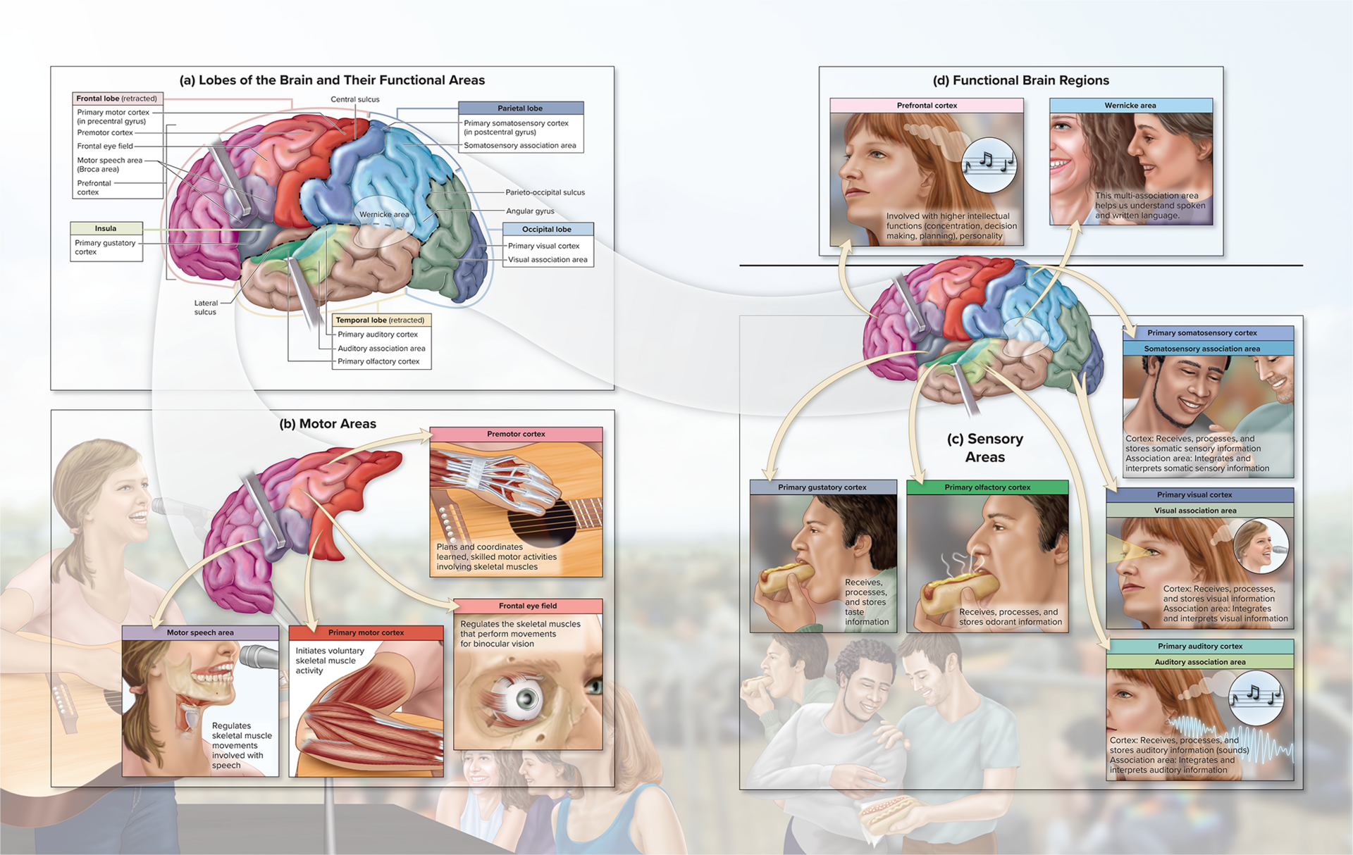
Frontal Lobe:
- Primary motor cortex (in precentral gyrus)
- initiates voluntary skeletal muscle activity
- Premotor cortex
- plans and coordinates learned, skilled motor activities involving skeletal muscles
- Frontal eye field
- regulates the skeletal muscles that perform movements for binocular vision
- Motor speech area (Broca’s Area)
- regulates skeletal muscle movement involved with speech
- Prefrontal Cortex
- involved with higher order intellectual functions (concentration, decision making, planning, personality)
Parietal Lobe:
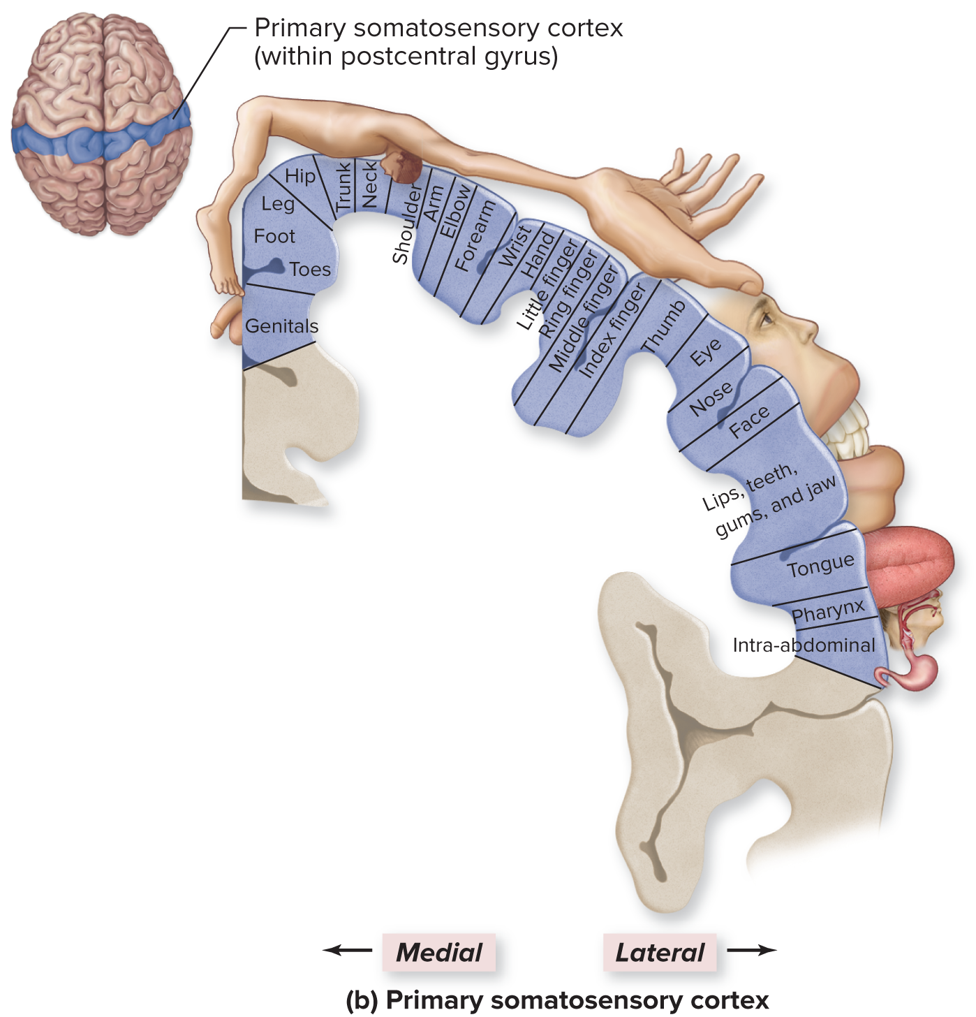
- Primary somatosensory cortex (in postcentral gyrus)
- receives, processes, and stores somatic sensory information
- Somatosensory association area
- integrates and interprets somatic sensory information
Temporal Lobe:
- Primary auditory cortex
- receives, processes, and stores auditory information
- Auditory association area
- integrates and interprets auditory information
- Primary olfactory cortex
- receives, processes, and stores odorant information
Occipital Lobe:
- Primary visual cortex
- receives, processes, ans stores visual information
- Visual association area
- integrates and interprets visual information
Insula:
- Primary gustatory cortex
- receives, processes, and stores taste information
Motor Areas of The Brain:
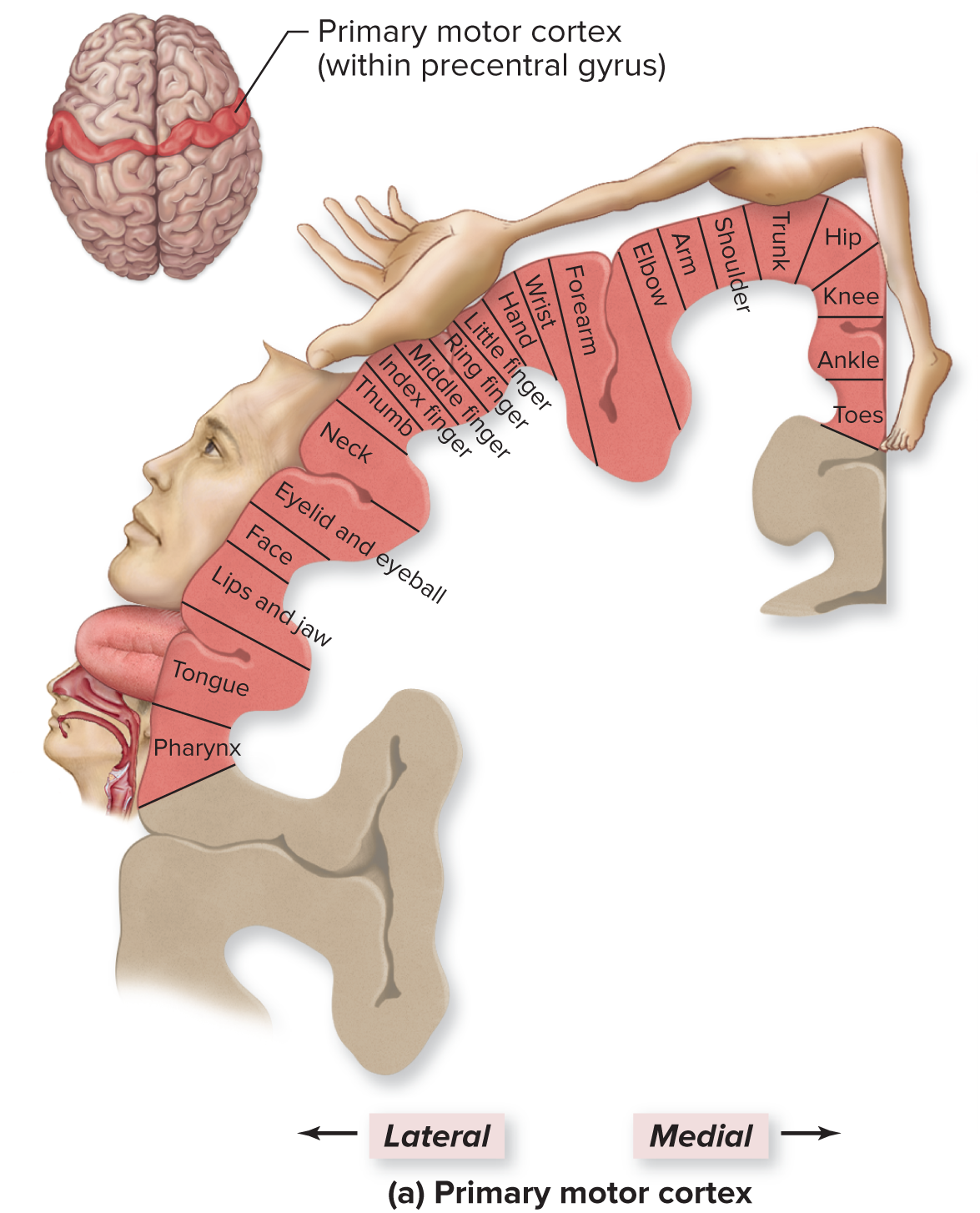
- Specifically located within the precentral gyrus of the frontal lobe
- Neurons in this area control voluntary skeletal muscle activity
- Axons project to the opposite side with either the brainstem or the spinal cord
Motor speech area:
- region responsible for controlling the muscular movements necessary for vocalization
Frontal eye field:
- controls and regulates the eye movements needed for reading and coordinating binocular vision
Premotor cortex:
- responsible for coordinating learned, skilled motor activities, such as moving the eyes in a coordinated fashion when reading a book or playing the guitar
Sensory Areas of The Cortex and Association Areas:
- The cortical areas within the parietal, temporal, and occipital lobes and the insula are involved with conscious awareness of sensation
Primary somatosensory cortex:
- Responsible for receiving, processing, and storing somatic sensory information (touch, pressure, pain, temperature)
- Proprioceptors from the joints and muscles regarding the conscious interpretation of body position
Somatosensory association area:
- Integrates sensory information and interprets sensations to determine the texture, temperature, pressure, and shape of objects
- Allows us to identify known objects without seeing them
Functional Brain Regions:
- Acts as a multi-association area between lobes for integrating information from individual association areas
Prefrontal cortex:
- Associated with many higher intellectual functions such as complex thought, judgment, expression of personality, planning future behaviors, and decision making
- Continues to develop into our teens and 20s, as the axons continue to myelinate in this region and unnecessary synapses are remove
Wernicke’s Area:
- Involved in recognizing, understanding, and comprehending spoken or written language
Central White Matter Tracts:
- Lies deep to the gray matter of the cerebral cortex, composed of primarily myelinated axons (bundles-tracts)
Association tracts:
- Connect different regions of the cerebral cortex within the same hemisphere
- Arcuate fibers: short association tracts that connect the neighboring gyri within the same lobe; ex. Tract that connects the premotor cortex with the primary motor cortex
- Longitudinal fasciculi: connect gyri in different lobes of the same hemisphere; ex. Wernicke’s area to Broca’s area
Commissural tracts:
- Extend between the cerebral hemispheres through axonal bridges called commissures
- Corpus callosum, and the smaller anterior and posterior commissure
- link the right and left cerebral hemispheres
Projection tracts:
- Link the cerebral cortex to both the inferior brain regions of the spinal cord
- Ex. the corticospinal tracts that transmit motor signals from the cerebrum to the brainstem and spinal cord
- Internal capsule: the packed group of axons in these tracts passing in between the cerebral nuclei and the gray matter of the thalamus
Cerebral Lateralization
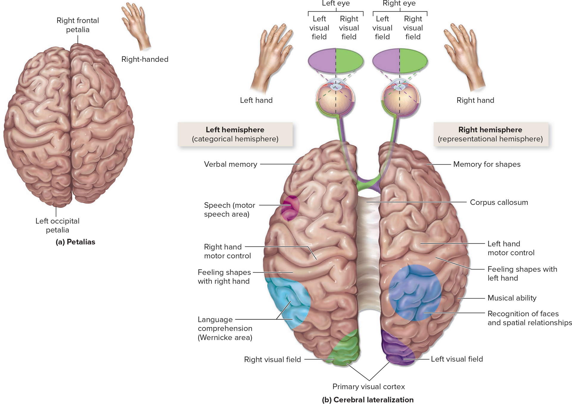
- Humans tend to have shape asymmetries of the frontal and occipital lobes of the brain, called petalias
- Right handed people typically have right frontal petalias, and left occipital petalias, left handed people have the reverse pattern
- Each hemisphere tends to be specialized for certain tasks
Left (categorical) hemisphere:
- Wernicke’s and motor speech area
- Analytical reasoning tasks
- Function in categorization and identification
Right (representational) hemisphere:
- Concerned with visuospatial relationships and analyses
- Imagination and insight, musical and artistic skill, perception of patterns
- Comparison of sights, sounds, smells, and tastes
Both the hemispheres remain in constant communication through commissures, especially through the corpus callosum
Cerebral Nuclei

- Paired, irregular masses of gray matter buried deep within the central white matter in the deepest region of the cerebral hemispheres inferior to the floor of the lateral ventricle
- Deep within the cerebrum
Caudate nucleus:
- has an enlarged head and a slender, arching tail that parallels the curve of the lateral ventricle
- When a person walks, the neurons in this nucleus stimulate the appropriate skeletal muscles to produce the pattern and rhythm of arm and leg movements
Lentiform nucleus:
- A compact, triangular mass, made up of both the putamen and globus pallidus (two masses of gray matter)
- Putamen controls skeletal muscular movement at the subconscious level
- Globus pallidus can either excite or inhibit the anterior group nuclei of the thalamus to help regulate skeletal muscle tone
Claustrum:
- A thin sliver of gray matter formed by a layer of neurons located immediately internal to the insula
- has extensive connections with many cerebral cortex areas like the primary visual and auditory cortices
- Ex. you can perceive a flower because the claustrum integrates information from viewing the flower, touching its petals, and smelling its fragrance
Amygdaloid: “amygdala”
- Participates in the expression of emotions, control of behavioral activities, and development of moods
Diencephalon:
Epithalamus:
- Partially forms the posterior roof of the diencephalon and covers the third ventricle
- Pineal gland
- Habenular nuclei: relay signals from the limbic system to the midbrain and are involved in visceral and emotional responses to odors
Thalamus:

- Composed of paired oval masses of gray matter, which form the superolateral walls of the third ventricle
- The principal and final relay point for incoming sensory information that is processed and then projected to the appropriate lobe of the cerebral cortex
- Only a relatively small portion of the sensory information that arrives at the thalamus is forwarded to the cerebrum because the thalamus acts as an information filter
- Ex. it is responsible for filtering out the sounds and sights in a crowded area when you are trying to study
- The thalamus lets the cerebrum know that sensory information it receives came from the eye, indicating that the information is visual
Hypothalamus:
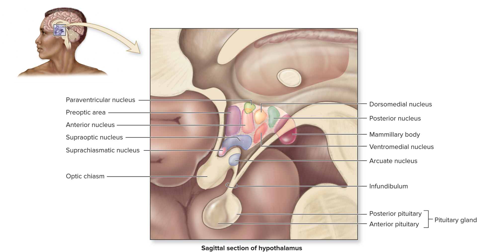
- The anteroinferior region of the diencephalon
Master control of the autonomic nervous system:
- The major autonomic integration center
- It projects descending axons to autonomic nuclei in the brainstem that influence activities such as heart rate, blood pressure, digestive activities, and respiration
Master control of the endocrine system:
- Secretes hormones that control secretory activities in the anterior pituitary gland (6) and posterior pituitary (ADH and OT)
Regulation of body temperature:
- Neurons in the preoptic area detect altered blood temperatures and signal other hypothalamic nuclei, which control the mechanisms that heat or cool the body
Control of food intake:
- Neurons within the ventromedial nucleus monitor levels of nutrients such as glucose and amino acids in the blood and produces sensations of hunger
Control of water intake:
- Specific neurons within the anterior nucleus of the hypothalamus continuously monitor the concentration of dissolved substances in the blood to regulate the sensation of thirst
- Nerve signals are relayed to the cerebral cortex and our sensation of thirst is stimulated
Regulation of circadian rhythms:
- The suprachiasmatic nucleus directs the pineal gland of the epithalamus to secrete melatonin at certain times of the day
Control of emotional behavior:
- Located at the center of the limbic system, the part of the brain that controls emotional responses, such as pleasure, aggression, fear, rage, contentment, and the sex drive
Nucleus or Hypothalamic Region | Function |
|---|---|
Anterior nucleus | “Thirst center”(stimulates fluid intake); autonomic control center |
Arcuate nucleus | Regulates appetite, release of GnRH, GHRh, and PIH |
Mammillary body | Directs sensations related to olfaction; controls swallowing |
Paraventricular nucleus | Produces oxytocin primarily |
Preoptic area | “Thermostat” (regulates body temperature) |
Suprachiasmatic nucleus | Circadian rhythms |
Supraoptic nucleus | Produces ADH |
Ventromedial nucleus | “Safety center” (produces hunger and satiety sensations) |
Brainstem:
- Connects the cerebrum, diencephalon, and cerebellum to the spinal cord
- A bidirectional passageway for all tracts extending between the major regions of the brain and spinal cord
- Contains autonomic centers and reflex centers required for regulating body functions necessary for survival
- Reticular formation extends through all the three regions
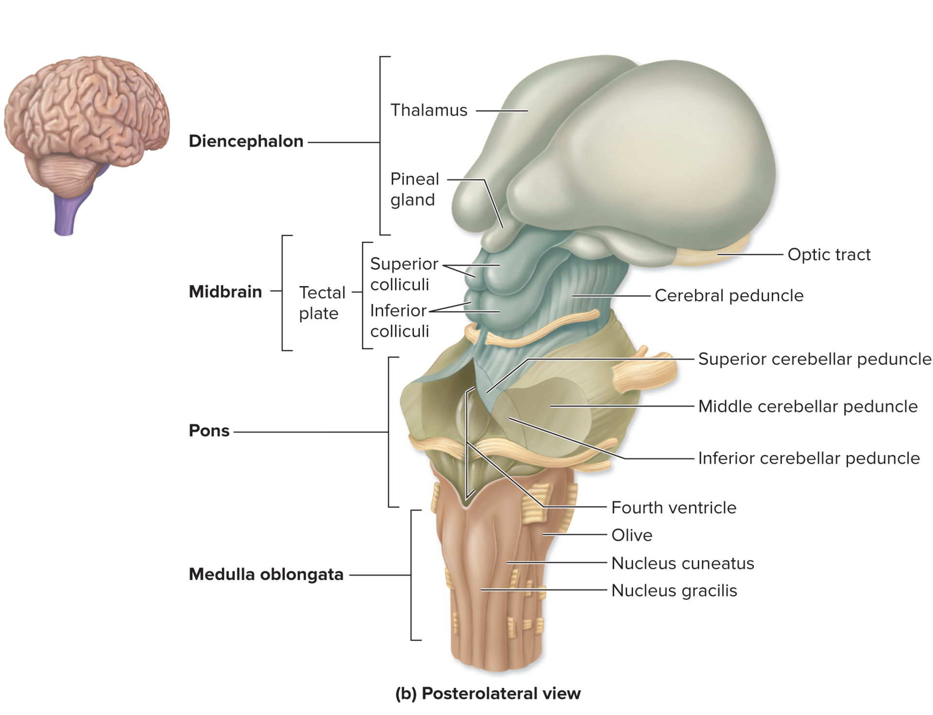
Midbrain:

- The superior portion of the brainstem
- The tracts (bundles of myelinated axons) associated with the midbrain nuclei (gray matter) are described based on their position from anterior to posterior
Cerebral peduncles
- Motor tracts located on the anterolateral surfaces of the midbrain
- Descending axon bundles of the pyramidal system project through the cerebral peduncles and relay voluntary motor commands from the primary motor cortex to each cerebral hemisphere
- Superior cerebellar peduncles connect midbrain to cerebellum
Substantia nigra
- Consists of bilaterally symmetric nuclei within the midbrain
- Houses clusters of neurons that produce the neurotransmitter dopamine, which affects the brains processes to control movement, emotional response, and ability to experience pleasure and pain
- Parkinson's Disease
Tegmentum
- Sandwiched between the nuclei of the substantia nigra and periaqueductal gray matter
- Contains the pigmented red nuclei and reticular formation
- Integrates information from the cerebrum and cerebellum and issues involuntary motor commands to the erector spinae muscles of the back
Cerebral aqueduct
- Connects third and fourth ventricles
- Surrounded by a region called the periaqueductal gray matter
- Oculomotor nerve (CN III) and the trochlear nerve (CN IV) are housed in the midbrain
Tectum
- Most posterior region of the midbrain
- Contains two pairs of sensory nuclei, the superior and inferior colliculi, collectively called the tectal plates
- These nuclei are relay stations in the processing pathway of visual and auditory sensory input
- Superior colliculi: VISUAL REFLEX CENTERS, they help visually track moving objects and control reflexes such as turning the eyes and head in response to a visual stimulus
- Inferior colliculi: AUDITORY REFLEX CENTERS, they control reflexive turning of the head and eyes in the direction of a sound
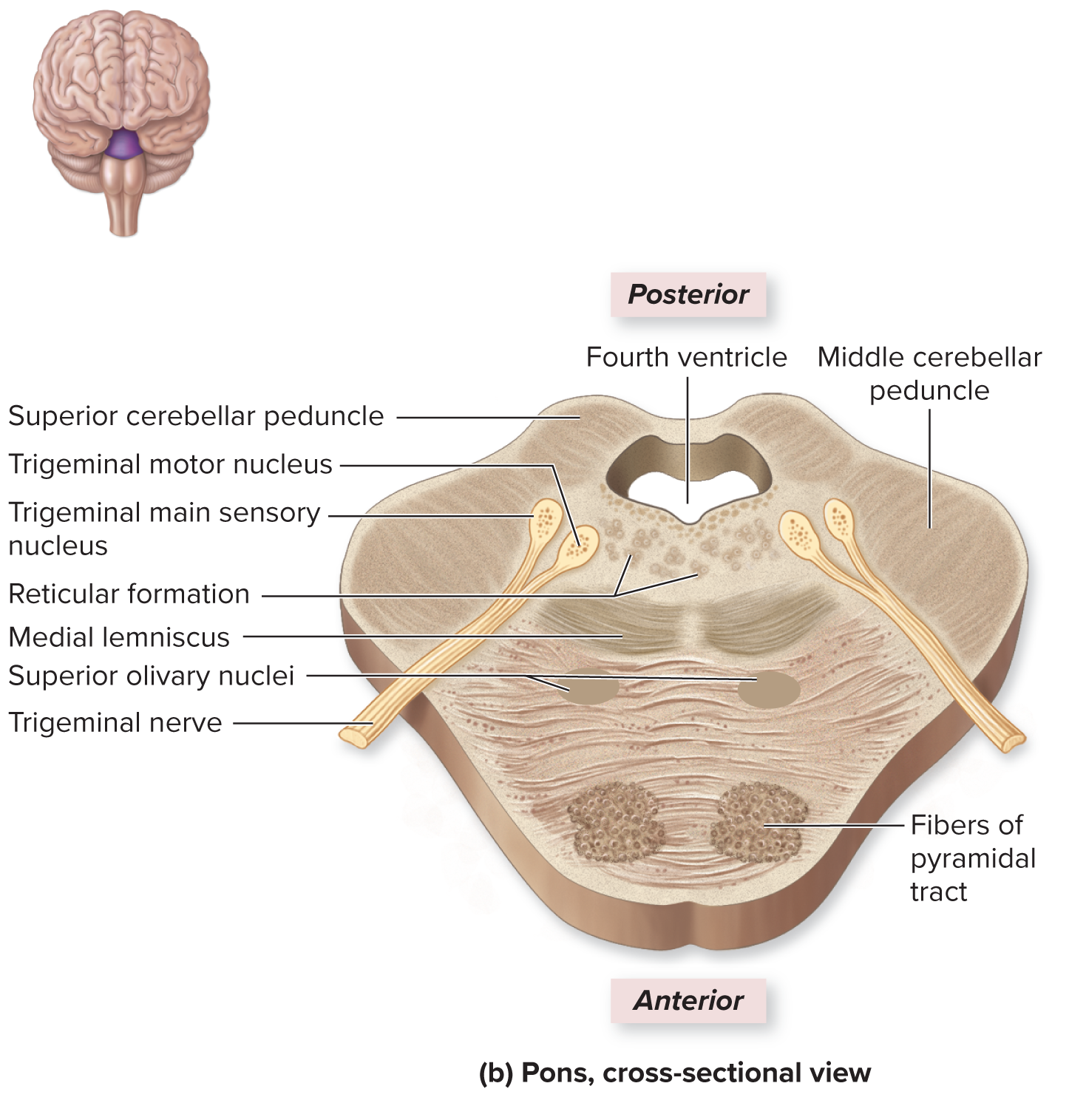
Pons

- A bulging region on the anterior part of the brainstem
- Middle cerebellar peduncles are transverse axons that connect the pons to the cerebellum
- Houses autonomic nuclei in the pontine respiratory center, which regulate muscles of respiration along with the medullary respiratory center of the medulla oblongata
- It regulates a smooth transition between inhalation and expiration
- Superior olivary nuclei are located in the inferior portion of the pons; they receive auditory input and is involved in the pathway for sound localization
- Houses sensory and motor cranial nerve nuclei for trigeminal (CN V), abducens (CN VI), and facial (CN VII) cranial nerves; additionally some of the nuclei for the vestibulocochlear cranial nerve (CN VIII)
Medulla Oblongata

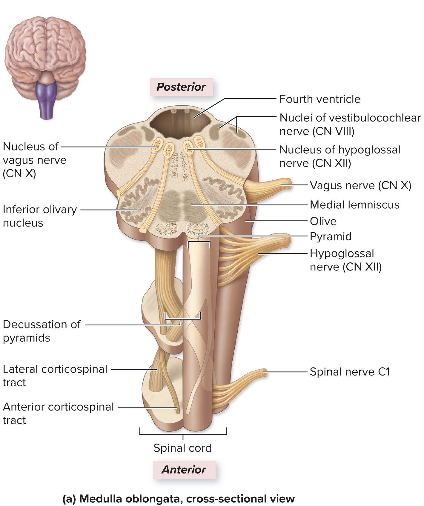
- All communication between the brain and spinal cord involves tracts that ascend or descend through the medulla oblongata
- The anterior surface exhibits two longitudinal ridges called the pyramids, which house the motor projection tracts called the corticospinal tracts that extend through the medulla
Decussation of pyramids:
- most of the actions of the pyramidal tracts cross to the opposite side of the brain at a point
- as a result of this crossover, each cerebral hemisphere controls the voluntary movements of the opposite side of the body
Olive (bulge):
- Contains a large fold of gray matter called the inferior olivary nucleus
- This relays ascending sensory nerve signals, especially proprioceptive information to the cerebellum
Inferior cerebellar peduncles
- Tracts that connect the medulla oblongata to the cerebellum
Autonomic nuclei:
Cardiovascular center
- Cardiac center: regulates both the heart’s rate and its force of contraction to alter cardiac output
- Vasomotor center: controls the contraction and relaxation of smooth muscle within the walls of arterioles, that alter its diameter
- Both influence blood pressure
Medullary respiratory center
- Regulates the respiratory rate
- Composed of a ventral respiratory group and a dorsal respiratory group, being influenced by the pontine respiratory center
- Rhythmically initiate nerve signals that cause contraction of breathing muscles
Other nuclei in the medulla, which are involved in coughing, sneezing, salivation, swallowing, gagging, and vomiting reflexes
Cranial nerves in the Medulla
- (CN VIII) Vestibulocochlear
- (CN IX) Glossopharyngeal
- (CN X) Vagus
- (CN XI) Accessory
- (CN XII) hypoglossal
- Nucleus cutaneous and nucleus gracilis, which relay somatic sensory information to the thalamus
Cerebellum
- Coordinates fine control over skeletal muscle actions and stores memories of movement patterns
Structural Components:

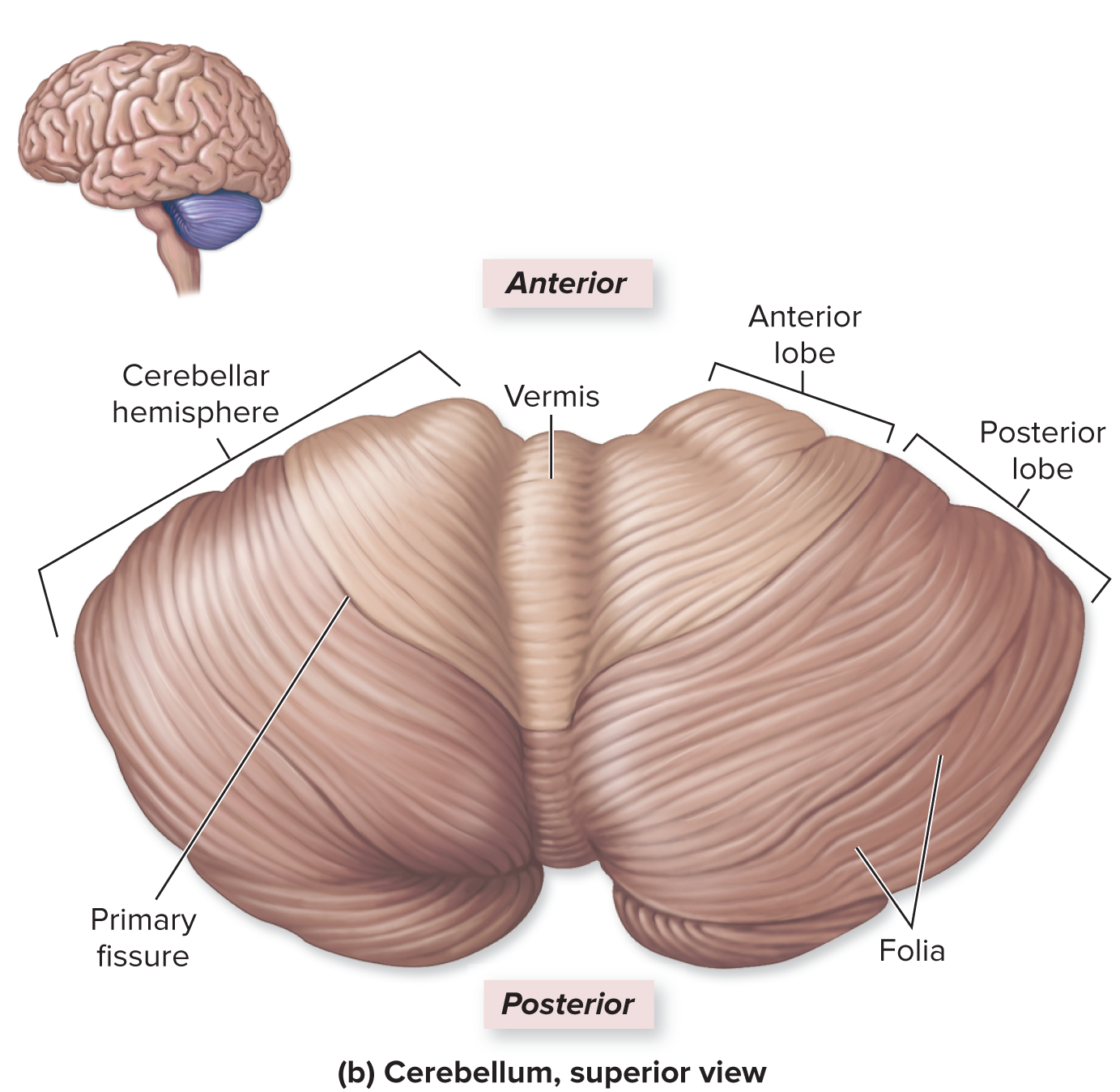
- Composed of left and right cerebellar hemispheres
- Has an anterior and posterior lobe, separated by a primary fissure
- The vermis lies along the midline between the left and right cerebellar lobes
- folia=folds
Partitioned into three regions:
- Cerebellar cortex (outer gray matter)
- Arbor vitae (internal region of white matter) - “tree branches”
- deepest gray matter layer composed of cerebellar nuclei
Cerebellar peduncles
- Connect cerebellum with the brainstem
- Superior: connect cerebellum to midbrain
- Middle: connect cerebellum to pons
- Inferior: connect cerebellum to medulla oblongata
Functions:
- Does not initiate skeletal muscle movement, it fine tunes skeletal muscle movements that were initiated by the cerebrum = smooth and rhythmic movements
- Stores memories of previously learned movement patterns, by regulating activity along both the voluntary and involuntary motor pathways at the cerebral cortex, cerebral nuclei, and motor centers in the brainstem
- Initiates a movement and sends a “rough draft” of the movement to the cerebellum, which then coordinates and adjusts it
- Receives proprioceptive information from the muscles and joints and uses this information to regulate the body’s position
Cerebellar Pathways:

Limbic System
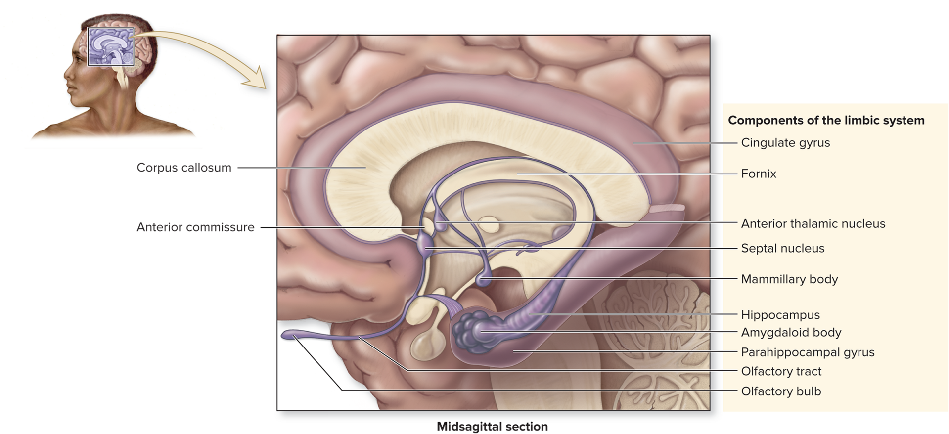
- Composed of multiple cerebral and diencephalic structures that collectively process and experience emotions
- “Emotional brain”
- Form a ring around the diencephalon
Cingulate gyrus
- Receives input from the other components of the limbic system
Parahippocampal gyrus
- Associated with hippocampus
Hippocampus
- Essential in storing memories and forming long-term memory
Amygdaloid body
- Involved in several aspects of emotion, especially fear
- Helps store and code memories based on how a person emotionally perceives them
Olfactory bulbs, tracts, and cortex
- Particular odors can provoke certain emotions or be associated with certain memories
Fornix
- Thin tract of white matter that connects hippocampus with limbic system structures of the diencephalon
Nuclei in the diencephalon:
- Anterior thalamic nuclei (epithalamus)
- Habenular nuclei (epithalamus)
- Septal nuclei (hypothalamus)
- Mammillary bodies (hypothalamus)
Reticular Formation
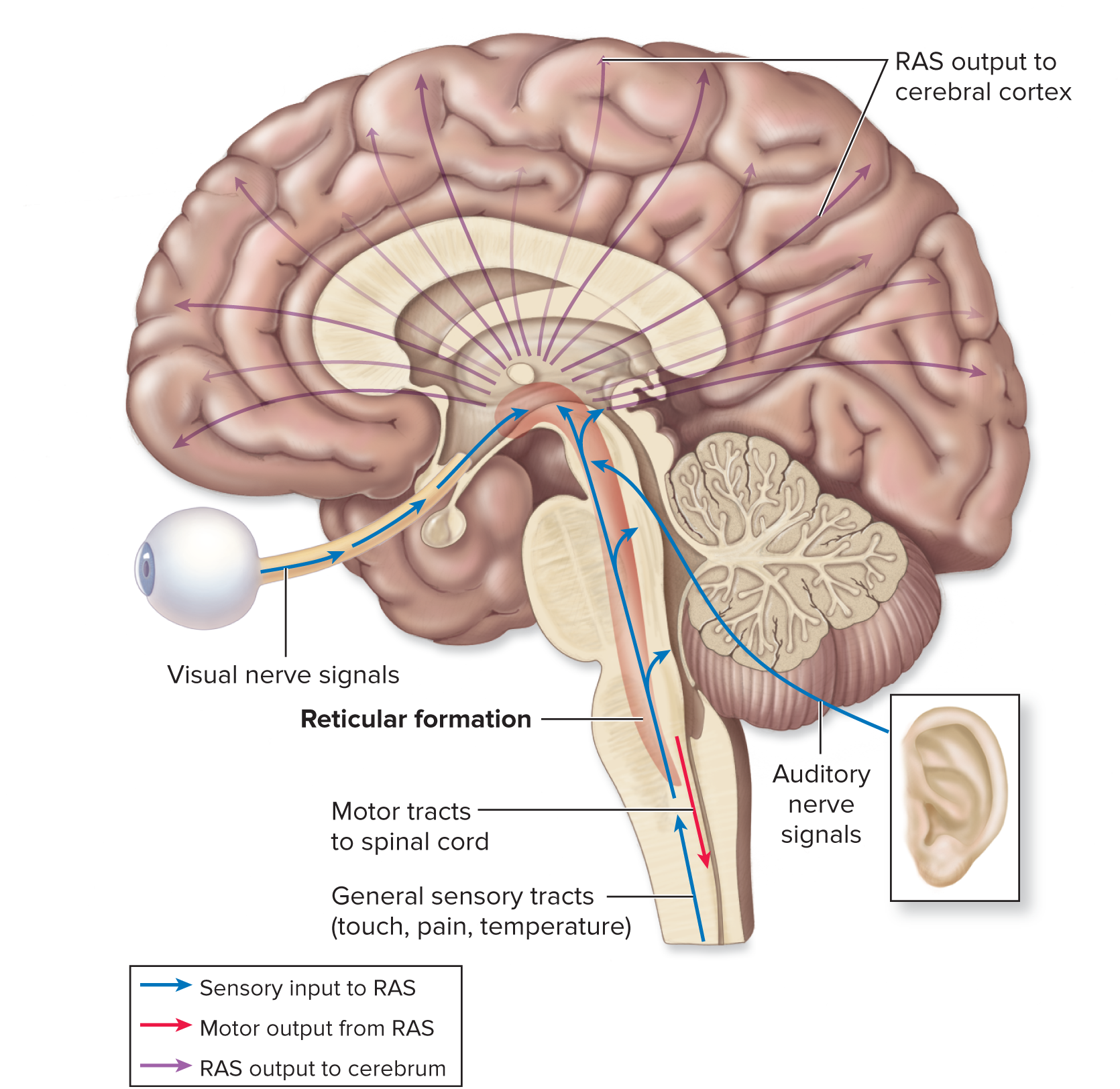
Processes various types of input from sensory receptors (blue)
- Responsible for alerting the cerebrum to incoming sensory information
- Reticular activating system: contains sensory axons that project to the cerebral cortex; processes visual, auditory, and touch stimuli and uses this information to keep us in a state of mental alertness
- Arouses us from sleep
Participates in cyclic activities such as arousing the cortex to consciousness, and circadian rhythm (purple)
- Involves the simultaneous activity of large areas of the cerebral cortex
Some motor output from the reticular formation influences muscle activity (red)
- Communicates with spinal cord and is responsible for regulating skeletal muscle tone
- Assists in reflexive motor functions (respiration, blood pressure, and heart rate, by working with the autonomic centers in the medulla and pons)
Higher Order Brain Functions
- Include memory, learning, and reasoning
- Occurs within the cerebral cortex and involve multiple brain regions connected by complicated networks and arrays of axons
Development
- Myelinated of many CNS axons continues throughout the first 2 years
- Brain grows rapidly in size and complexity so that byu the age of 5, brain growth is 95% complete
- Some neurons expand their number of connections
- The brain will “prune” various synaptic connections
The 12 Cranial Nerves
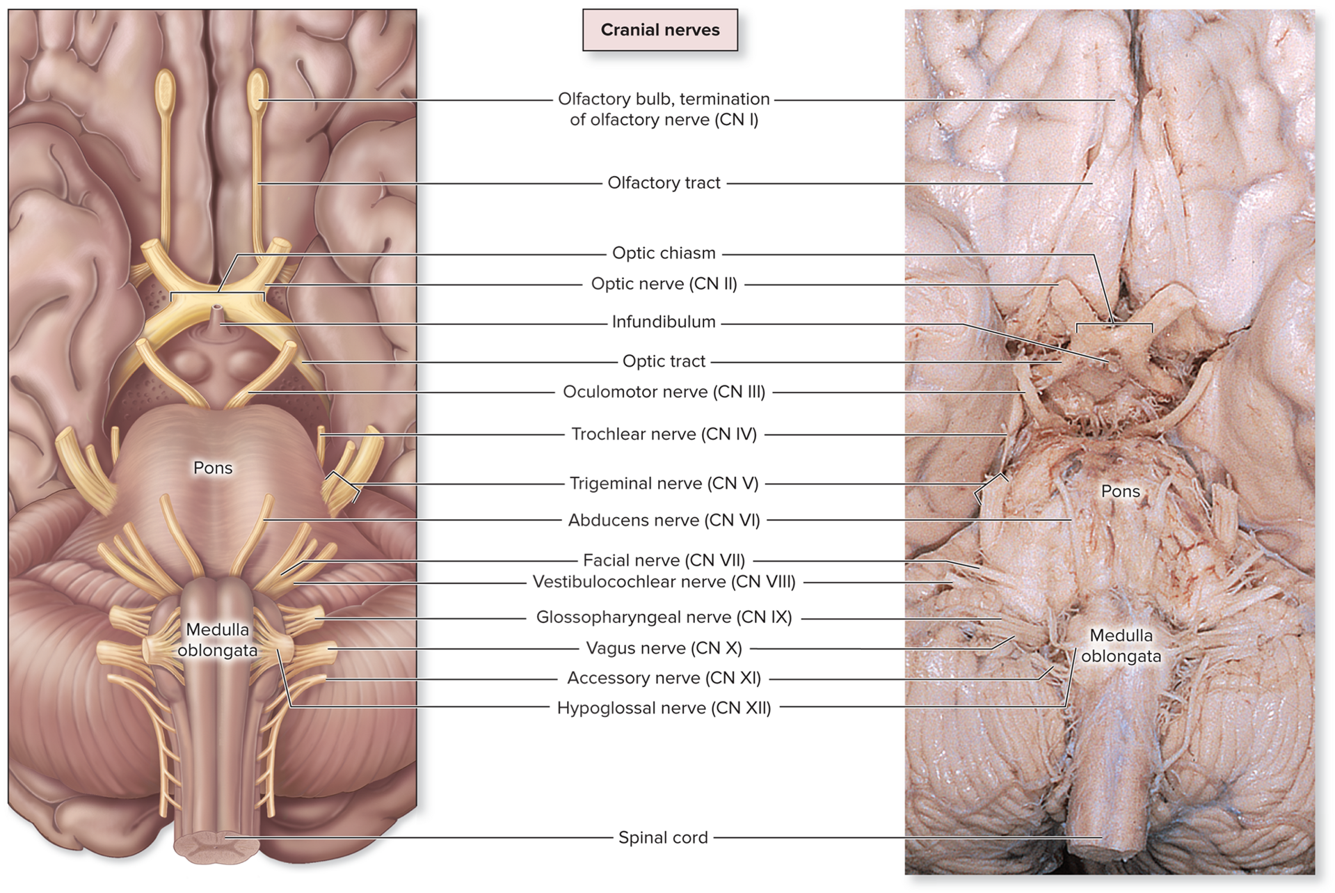
Primary Functions of Cranial Nerves
Cranial Nerve | Sensory Function | Somatic Motor Function | Autonomic Function |
|---|---|---|---|
I - olfactory | Olfaction (smell) | None | none |
II - optic | Vision | None | none |
III - oculomotor | None | Four extrinsic eye muscles; levator palpebrae; superioris muscle | Innervates sphincter pupillae muscle in eye to make pupil constrict; contracts ciliary muscles to make lens of eye more rounded |
IV - trochlear | None | Superior oblique extrinsic eye muscle | none |
V - trigeminal | General sensory from anterior scalp, nasal cavity, nasopharynx, entire face, most of oral cavity, teeth, anterior two-thirds of tongue; part of auricle of ear; meninges | Muscles of mastication, mylohyoid, digastric (ant. belly), tensor tympani, tensor veli palatini | None |
VI - abducens | None | Lateral rectus extrinsic eye muscle | none |
VII - facial | Taste from anterior two-thirds of tongue | Muscle of facial expression, digastric (pos. belly), stylohyoid, stapedius | Increases secretion from lacrimal gland of eye, submandibular and sublingual salivary glands |
VIII - vestibulocochlear | Hearing (cochlea): equilibrium (vestibular notch) | None | none |
IX - glossopharyngeal | General sensory and taste from posterior one-third of tongue, general sensory from part of pharynx, visceral sensory from carotid bodies | One pharyngeal muscle (stylopharyngeus) | Increases secretion from parotid salivary gland |
X - vagus | Visceral sensory information from heart, lungs, most abdominal organs. General sensory information from external acoustic meatus, tympanic membrane, part of pharynx, laryngopharynx, and larynx | Most pharyngeal muscles; all laryngeal muscles | Innervates smooth muscle and glands of heart, lungs, larynx, trachea, most abdominal organs |
XI - accessory | None | Trapezius and sternocleidomastoid | None |
XII - hypoglossal | None | Intrinsic and extrinsic tongue muscles | none |
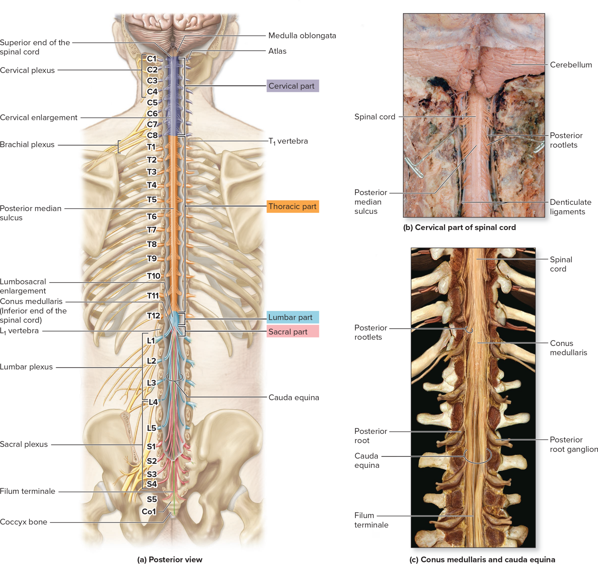
Anatomy of The Spinal Cord:
- Part of the CNS and has 31 pairs of spinal nerves that extend from it
- 8 pairs of cervical nerves
- 12 pairs of thoracic nerves
- 5 pairs of lumbar nerves
- And one pair of coccygeal nerves
General Functions:
- Provide an essential structural and functional link between the brain and the torso and limbs of the body
- The spinal nerves serve as a pathway for sensory and motor nerve signals and are responsible for reflexes
Gross Anatomy:
- The adult spinal cord is within the vertebral canal and typically ends at the ends at the level of the L1 vertebra
Spinal Cord Protection and Support

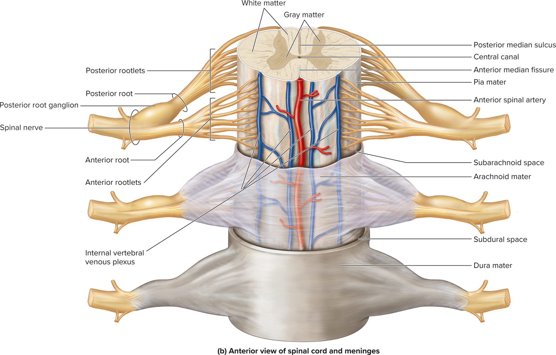
- Protection and support of the spinal cord includes the bony vertebral column, meninges, and CSF
- The meninges include the pia mater, arachnoid mater, and dura mater
- A potential subdural space lies between the dura mater and arachnoid mater, the epidural space is external to the dura mater and contains adipose connective tissue and blood vessels, and the subarachnoid space lies between the arachnoid mater and pia mater and contains CSF
Vertebral column:
- Formed by 26 stacked vertebrae and the intervertebral discs between each vertebrae
Sectional Anatomy of the Spinal Cord and Spinal Roots
- Gray matter is centrally located and composed of neuron cell bodies, dendrite, unmyelinated axons, and glial cells
- White matter is peripherally located and composed of myelinated axons
Distribution of gray matter:
- Has three horns
- Posterior: contains sensory axons and cell bodies of interneurons
- Anterior: contains cell bodies of somatic motor neurons
- Lateral: contains cell bodies of autonomic motor neurons
Distribution of white matter:
- Organized into three pairs of funiculi, most of which contains sensory (ascending) tracts and motor (descending) tracts
Sensory and Motor Pathways
- CNS communicates with the body through conduction pathways that extend through the spinal cord
Conduction pathways:
- Sensory pathways transmit ascending information from sensory receptors to the CNS, whereas motor pathways transmit descending information from the brain to muscles and glands
Sensory pathways:
- Ascending pathways
- Use primary, secondary, and tertiary neurons
- The posterior funiculus-medial lemniscal pathway transmits stimuli of conscious proprioception and discriminative touch to the cerebrum (parietal lobe)
- The anterolateral pathway transmits stimuli related to crude touch, pressure, pain, and temperature to the cerebrum (parietal lobe)
- The spinocerebellar pathway transmits stimuli from proprioceptors to the cerebellum for subconscious perception
Motor pathways:
- Descending pathways
- Use upper and lower motor neurons
- Somatic motor pathways include:
- Direct system: conscious control of skeletal muscle; extends through the spinal cord consists of the corticospinal tracts
- Indirect system: for unconscious control of skeletal muscle; consists of lateral pathway (rubrospinal tracts) and the medial pathway (reticulospinal, tectospinal, and vestibulospinal tracts)
Spinal Nerves:
- Formed from the union of an anterior root and a posterior root
Distribution:
- Two branches:
- Posterior ramus: innervates deep muscles of the back
- Anterior ramus: innervates skin and muscle of the anterior and lateral portions of the trunk and the limbs
Nerve Plexuses:
- A network of interwoven anterior rami
- Occur in pairs on either side of the body
Intercostal nerves:
- Anterior rami of spinal nerves T1-T11 do not form a plexus, but rather form the intercostal nerves
- Nerve T12 is called the subcostal nerve
Cervical plexuses:
- Each cervical plexus is formed from the anterior rami of C1-C4 spinal nerves
- Innervates the anterior neck muscles and the skin along the neck and shoulder
- Phrenic nerve is formed, in part, from branches of this plexus, but is not considered of the plexus
Brachial Plexuses:
- Each brachial plexus is formed from the anterior rami of spinal nerves C5-T1.
- Innervates an upper limb
Lumbar Plexuses:
- Each lumbar plexus is formed from the anterior rami of spinal nerves L1-L4
- Innervate the anterior and medial thigh, lower abdominal wall, skin of medial leg
Sacral Plexuses:
- Each sacral plexus is formed from the anterior rami of spinal nerves L4-S4
- Innervates most of the lower limb as well as the perineum
Reflex Arc:
- A reflex is a rapid, preprogrammed response of muscles or glands to a stimulus
Components:
- Activation of a receptor by a stimulus
- Nerve signal propagation along a sensory neuron to the CNS
- Integration and processing of information by interneurons
- Nerve signal propagation along a motor neuron
- Effector response
Somatic and Autonomic Nervous Systems
Functional Organization:
- Somatic: Includes sensory input from special senses, skin, muscles and joints, and motor output to control skeletal muscle
- Autonomic: includes involuntary motor output to cardiac muscle, smooth muscle, and glands; responds to sensory input from visceral sensory components
Lower Motor Neurons:
- A single motor neuron innervates skeletal muscles fibers in the SNS
- ANS has a two-neuron pathway consisting of preganglionic neurons in the CNS and ganglionic neurons in the PNS
CNS control of ANS:
- Regulated by hypothalamus, brainstem, and spinal cord
Sympathetic and Parasympathetic Nervous Systems + Anatomical Differences
- Parasympathetic preganglionic neurons reside in the brainstem and sacral regions of the spinal cord
- Sympathetic preganglionic axons reside in the thoracic and lumbar regions of the spinal cord
Cranial Components of Parasympathetic Division
- Parasympathetic preganglionic axons extend through the oculomotor, facial, glossopharyngeal, and vagus cranial nerves
Trunks and Ganglia of Sympathetic Division
- Preganglionic neurons reside in the T1-L2 segments of the spinal cord
- Preganglionic neuronal cell bodies are housed within the lateral gray horn of the spinal gray matter and their axons extend through white rami communicantes to the sympathetic trunk
- Gray rami communicantes are composed of postganglionic sympathetic axons from the sympathetic trunk to the spinal nerve
- Some preganglionic axons pass through the sympathetic trunk without synapsing and form splanchnic nerves that project to the prevertebral ganglia
- Postganglionic axons extend from the prevertebral ganglia to the effector
Sympathetic Pathways
Spinal nerve pathway:
- Postganglionic axon enters the spinal nerve through gray ramus and extends to effectors
- Blood vessels and glands of the skin of neck, torso, and limbs
Postganglionic sympathetic nerve pathway:
- Postganglionic axons extends from the sympathetic trunk and projects directly to the effectors
- Head, neck, viscera, and thoracic viscera
Splanchnic nerve pathway:
- Preganglionic axon passes through the sympathetic trunk and travels to the prevertebral ganglia, where it synapses with a ganglionic neuron
- Effectors: most abdominal and pelvic viscera
Adrenal medulla pathway:
- Preganglionic axon extends through both a sympathetic trunk ganglion and a prevertebral ganglion without synapsing
- Synapses on secretory cells in the adrenal medulla that releases epinephrine and norepinephrine
Interactions Between Parasympathetic and Sympathetic Divisions
- Most organs are innervated by both divisions of the ANS
Autonomic Tone:
- Both ANS divisions maintain some continual activity, which is called the autonomic tone for that division
Dual Innervation:
- Many visceral effectors have dual innervation, meaning they are innervated by both ANS divisions
- The actions of these divisions mostly are antagonistic, but a few are cooperative
ONLY THE SYMPATHETIC DIVISION:
- ANS helps maintain homeostasis through autonomic reflexes, which are also called visceral reflexes
Stretch Reflex, Golgi Reflex, Withdrawal and Crossed-extensor Reflex
- A stretch reflex is monosynaptic and contracts the muscle in response to increased stretch in a muscle spindle
- A tendon reflex is polysynaptic and prevents muscles from tensing excessively
- A withdrawal reflex is polysynaptic and activates flexor muscles to immediately remove a body part from a painful stimulus
- A crossed-extensor reflex stimulates the extensor muscles in the opposite limb in response to the withdrawal reflex
Neurotransmitters and Receptors in ANS
- Acetylcholine is the neurotransmitter released by cholinergic neurons, which include all preganglionic neurons as well as ganglionic parasympathetic neurons
- ACH also is used by some ganglionic sympathetic neurons to sweat glands
- Norepinephrine is the neurotransmitter released from adrenergic neurons, which include all other sympathetic postganglionic neurons
Cholinergic Receptors:
- Nicotinic receptors are located on all ganglionic neurons and cells in the adrenal medulla, and they are always excitatory
- Muscarinic receptors are located on all parasympathetic target cells and on all sweat glands in the skin; may be excitatory or inhibitory depending upon the muscarinic receptor subtype