Integumentary System- Chapter 3.2
integumentary system- largest organ of the body that forms a physical barrier between the external environment and the internal environment that it serves to protect and maintain (skin)
Functions:
Skin is also critically important in regulating body temperature, due to the extensive array of tiny capillaries and sweat glands that lie near the surface of the skin.
It is also involved in certain chemical processes in the body.
Keratin:
keratin- a tough protein also found in hair and nails that adds structural strength, helps to protect the skin against damage
keratinocytes- produce keratin
Keratin and naturally occurring oils in the skin assist in serving as a water barrier, and keratin prevents water from entering the body during bathing or swimming.
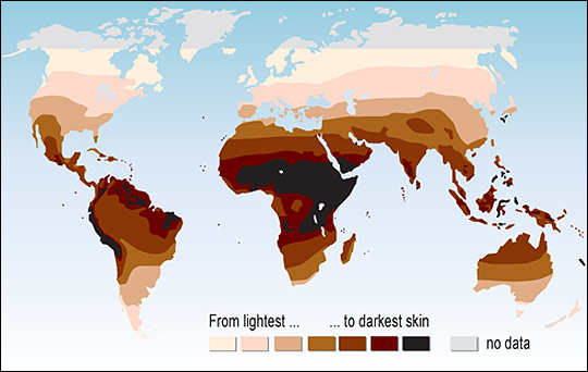
Melanin:
Melanocytes- produce melanin, a pigment that protects the body from harmful UV radiation
Melanin- primarily responsible for human skin color
Exposure to sunlight causes melanocytes to produce more melanin.
Freckles- clusters of melanocytes
Albinism- prevents the normal production of melanin, and produces very little pigment in the skin, hair, and eyes.
Light skin is good for rapid Vitamin D production, and dark skin is better for protection from Atoo much UV.
Appendages of the Skin:
Sweating:
Process of sweating- the body eliminates chemical waste products, including urea, uric acid, and salts
sudoriferous glands- distributed in the dermis over the entire body (sweat glands)
eccrine glands- the major sweat glands of the body, cover most of the body, open directly onto the skin
fluid secreted by the sweat glands is acidic, and helps to protect the body against bacterial infections
Sebum:
sebaceous glands- located all over the body except for the palms of the hands and soles of the feet, produce an oily substance called sebum
sebum- an oily and slightly waxy substance found on the skin, helps to keep the skin and hair softbut also contains chemicals that kill bacteria
Anatomy of the Skin:
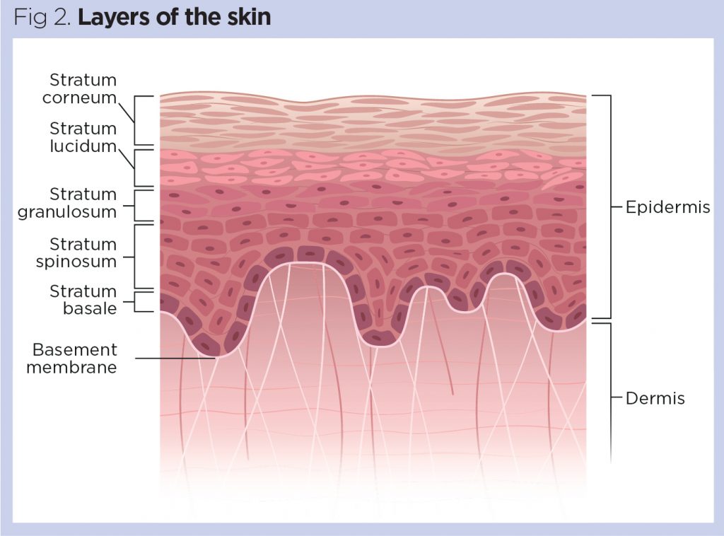
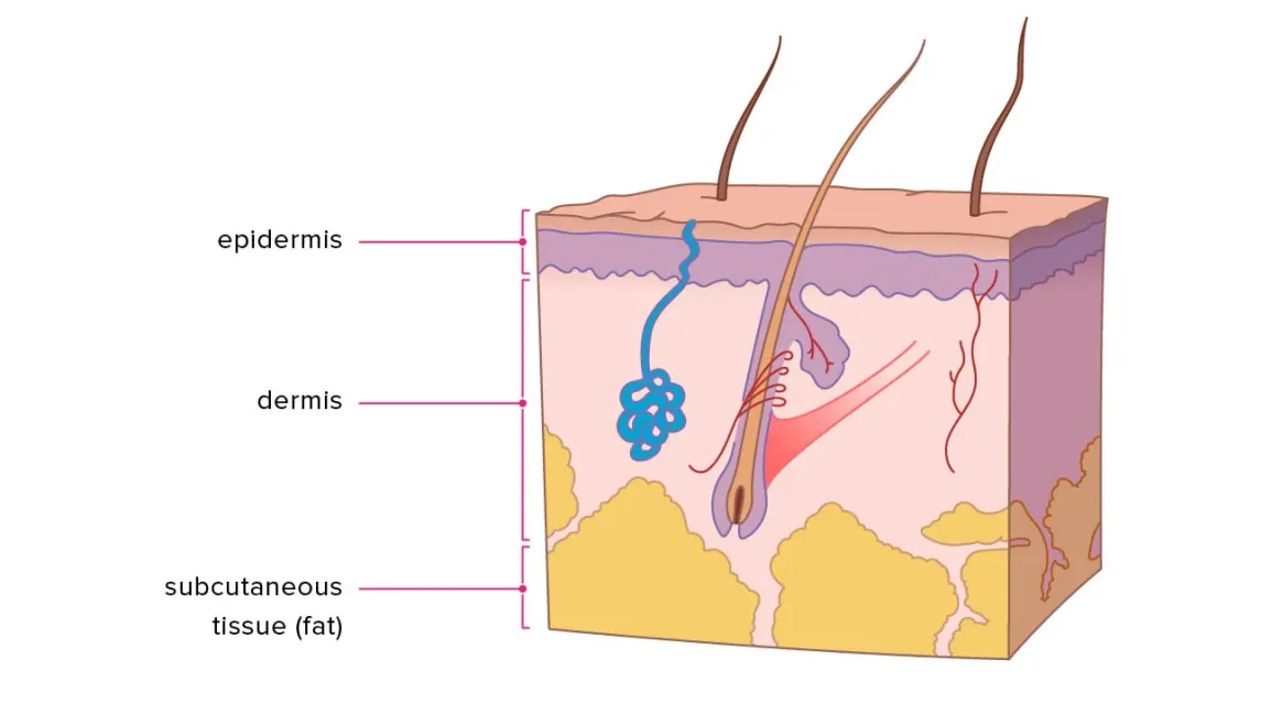
Epidermis:
Epidermis- the surface epithelium of the skin, overlying the dermis.
The epidermis contains five layers of tissue.
From superficial to deep (the outside going in), these are the stratum corneum, stratum lucidum, stratum granulosum, stratum spinosum, and stratum basale.
The epidermis completely replaces itself every 25 to 45 days.
Layers of the Epidermis:
Stratum Corneum- the oldest layer of cells
dust- (dead) cells that shed from the stratum corneum
Stratum Lucidum- clear layer of dead, keratinized cells
This is only present in areas of the body with thick skin.
Epidermal Ridges- increases surface area for oxygen, diffusion from the dermis
This causes finger and footprints to be made.
Stratum Granulosum- produces a large amount of keratin, die eventually due to lack of oxygen
Stratum Spinosum- cells begin forming desmosomes to attach to each other in layers.
Stratum Basale- deepest layer with melanocytes
Merkel cells- located in the stratum basale, function as touch receptors
Cells in the Epidermis:
Epidermal dendritic cells- respond to the presence of foreign bacteria or viruses by initiating an immune system response
Dermis:
Dermis- thick layer of living tissue below the epidermis which forms the true skin, containing blood capillaries, nerve endings, sweat glands, hair follicles, and other structures
Papillary Layer- dermal papillae that protrude from the surface up into the epidermis
Reticular Layer- collagen and elastic fibers in this region have an irregular arrangement, it includes blood and lymphatic vessels, sweat and oil glands, involuntary muscles, hair follicles, and nerve endings
When the two separate, they form blisters, or a “fluid filled pocket".
Some of the dermal papillae contain capillaries that supply nutrients to the epidermis. Other dermal papillae contain nerve endings involved in sensing touch and pain.
Hypodermis:
Hypodermis (subcutaneous fascia)- a storage repository for fat
not a part of the skin, but connects the skin to the underlying tissues
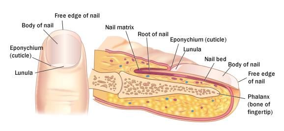
 Nails:
Nails- a more compacted, keratinized version of the stratum corneum
Cuticle- overlap of the stratum corneum, keeps bacteria away from nail root
Bed- contains tissue from stratum basale and spinosum
Matrix- contains cells that constantly divide