
Chapter 8- Special Senses
Special Senses
Special senses include:
Smell
Taste
Sight
Hearing
Equilibrium
Special sense receptors
Large, complex sensory organs
Localized clusters of receptors
The Eye and Vision
70% of all sensory receptors are in the eyes
Each eye has over 1 million nerve fibers carrying information to the brain
Anatomy of the Eye
Accessory structures include the:
Extrinsic eye muscles (operating from the outside)
Eyelids
Conjunctiva
Lacrimal apparatus
External and Accessory Structures
Eyelids
Meet at the medial and lateral commissure (canthus)
Eyelashes
Tarsal glands produce an oily secretion that lubricates the eye
Ciliary glands are located between the eyelashes
Conjunctiva
Membranes that lines the eyelids and eyeball
Connects with the transparent cornea
Secretes to lubricate the eye and keep it moist
Lacrimal apparatus = lacrimal gland + ducts
Lacrimal gland—produces lacrimal fluid (tears); situated on lateral end of each eye
Tears drain across the eye into the lacrimal canaliculi, then the lacrimal sac, and into the nasolacrimal duct-, which empties into the nasal cavity
Tears contain:
Dilute salt solution (saline)
Mucus
Antibodies
Lysozyme (enzyme that destroys bacteria)
Function of tears
Cleanse, protect, moisten, lubricate the eye
Extrinsic eye muscles
6 muscle attach attach to the outer surface of the eye
Produce gross eye movements
Internal Structures: The Eyeball
Three layers, or tunics, form the wall of the eyeball
Fibrous layer: outside layer
Vascular layer: middle layer
Sensory layer: inside layer
Humors are fluids that fill the interior of the eyeball
Lens divides the eye into two chambers
Fibrous layer = sclera + cornea
Sclera
White connective tissue layer ”white of the eye”
Cornea
Transparent, central anterior portion
Allows for light to pass through
Repairs itself easily
The only human tissue that can be transplanted without fear of rejection
Vascular layer
Choroid is a blood-rich nutritive layer that contains a pigment(prevents light from scattering) & is modified anteriorly into two smooth muscle structures
Ciliary body
Iris -—regulates amount of light entering eye
Pigmented layer—gives eye color
Pupil—rounded opening in the iris
Sensory layer
Retina contains two layers
Outer pigmented layer absorbs light and prevents it from scattering
Inner neural layer contains receptor cells (photoreceptors)
Rods
Cones
Electrical signals pass from photoreceptors via a two-neuron chain
Bipolar neuronsGanglion cells
Signals leave the retina toward the brain through the optic nerve
Optic disc- (blind spot) is where the optic nerve leaves the eyeball
Cannot see images focused on the optic disc
Rods
Most are found toward the edges of the retina
Allow vision in dim light and peripheral vision
All perception is in gray tones
Cones
Allow for detailed color vision
Densest in the center of the retina
Fovea centralis–lateral to blind spot
Area of the retina with only cones
Visual acuity(sharpest vision) is here
No photoreceptor cells are at the optic disc, or blind spot
Cone sensitivity
Three types of cones
Each cone type is sensitive to different wavelengths of visible light
Lens
Flexible, biconvex (convex on both sides) crystal-like structure
Held in place by a suspensory ligament attached to the ciliary body
Lens divides the eye into two chambers
Anterior (aqueous) segment
Anterior to the lens
Contains aqueous humor, a clear, watery fluid
Posterior (vitreous) segment
Posterior to the lens
Contains vitreous humor, a gel-like substance
Aqueous humor
Watery fluid found between lens and cornea
Similar to blood plasma
Helps maintain intraocular pressure
Provides nutrients for the lens and cornea
Reabsorbed into venous blood through the scleral venous sinus, or canal of Schlemm
Vitreous humor
Gel- like substance posterior to the lens
Prevents the eye from collapsing
Helps maintain intraocular pressure
Ophthalmoscope
Instrument used to illuminate the interior of the eyeball and fundus (posterior wall)
Can detect diabetes, arteriosclerosis, degeneration of the optic nerve and retina
Physiology of Vision
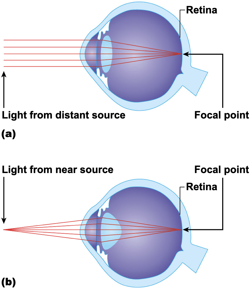
Path of light through eye & light refraction
Light must be focused to a point on the retina for optimal vision
Light is bent, or refracted, by the cornea, aqueous humor, lens, and vitreous humor
The eye is set for distant vision (over 20 feet away)
Accommodation—the lens must change shape to focus on closer objects (less than 20 feet away)
Pathway of light through the eye and light refraction (continued)
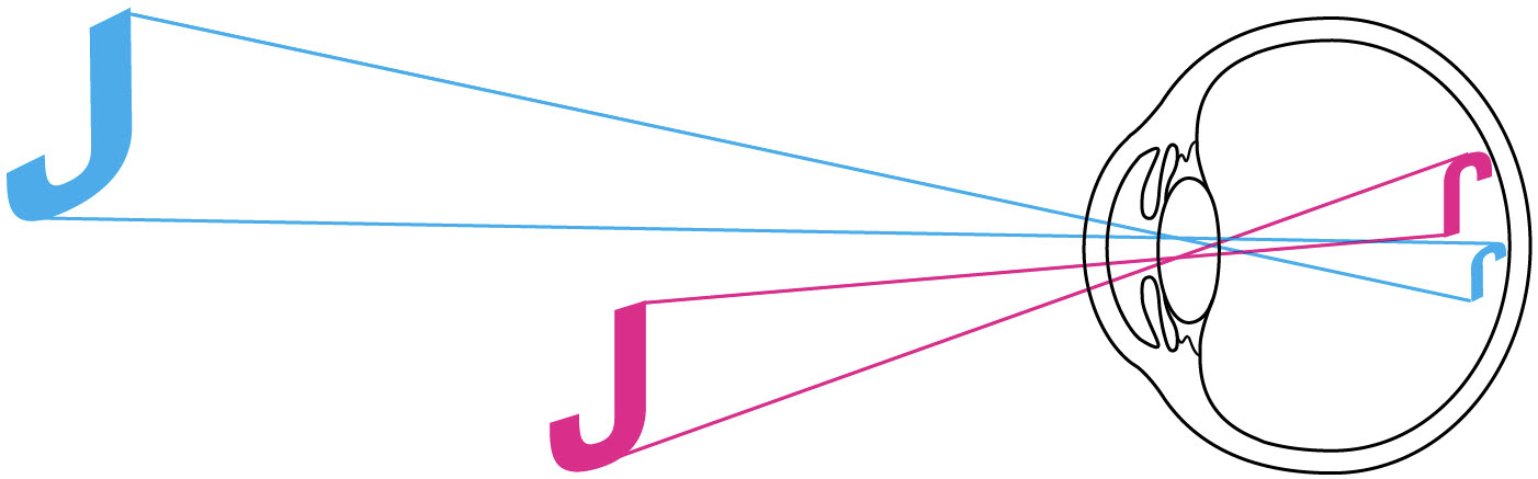
Image formed on the retina is a real image
Real images are:
Reversed from left to right
Upside down
Smaller than the object
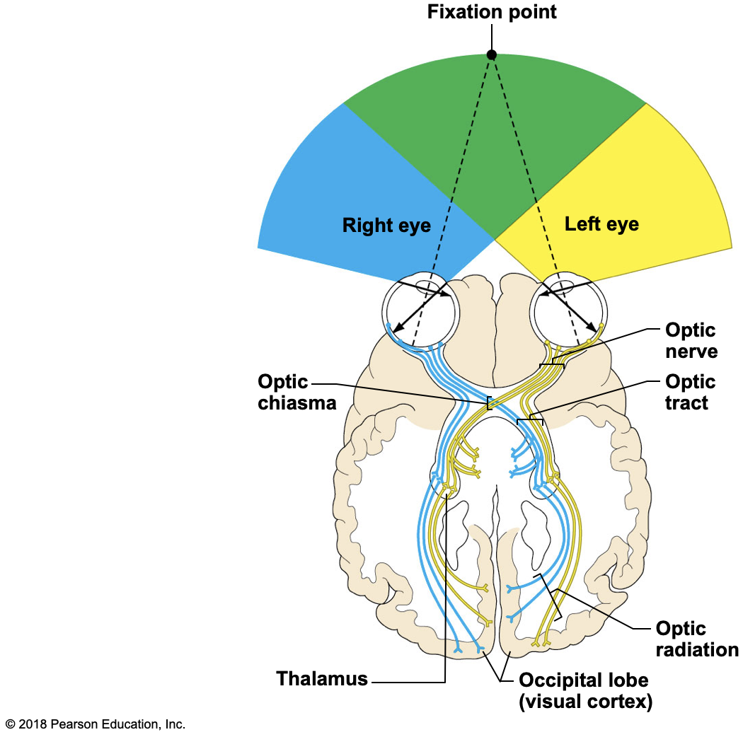
Visual fields and visual pathways to brain
Optic nerve
Bundle of axons that exit the back of the eye carrying impulses from the retina
Optic chiasma
Location where the optic nerves cross
Fibers from the medial side of each eye cross over to the opposite side of the brain
Visual fields & visual pathways to the brain
Optic tracts
Contain fibers from the lateral side of the eye on the same side and the medial side of the opposite eye
Synapse with neurons in the thalamus- (relaying of sensory signals, including motor signals, to the cerebral cortex, and the regulation of consciousness, sleep, and alertness)
Optic radiation
Axons from the thalamus run to the occipital lobe
Synapse with cortical cells, and vision interpretation (seeing) occurs
Summary of the pathway of impulses from the retina to the point of visual interpretation
Optic nerve
Optic chiasma
Optic tract
Thalamus
Optic radiation
Optic cortex in occipital lobe of brain
Visual fields
Each eye “sees” a slightly different view
Field of view overlaps for each eye
Binocular vision results and provides:
Depth perception (three-dimensional vision)
A Closer Look
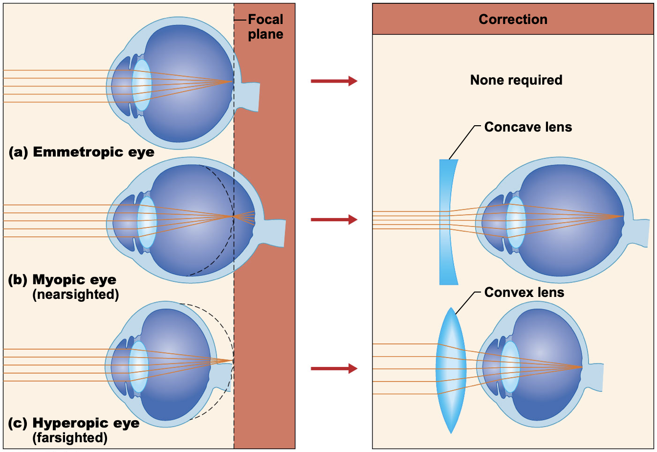
Emmetropia—eye focuses images correctly on the retina
Myopia (nearsightedness)
Distant objects appear blurry
Light from those objects fail to reach the retina and are focused in front of it
Results from an eyeball that is too long
Hyperopia (farsightedness)
Near -objects are blurry, whereas distant objects are clear
Distant objects are focused behind the retina
Results from an eyeball that is too short or from a “lazy lens”
Astigmatism
Images are blurry
Results from light focusing as lines, not points, on the retina because of unequal curvatures of the cornea or lens
Convergence: reflexive movement of the eyes medially when we focus on a close object
Photopupillary reflex: bright light causes pupils to constrict
Accommodation pupillary reflex: viewing close objects causes pupils to constrict
The Ear: Hearing and Balance
Ear houses two senses
Hearing
Equilibrium (balance)
Receptors are mechanoreceptors (respond to touch or feel)
Different organs house receptors for each sense
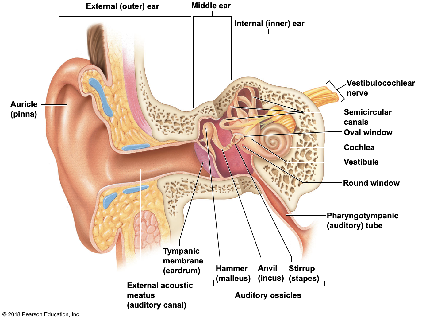
Anatomy of the Ear
The ear is divided into three areas
External (outer) ear
Middle ear
Internal (inner) ear
External (outer) ear
Auricle (pinna)
Ext. acoustic meatus (auditory canal)
Narrow chamber in the temporal bone
Lined with skin and ceruminous (earwax) glands
Ends at the tympanic membrane (eardrum)
External ear is involved only in collecting sound waves
Middle ear cavity (tympanic cavity)
Air filled, mucosa-lined cavity within the temporal bone
Involved only in the sense of hearing
Located between tympanic membrane and oval window and round window
Pharyngotympanic tube (auditory tube)
Links middle ear cavity with the throat
Equalizes pressure in the middle ear cavity so the eardrum can vibrate
Middle ear cavity (tympanic cavity)
Three bones (ossicles) span the cavity
Malleus(hammer), Incus(anvil), Stapes(stirrup)
Function
Transmit vibration from tympanic membrane to the fluids of the inner ear
Vibrations travel: hammer -> anvil -> stirrup -> oval window of inner ear
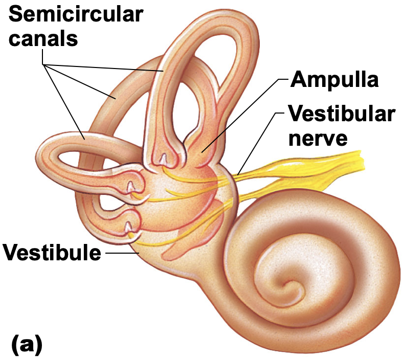
Internal (inner) ear
Sense organs for hearing and balance
Bony labyrinth (osseous labryrinth) consists of:
Cochlea, vestibule, semicircular canals
Bony labyrinth is filled with perilymph
Membranous labyrinth is suspended in perilymph and contains endolymph
Equilibrium
Equilibrium receptors of the inner ear are called the vestibular apparatus
Vestibular apparatus has two functional parts
Static equilibrium
Dynamic equilibrium
Static Equilibrium
Maculae—receptors in the vestibule
Report on the position of the head
Help us keep our head erect
Send information via the vestibular nerve (division of cranial nerve VIII) to the cerebellum of the brain
Anatomy of the maculae
Hair cells are embedded in the otolithic membrane
Otoliths (tiny stones) float in a gel around hair cells
Movements cause otoliths to roll and bend hair cells
Dynamic Equilibrium
Crista ampullaris
Responds to angular or rotational of the head
In ampulla of each semicircular canal
Tuft of hair cells covered with cupula (gelatinous cap)
If the head moves, the cupola drags against the endolymph
Hair cells are stimulated, impulse travels vestibular n. to the cerebellum
Hearing
Spiral organ of Corti
Located within the cochlear duct
Receptors = hair cells on the basilar membrane
Gel-like tectorial membrane is capable of bending hair cells
Cochlear nerve attached to hair cells transmits nerve impulses to auditory cortex on temporal lobe
Pathway of vibrations from sound waves
Ear drumossiclesoval window
Sound is amplified by the ossicles
Pressure waves cause vibrations in the basilar membrane in the organ of Corti
Hair cells of the tectorial membrane are bent when the basilar membrane vibrates against it
An action potential starts in the cochlear nerve (cranial nerve VIII), and the impulse travels to the temporal lobe
High pitched sounds disturb the short, stiff fibers of the basilar membrane
Receptor cells close to the oval window are stimulated
Low pitched sounds disturb the long, floppy fibers of the basilar membrane
Specific hair cells further along the cochlea are affected
Hearing and Equilibrium Deficits
Deafness is any degree of hearing loss
Conduction deafness results when the transmission of sound vibrations through the external and middle ears is hindered
Sensorineural deafness results from damage to the nervous system structures involved in hearing
Meniere’s affects inner ear and causes progressive deafness and perhaps vertigo (sensation of spinning)
Chemical Senses: Smell & Taste
Chemoreceptors
Stimulated by chemicals in solution
Taste has five types of receptors
Smell can differentiate a wider range of chemicals
Both senses complement each other and respond to many of the same stimuli
Olfactory Receptors/Sense of Smell
Olfactory receptors in roof of nasal cavity
Olfactory receptor cells (neurons) with long cilia (olfactory hairs) detect chemicals
Chemicals must be dissolved in mucus for detection by chemoreceptors called olfactory receptors
Impulses are transmitted via the olfactory filaments to the olfactory nerve (I)
Smells interpreted in the olfactory cortex
Taste Buds and Sense of Taste
Taste buds house the receptor organs
Locations of taste buds
Most are on the tongue
Soft palate
Superior part of the pharynx
Cheeks
The tongue is covered with projections called papillae that contain taste buds
Vallate (circumvallate) papillae
Fungiform papillae
Filiform papillae
Gustatory cells are the taste receptors
Possess gustatory hairs (long microvilli)
Gustatory hairs protrude through a taste pore
Hairs are stimulated by chemicals dissolved in saliva
Impulses are carried to the gustatory complex by several cranial nerves because taste buds are found in different areas
Facial nerve (cranial nerve VII)
Glossopharyngeal nerve (cranial nerve IX)
Vagus nerve (cranial nerve X)
Taste buds are replaced frequently by basal cells
Five basic taste sensations
Sweet receptors respond to sugars, saccharine, some amino acids
Sour receptors respond to H+ ions or acids
Bitter receptors respond to alkaloids
Salty receptors respond to metal ions
Umami receptors respond to the amino acid glutamate or the beefy taste of meat
Developmental Aspects of the Special Senses
Special sense organs are formed early in embryonic development
Maternal infections during the first 5 or 6 weeks of pregnancy may cause visual abnormalities as well as sensorineural deafness in the developing child
Vision requires the most learning
The infant has poor visual acuity (is farsighted) and lacks color vision and depth perception at birth
The eye continues to grow and mature until age 8 or 9
Age-related eye issues
Presbyopia—“old vision” results from decreasing lens elasticity that accompanies aging
Difficulty to focus for close vision
Lacrimal glands become less active
Lens becomes discolored
Dilator muscles of iris become less efficient, pupils remain constricted
The newborn infant can hear sounds, but initial responses are reflexive
By the toddler stage, the child is listening critically and beginning to imitate sounds as language development begins
Age-related ear problems
Presbycusis—type of sensorineural deafness that may result from otosclerosis (ear ossicles fuse)
Congenital ear problems usually result from missing pinnas and closed or missing external acoustic meatuses
Taste and smell are most acute at birth and decrease in sensitivity after age 40 as the number of olfactory and gustatory receptors decreases