


Chapter 7: Axial Skeleton
Divisions of the Skeletal System
The skeletal system includes all of the bones, cartilages, and ligaments of the body that support and give shape to the body and body structures.
The skeleton consists of the bones of the body.
For adults, there are 206 bones in the skeleton.
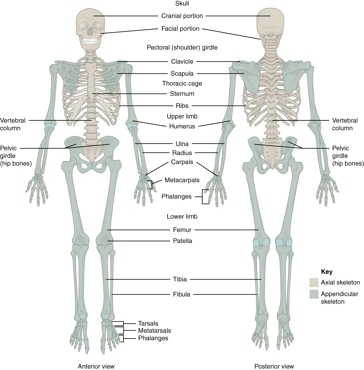
The skeleton is subdivided into two major divisions—the axial and appendicular.
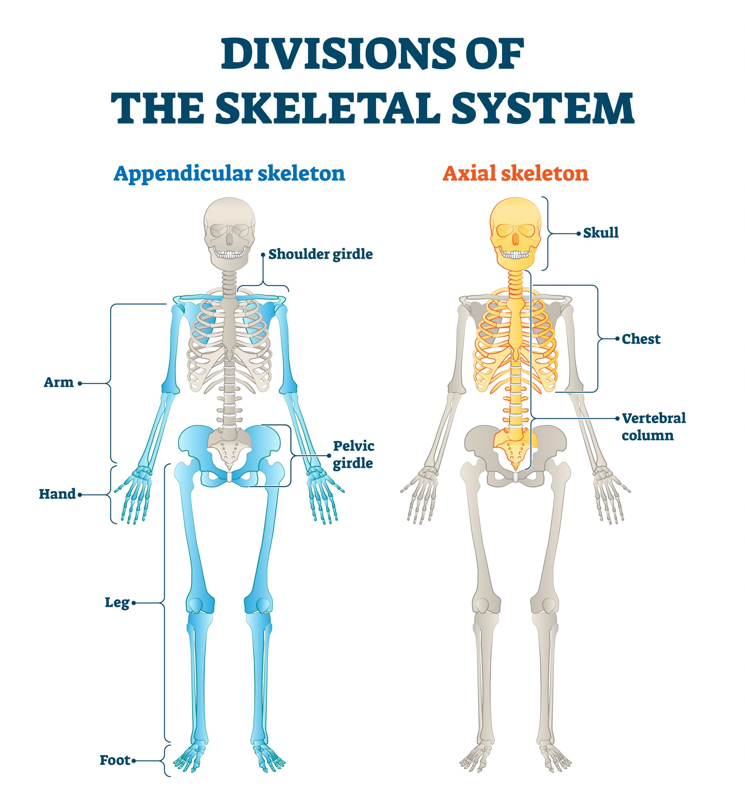
The Axial Skeleton
The axial skeleton forms the vertical, central axis of the body and includes all bones of the head, neck, chest, and back.
The axial skeleton of the adult consists of 80 bones, including the skull, the vertebral column, and the thoracic cage.
Also associated with the head are an additional seven bones, including the hyoid bone and
The ear ossicles (three small bones found in each middle ear).
The vertebral column consists of 24 bones, each called a vertebra, plus the sacrum and coccyx.
The thoracic cage includes the 12 pairs of ribs, and the sternum, the flattened bone of the anterior chest.
The Appendicular Skeleton
The appendicular skeleton includes all bones of the upper and lower limbs, plus the bones that attach each limb to the axial skeleton.
There are 126 bones in the appendicular skeleton of an adult.
The Skull
The cranium (skull) is the skeletal structure of the head that supports the face and protects the brain.
It is subdivided into the facial bones and the brain case, or cranial vault.
The 22nd bone is the mandible (lower jaw), which is the only moveable bone of the skull.
Anterior View of Skull
The anterior skull consists of the facial bones and provides the bony support for the eyes and structures of the face.
The orbit is the bony socket that houses the eyeball and muscles that move the eyeball or open the upper eyelid.
The upper margin of the anterior orbit is the supraorbital margin.
Located near the midpoint of the supraorbital margin is a small opening called the supraorbital foramen.
This provides for passage of a sensory nerve to the skin of the forehead.
The infraorbital foramen, which is the point of emergence for a sensory nerve that supplies the anterior face below the orbit.
Inside the nasal area of the skull, the nasal cavity is divided into halves by the nasal septum.
The upper portion of the nasal septum is formed by the perpendicular plate of the ethmoid bone and the lower portion is the vomer bone.
The larger of these is the inferior nasal concha, an independent bone of the skull.
Located just above the inferior concha is the middle nasal concha, which is part of the ethmoid bone.
A third bony plate, also part of the ethmoid bone, is the superior nasal concha.
Lateral View of Skull
The zygomatic arch is the bony arch on the side of skull that spans from the area of the cheek to just above the ear canal.
It is formed by the junction of two bony processes: a short anterior component, the temporal process of the zygomatic bone (the cheekbone) and a longer posterior portion, the zygomatic process of the temporal bone, extending forward from the temporal bone.
On the lateral side of the brain case, above the level of the zygomatic arch, is a shallow space called the temporal fossa.
Below the level of the zygomatic arch and deep to the vertical portion of the mandible is another space called the infratemporal fossa.
Bones of the Brain Case
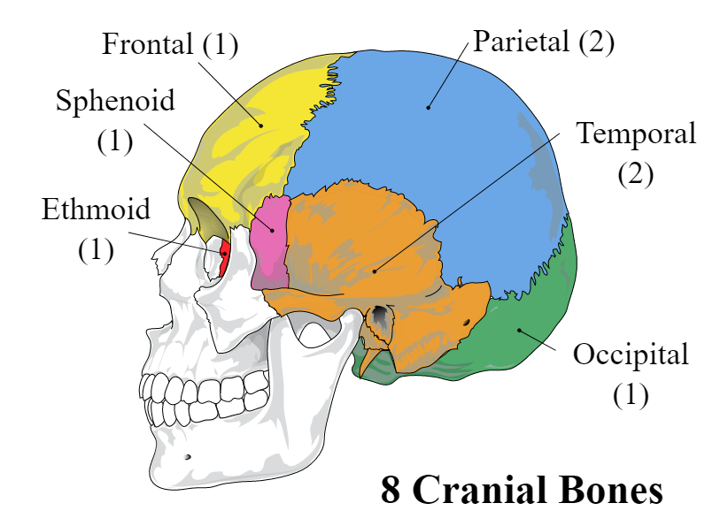
The brain case contains and protects the brain.
The interior space that is almost completely occupied by the brain is called the cranial cavity.
This cavity is bounded superiorly by the rounded top of the skull, which is called the calvaria (skullcap), and the lateral and posterior sides of the skull.
This is a complex area that varies in depth and has numerous openings for the passage of cranial nerves, blood vessels, and the spinal cord. Inside the skull, the base is subdivided into three large spaces, called the anterior cranial fossa, middle cranial fossa, and posterior cranial fossa (fossa = “trench or ditch”).
Parietal Bone: The parietal bone forms most of the upper lateral side of the skull.
Temporal Bone
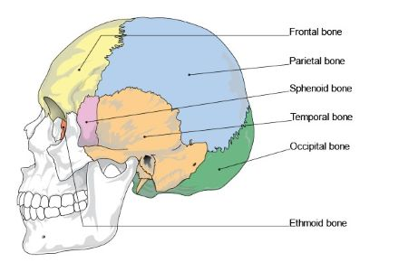
The temporal bone forms the lower lateral side of the skull.
Common wisdom has it that the temporal bone (temporal = “time”) is so named because this area of the head (the temple) is where hair typically first turns gray, indicating the passage of time.
Projecting inferiorly from this region is a large prominence, the mastoid process, which serves as a muscle attachment site.
On the interior of the skull, the petrous portion of each temporal bone forms the prominent, diagonally oriented petrous ridge in the floor of the cranial cavity.
External acoustic meatus (ear canal)—This is the large opening on the lateral side of the skull that is associated with the ear.
Internal acoustic meatus—This opening is located inside the cranial cavity, on the medial side of the petrous ridge.
Mandibular fossa—This is the deep, oval-shaped depression located on the external base of the skull, just in front of the external acoustic meatus.
Articular tubercle—The smooth ridge located immediately anterior to the mandibular fossa.
Styloid process—Posterior to the mandibular fossa on the external base of the skull is an elongated, downward bony projection called the styloid process, so named because of its resemblance to a stylus (a pen or writing tool).
Stylomastoid foramen—This small opening is located between the styloid process and mastoid process.
Carotid canal—The carotid canal is a zig-zag shaped tunnel that provides passage through the base of the skull for one of the major arteries that supplies the brain.
Frontal Bone
The frontal bone is the single bone that forms the forehead.
At its anterior midline, between the eyebrows, there is a slight depression called the glabella.
Occipital Bone
The occipital bone is the single bone that forms the posterior skull and posterior base of the cranial cavity.
On its outside surface, at the posterior midline, is a small protrusion called the external occipital protuberance, which serves as an attachment site for a ligament of the posterior neck.
Lateral to either side of this bump is a superior nuchal line (nuchal = “nape” or “posterior neck”).
On the base of the skull, the occipital bone contains the large opening of the foramen magnum, which allows for passage of the spinal cord as it exits the skull.
On either side of the foramen magnum is an oval-shaped occipital condyle.
Sphenoid Bone
The sphenoid bone is a single, complex bone of the central skull.
It serves as a “keystone” bone, because it joins with almost every other bone of the skull.
Inside the cranial cavity, the right and left lesser wings of the sphenoid bone, which resemble the wings of a flying bird, form the lip of a prominent ridge that marks the boundary between the anterior and middle cranial fossae.
The sella turcica (“Turkish saddle”) is located at the midline of the middle cranial fossa.
The rounded depression in the floor of the sella turcica is the hypophyseal (pituitary) fossa, which houses the pea-sized pituitary (hypophyseal) gland.
The greater wings of the sphenoid bone extend laterally to either side away from the sella turcica, where they form the anterior floor of the middle cranial fossa.
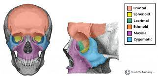
Ethmoid Bone
The ethmoid bone is a single, midline bone that forms the roof and lateral walls of the upper nasal cavity, the upper portion of the nasal septum, and contributes to the medial wall of the orbit.
The crista galli (“rooster’s comb or crest”) is a small upward bony projection located at the midline.
To either side of the crista galli is the cribriform plate (cribrum = “sieve”), a small, flattened area with numerous small openings termed olfactory foramina.
Sutures of the Skull
A suture is an immobile joint between adjacent bones of the skull.
The coronal suture runs from side to side across the skull, within the coronal plane of section.
The sagittal suture extends posteriorly from the coronal suture, running along the midline at the top of the skull in the sagittal plane of section.
The lambdoid suture extends downward and laterally to either side away from its junction with the sagittal suture.
The squamous suture is located on the lateral skull.
At the intersection of four bones is the pterion, a small, capital-H-shaped suture line region that unites the frontal bone, parietal bone, squamous portion of the temporal bone, and greater wing of the sphenoid bone.
Facial Bones of the Skull
The facial bones of the skull form the upper and lower jaws, the nose, nasal cavity and nasal septum, and the orbit.
The facial bones include 14 bones, with six paired bones and two unpaired bones.
Maxillary Bone
The maxillary bone, often referred to simply as the maxilla (plural = maxillae), is one of a pair that together form the upper jaw, much of the hard palate, the medial floor of the orbit, and the lateral base of the nose.
The curved, inferior margin of the maxillary bone that forms the upper jaw and contains the upper teeth is the alveolar process of the maxilla.
On the inferior skull, the palatine process from each maxillary bone can be seen joining together at the midline to form the anterior three-quarters of the hard palate.
The hard palate is the bony plate that forms the roof of the mouth and floor of the nasal cavity, separating the oral and nasal cavities.
Palatine Bone
The palatine bone is one of a pair of irregularly shaped bones that contribute small areas to the lateral walls of the nasal cavity and the medial wall of each orbit.
The largest region of each of the palatine bone is the horizontal plate.
Zygomatic Bone
The zygomatic bone is also known as the cheekbone.
Each of the paired zygomatic bones forms much of the lateral wall of the orbit and the lateral-inferior margins of the anterior orbital opening.
Nasal Bone: The nasal bone is one of two small bones that articulate (join) with each other to form the bony base (bridge) of the nose.
Lacrimal Bone
Each lacrimal bone is a small, rectangular bone that forms the anterior, medial wall of the orbit.
The anterior portion of the lacrimal bone forms a shallow depression called the lacrimal fossa, and extending inferiorly from this is the nasolacrimal canal.
Mandible
The mandible forms the lower jaw and is the only moveable bone of the skull.
Each side of the mandible consists of a horizontal body and posteriorly, a vertically oriented ramus of the mandible (ramus = “branch”).
The outside margin of the mandible, where the body and ramus come together is called the angle of the mandible.
The more anterior projection is the flattened coronoid process of the mandible, which provides attachment for one of the biting muscles.
The posterior projection is the condylar process of the mandible, which is topped by the oval-shaped condyle.
The broad U-shaped curve located between the coronoid and condylar processes is the mandibular notch.
Alveolar process of the mandible—This is the upper border of the mandibular body and serves to anchor the lower teeth.
Mental protuberance—The forward projection from the inferior margin of the anterior mandible that forms the chin (mental = “chin”).
Mental foramen—The opening located on each side of the anterior-lateral mandible, which is the exit site for a sensory nerve that supplies the chin.
Mylohyoid line—This bony ridge extends along the inner aspect of the mandibular body.
Mandibular foramen—This opening is located on the medial side of the ramus of the mandible.
Lingula—This small flap of bone is named for its shape (lingula = “little tongue”).
The Orbit
The orbit is the bony socket that houses the eyeball and contains the muscles that move the eyeball or open the upper eyelid.
At the posterior apex of the orbit is the opening of the optic canal, which allows for passage of the optic nerve from the retina to the brain.
The Nasal Septum and Nasal Conchae
The nasal septum consists of both bone and cartilage components.
The anterior nasal septum is formed by the septal cartilage, a flexible plate that fills in the gap between the perpendicular plate of the ethmoid and vomer bones.
Attached to the lateral wall on each side of the nasal cavity are the superior, middle, and inferior nasal conchae (singular = concha), which are named for their positions.
Middle Cranial Fossa
Optic canal—This opening is located at the anterior lateral corner of the sella turcica. It provides for passage of the optic nerve into the orbit.
Superior orbital fissure—This large, irregular opening into the posterior orbit is located on the anterior wall of the middle cranial fossa, lateral to the optic canal and under the projecting margin of the lesser wing of the sphenoid bone.
Foramen rotundum—This rounded opening (rotundum = “round”) is located in the floor of the middle cranial fossa, just inferior to the superior orbital fissure.
Foramen ovale of the middle cranial fossa—This large, oval-shaped opening in the floor of the middle cranial fossa provides passage for a major sensory nerve to the lateral head, cheek, chin, and lower teeth.
Foramen spinosum—This small opening, located posterior-lateral to the foramen ovale, is the entry point for an important artery that supplies the covering layers surrounding the brain.
Carotid canal—This is the zig-zag passageway through which a major artery to the brain enters the skull.
Foramen lacerum—This irregular opening is located in the base of the skull, immediately inferior to the exit of the carotid canal.
Posterior Cranial Fossa
The posterior cranial fossa is the most posterior and deepest portion of the cranial cavity. It contains the cerebellum of the brain.
Located at the anterior-lateral margin of the foramen magnum is the hypoglossal canal.
Immediately inferior to the internal acoustic meatus is the large, irregularly shaped jugular foramen.
Paranasal Sinuses
The paranasal sinuses are hollow, air-filled spaces located within certain bones of the skull.
The frontal sinus is located just above the eyebrows, within the frontal bone.
The largest sinus is the maxillary sinus.
The sphenoid sinus is a single, midline sinus.
Each of these spaces is called an ethmoid air cell.
The Vertebral Column
The vertebral column is also known as the spinal column or spine.
It consists of a sequence of vertebrae (singular = vertebra), each of which is separated and united by an intervertebral disc.
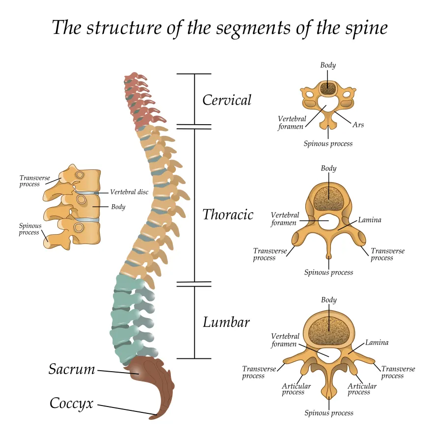
Curvatures of the Vertebral Column
In the adult, this fetal curvature is retained in two regions of the vertebral column as the thoracic curve, which involves the thoracic vertebrae, and the sacrococcygeal curve, formed by the sacrum and coccyx.
Each of these is thus called a primary curve because they are retained from the original fetal curvature of the vertebral column.
A secondary curve develops gradually after birth as the child learns to sit upright, stand, and walk.
The cervical curve of the neck region develops as the infant begins to hold their head upright when sitting.
As the child begins to stand and then to walk, the lumbar curve of the lower back develops.
Disorders associated with the curvature of the spine include kyphosis (an excessive posterior curvature of the thoracic region), lordosis (an excessive anterior curvature of the lumbar region), and scoliosis (an abnormal, lateral curvature, accompanied by twisting of the vertebral column).
General Structure of a Vertebra
The vertebral arch forms the posterior portion of each vertebra.
Each pedicle forms one of the lateral sides of the vertebral arch.
Each lamina forms part of the posterior roof of the vertebral arch.
The large opening between the vertebral arch and body is the vertebral foramen, which contains the spinal cord.
In the intact vertebral column, the vertebral foramina of all of the vertebrae align to form the vertebral (spinal) canal, which serves as the bony protection and passageway for the spinal cord down the back.
When the vertebrae are aligned together in the vertebral column, notches in the margins of the pedicles of adjacent vertebrae together form an intervertebral foramen, the opening through which a spinal nerve exits from the vertebral column
Each paired transverse process projects laterally and arises from the junction point between the pedicle and lamina.
The single spinous process (vertebral spine) projects posteriorly at the midline of the back.
A superior articular process extends or faces upward, and an inferior articular process faces or projects downward on each side of a vertebrae.
Cervical Vertebrae
Typical cervical vertebrae, such as C4 or C5, have several characteristic features that differentiate them from thoracic or lumbar vertebrae.
Each transverse process also has an opening called the transverse foramen.
The first cervical (C1) vertebra is also called the atlas, because this is the vertebra that supports the skull on top of the vertebral column.
Instead, it is ring-shaped, consisting of an anterior arch and a posterior arch.
The second cervical (C2) vertebra is called the axis, because it serves as the axis for rotation when turning the head toward the right or left.
The axis resembles typical cervical vertebrae in most respects, but is easily distinguished by the dens (odontoid process), a bony projection that extends upward from the vertebral body.
Thoracic Vertebrae
The bodies of the thoracic vertebrae are larger than those of cervical vertebrae.
Thoracic vertebrae have several additional articulation sites, each of which is called a facet, where a rib is attached.
Most thoracic vertebrae have two facets located on the lateral sides of the body, each of which is called a costal facet (costal = “rib”).
Lumbar Vertebrae
Lumbar vertebrae carry the greatest amount of body weight and are thus characterized by the large size and thickness of the vertebral body.
Sacrum and Coccyx
On the posterior surface, running down the midline, is the median sacral crest, a bumpy ridge that is the remnant of the fused spinous processes (median = “midline”; while medial = “toward, but not necessarily at, the midline”).
Similarly, the fused transverse processes of the sacral vertebrae form the lateral sacral crest.
The sacral promontory is the anterior lip of the superior base of the sacrum.
Passing inferiorly through the sacrum is a bony tunnel called the sacral canal, which terminates at the sacral hiatus near the inferior tip of the sacrum.
The anterior and posterior surfaces of the sacrum have a series of paired openings called sacral foramina (singular = foramen) that connect to the sacral canal.
Each of these openings is called a posterior (dorsal) sacral foramen or anterior (ventral) sacral foramen.
The superior articular process of the sacrum, one of which is found on either side of the superior opening of the sacral canal, articulates with the inferior articular processes from the L5 vertebra.
Intervertebral Disc
An intervertebral disc is a fibrocartilaginous pad that fills the gap between adjacent vertebral bodies.
The anulus fibrosus is the tough, fibrous outer layer of the disc.
Inside is the nucleus pulposus, consisting of a softer, more gel-like material.
It has a high water content that serves to resist compression and thus is important for weight bearing.
Ligaments of the Vertebral Column
The anterior longitudinal ligament runs down the anterior side of the entire vertebral column, uniting the vertebral bodies.
The supraspinous ligament is located on the posterior side of the vertebral column, where it interconnects the spinous processes of the thoracic and lumbar vertebrae.
In the posterior neck, where the cervical spinous processes are short, the supraspinous ligament expands to become the nuchal ligament (nuchae = “nape” or “back of the neck”).
The posterior longitudinal ligament is found anterior to the spinal cord, where it is attached to the posterior sides of the vertebral bodies.
Posterior to the spinal cord is the ligamentum flavum (“yellow ligament”).
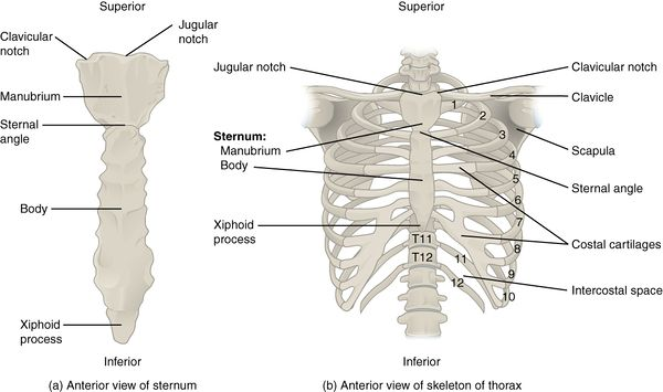
The Thoracic Cage
The thoracic cage (rib cage) forms the thorax (chest) portion of the body.
It consists of the 12 pairs of ribs with their costal cartilages and the sternum.
The thoracic cage protects the heart and lungs.
Sternum
The manubrium is the wider, superior portion of the sternum.
The top of the manubrium has a shallow, U-shaped border called the jugular (suprasternal) notch.
The clavicular notch is the shallow depression located on either side at the superior-lateral margins of the manubrium.
The manubrium and body join together at the sternal angle, so called because the junction between these two components is not flat, but forms a slight bend.
The inferior tip of the sternum is the xiphoid process.
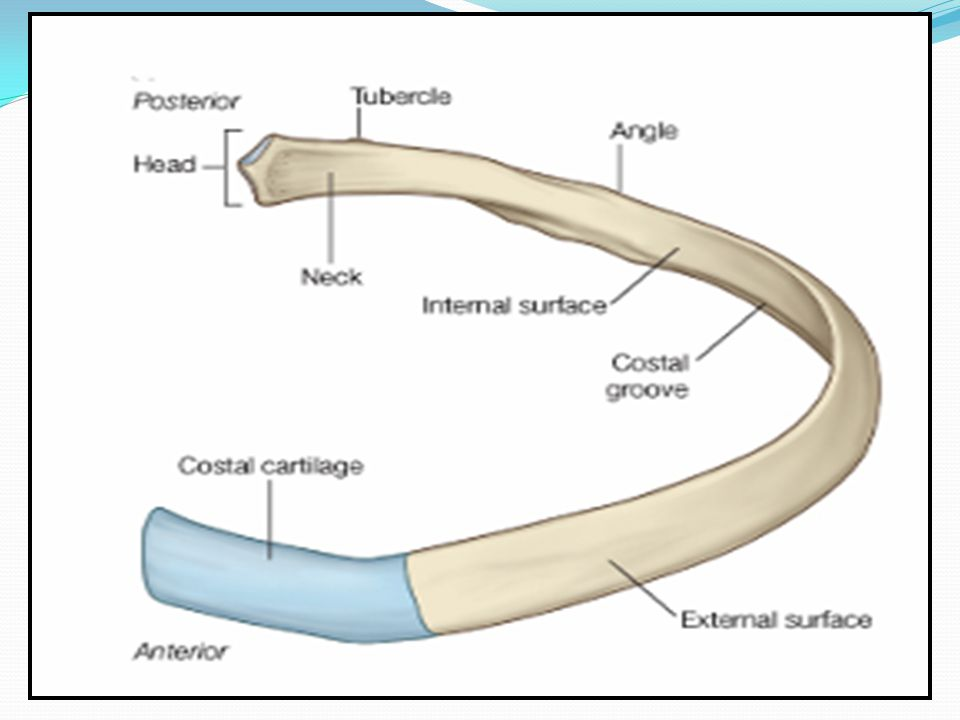
Parts of a Typical Rib
The posterior end of a typical rib is called the head of the rib.
Lateral to the head is the narrowed neck of the rib.
A small bump on the posterior rib surface is the tubercle of the rib, which articulates with the facet located on the transverse process of the same numbered vertebra.
The remainder of the rib is the body of the rib (shaft).
Just lateral to the tubercle is the angle of the rib, the point at which the rib has its greatest degree of curvature.
A shallow costal groove for the passage of blood vessels and a nerve is found along the inferior margin of each rib.
Rib Classifications
The bony ribs do not extend anteriorly completely around to the sternum. Instead, each rib ends in a costal cartilage.
Ribs 1–7 are classified as true ribs (vertebrosternal ribs).
Ribs 8–12 are called false ribs (vertebrochondral ribs).
The last two false ribs (11–12) are also called floating ribs (vertebral ribs).
Development of the Skull
During the third week of embryonic development, a rod-like structure called the notochord develops dorsally along the length of the embryo.
By the fourth week, mesoderm tissue located on either side of the notochord thickens and separates into a repeating series of block-like tissue structures, each of which is called a somite.
The most medial of these parts is called a sclerotome.
As the braincase bones grow in the fetal skull, they remain separated from each other by large areas of dense connective tissue, each of which is called a fontanelle.