Chapter 24: The Maintenance Systems
24.1 Respiratory System
The role of the respiratory system in gas exchange:
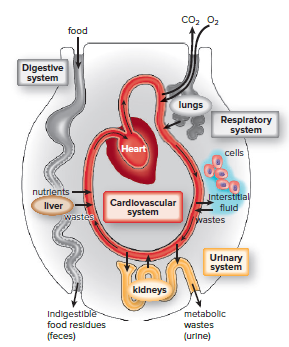
Cells are bathed in interstitial fluid.
Cells exchange oxygen, nutrients, and waste with interstitial fluid.
Interstitial fluid exchanges compound with blood.
Blood is refreshed through exchanges with the external environment.
The respiratory system exchanges gases in lungs.
Blood gives up CO2 and picks up O2 in lungs.
Respiration contributes to homeostasis.
Respiration requires breathing and external and internal exchange of gases.
Oxygen enters mitochondria for cellular respiration.
ATP is produced with oxygen delivery.
CO2 is a waste product of cellular respiration.

The Human Respiratory Tract
Includes structures that conduct air to and from the lungs.
Lungs are the major organ of gas exchange.
Air is filtered, warmed, and humidified as it moves through the respiratory tract.
Nose hairs and cilia filter air in the nose and respiratory passages.
Cilia beat upward, carrying mucus, dust, and particles to the throat to be swallowed or spit out.
Smoking destroys cilia, leading to lung disorders.
Air cools and loses moisture as it moves out of the respiratory tract.
Moisture deposits on the lining of the tract and may cause dripping from the nose.
Air still retains enough moisture to form a small cloud on a cold day when breathed out.
Upper Respiratory Tract
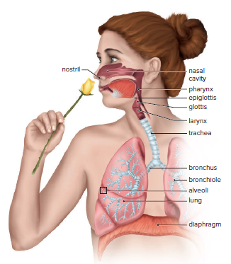
The upper respiratory tract consists of the nose, pharynx, and larynx.
The nose is the only external portion of the respiratory system.
The nose contains two nasal cavities, which are separated by a septum.
The nasal cavities are connected to the sinuses, which are air-filled spaces.
The nasal cavities and sinuses help to warm, moisten, and filter the air that we breathe.
The pharynx is a funnel-shaped passageway that connects the nasal cavity and mouth to the larynx.
The tonsils are a ring of lymphatic tissue at the junction of the mouth and pharynx.
Tonsillitis is an inflammation of the tonsils.
The epiglottis is a flap of cartilage that covers the glottis, the opening into the larynx, during swallowing.
The larynx contains the vocal cords, which vibrate to produce sound.
Laryngitis is an infection of the larynx that causes hoarseness.
Lower Respiratory Tract
The lower respiratory tract contains:
The respiratory tree.
Trachea (windpipe.)
Bronchi.
Bronchioles.
The trachea:
Connects the larynx to the bronchi.
Is held open by C-shaped, cartilaginous rings.
Allows food to pass down through the esophagus without damaging it.
Divides into two primary bronchi that enter the right and left lungs.
Bronchi:
Continue to branch until there are many smaller passages called bronchioles.
Resemble the trachea in structure.
Walls become thinner, rings of cartilage disappear, and the amount of smooth muscle increases as passages divide and subdivide.
Bronchioles:
Each terminates in an elongated space enclosed by alveoli (little air pockets that make up the lungs.)
Bronchitis:
Infection of the bronchi.
Nonproductive cough becomes a deep cough that produces mucus and perhaps pus.
Smokers with a deep cough have bronchitis and irritated respiratory tract.
Progression can reverse if smoking stops.
Chronic bronchitis is the second step toward emphysema and lung cancer caused by smoking cigarettes.
Lung cancer often begins in the bronchi and spreads to the lungs.
Respiration in other animals:
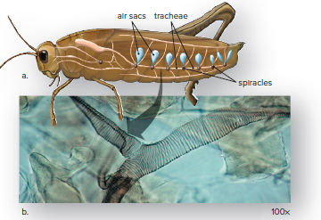
Insects have many tracheae, little air tubes supported by rings of chitin that branch into every part of the body.
Tracheal system begins at spiracles, openings that perforate the insect’s body wall, and ends in very fine, fluid-filled tubules.
Ventilation is assisted by the presence of air sacs that draw in the air.
No cell is very far from the site of gas exchange, so the bloodstream does not transport oxygen, and no oxygen-carrying pigment is required.
Breathing
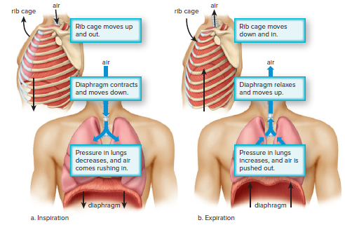
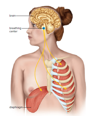
The diaphragm is a muscular, membranous partition in humans.
It divides the upper thoracic cavity from the lower abdominal cavity.
Muscle contractions lower the diaphragm and raise the ribs during breathing.
This increases the volume of the thoracic cavity and lungs.
The increased volume creates a negative pressure in the thoracic cavity and lungs.
Air flows into the lungs due to this negative pressure, which is called inspiration.
Air does not force the lungs open, they have already opened up.
When the rib and diaphragm muscles relax, the lungs recoil and air moves out.
This is due to increased pressure in the lungs, which is called expiration.
Breathing In Other Animals
Mammals and most reptiles have the same route for air in and out of their lungs.
Humans have residual air left in their lungs due to this.
Birds use a one-way ventilation mechanism.
Incoming air goes through the trachea and into abdominal air sacs.
Air then passes through the lungs into thoracic air sacs.
Fresh air never mixes with used air in the lungs of birds.
This greatly improves gas exchange efficiency.

Control of Breathing
Increased concentrations of H+ and CO2 in the blood increase breathing rate in humans.
Aortic bodies and carotid bodies are chemoreceptors that monitor the chemical content of the blood.
Aortic bodies and carotid bodies are sensitive to changes in H+ and CO2 concentrations but minimally sensitive to lower O2 concentrations.
The need to breathe comes from a buildup of CO2 in the bloodstream, not necessarily from a lack of O2.
Information from the chemoreceptors goes to the breathing center in the brain, which increases breathing rate when H+ and CO2 concentrations rise.
The breathing center is also directly sensitive to the chemical content of the blood, including its O2 content.
Lungs and External Exchange of Gases
Mammals and most reptiles have the same route for air in and out, leaving residual air in the lungs.
Birds use a one-way ventilation mechanism, which greatly improves gas exchange efficiency.
Frogs and salamanders use their moist skin for gas exchange in addition to their lungs.
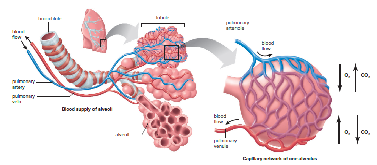
Human lungs have a total surface area at least 50 times the skin’s surface area due to the presence of alveoli.
Alveoli are lined with surfactant, which prevents the sides of the alveoli from sticking together during exhalation.
The respiratory membrane is formed by the alveolar epithelium and the capillary epithelium.
Emphysema is a serious lung condition that reduces the surface area for gas exchange, leading to low energy levels.
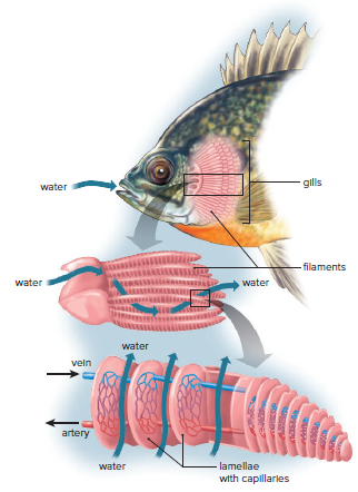
Gills of Fish
Fish and other aquatic animals use gills as their respiratory organ.
Water is drawn into the mouth and out from the pharynx across the gills.
The flow of blood in gill capillaries is opposite the flow of water across the gills.
Blood is always exposed to water having a higher oxygen content.
About 80-90% of the dissolved oxygen in the water is absorbed.
Transport and Internal Exchange of Gases
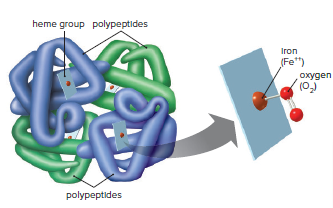
Hemoglobin molecules carry oxygen to the body's tissues.
Without hemoglobin, it would take 3 years for an oxygen molecule to move from lungs to toes by simple diffusion.
Each hemoglobin molecule contains 4 polypeptide chains: 2 α and 2 β chains.
Each polypeptide is folded around an iron-containing group called heme.
The iron in heme bonds with oxygen and carries it to the tissues.
Each red blood cell contains about 250 million hemoglobin molecules.
Each red blood cell can carry at least 1 billion molecules of oxygen.
Hemoglobin gives up oxygen in the tissues during an internal exchange.
Interstitial fluid always has a lower oxygen concentration than blood.
Cells take up and utilize oxygen during cellular respiration, causing the difference in oxygen concentration.
Hemoglobin also gives up oxygen due to warmer temperatures and lower pH in the tissues.
The warmer temperature and lower pH are caused by cellular respiration.
Cells give off heat and carbon dioxide as by-products during cellular respiration.
Carbon dioxide enters the blood during the internal exchange.
Interstitial fluid has a higher concentration of carbon dioxide.
Most of the carbon dioxide is transported as bicarbonate ions (HCO3-).
Carbon dioxide combines with water to form carbonic acid.
Carbonic acid dissociates to hydrogen ion (H+) and HCO3-.

H+ causes pH to lower, but only slightly due to absorption by globin portions of hemoglobin.
HCO3- is carried in plasma.
In the lungs, carbon dioxide diffuses out of the blood into alveoli as blood enters pulmonary capillaries.
Hemoglobin gives up H+ it has been carrying during this reaction.

Carbon dioxide diffuses out of the blood into the alveoli of the lungs.
If the blood level of H+ rises, the breathing center in the brain increases the breathing rate.
As more CO2 leaves the blood, the pH of the blood is corrected.
24.2 Urinary System
The urinary system in vertebrate animals, including humans, is important for homeostasis. The kidneys are the primary organs responsible for the following functions:
Excretion of nitrogenous wastes, such as urea and uric acid.
Maintenance of the water-salt balance of the blood.
Maintenance of the acid-base balance of the blood.
The kidneys in humans are bean-shaped, reddish-brown organs.
They are about the size of a fist and located on each side of the vertebral column, just below the diaphragm.
The kidneys are partially protected by the lower rib cage.
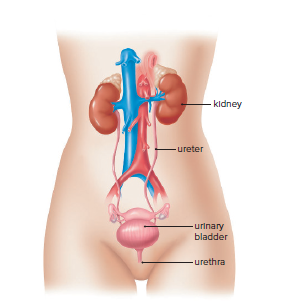
Urine made by the kidneys is conducted from the body by the other organs in the urinary system.
Each kidney is connected to a ureter, a tube that takes urine from the kidney to the urinary bladder.
Urine is stored in the urinary bladder until it is voided from the body through the single urethra.
In males, the urethra passes through the penis; in females, it opens in front of the opening of the vagina.
In females, there is no connection between the genital (reproductive) and urinary systems.
In males, there is a connection between the genital and urinary systems, since the urethra also carries sperm during ejaculation.
In amphibians, birds, reptiles, and some fishes, the bladder empties into the cloaca, a common chamber and outlet for the digestive, urinary, and genital tracts.
Human Kidney
When a kidney is sectioned longitudinally, three major parts can be distinguished:
Renal cortex: the outer region of the kidney with a granular appearance.
Renal medulla: consists of cone-shaped renal pyramids, located inside the renal cortex.
Renal pelvis: the innermost part of the kidney, which is a hollow chamber where urine collects.
Urine is carried from the renal pelvis to the bladder by a ureter.
The cortex and medulla of each kidney are composed of about 1 million tiny tubules called nephrons.
The nephrons of a kidney produce urine.
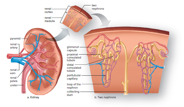
Nephrons
Each nephron is composed of several parts.
The glomerular capsule is formed by the closed end of a nephron.
Filtration of the blood occurs at the glomerular capsule.
The inner layer of the capsule is made up of specialized cells that allow easy passage of molecules.
The proximal convoluted tubule is a portion of the nephron that follows the capsule.
The proximal convoluted tubule is lined by cells with many mitochondria and tightly packed microvilli.
The nephron loop comes after the proximal convoluted tubule and has a descending and ascending limb.
The distal convoluted tubule follows the nephron loop.
Several distal tubules enter one collecting duct.
A collecting duct delivers urine to the renal pelvis.
The nephron loop and the collecting duct give the pyramids of the renal medulla their striped appearance.
Each nephron has its own blood supply.
Various exchanges occur between parts of the nephron and a blood capillary as urine forms.
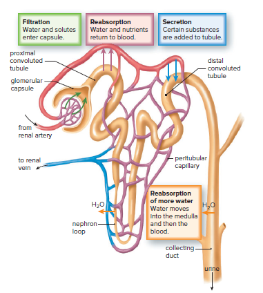
Urine Formation
Urine formation requires three steps: filtration, reabsorption, and secretion.
Filtration is the movement of small molecules from a blood capillary to the inside of the glomerular capsule as a result of blood pressure.
Small molecules such as water, nutrients, salts, and urea move to the inside of the capsule.
Plasma proteins and blood cells are too large to be part of this filtrate, so they remain in the blood.
If the composition of the filtrate were not altered in other parts of the nephron, death from loss of nutrients (starvation) and loss of water (dehydration) would quickly follow.
The next step, reabsorption, helps prevent this.
Reabsorption of Solutes:
Water and other substances move from the proximal convoluted tubule into the blood.
Nutrients such as glucose and amino acids also return to the blood.
Some molecules, such as glucose, are both passively and actively reabsorbed.
Cells of the proximal convoluted tubule have numerous microvilli and mitochondria.
Glucose in urine is a sign of diabetes mellitus.
Sodium ions (Na+) are actively pumped into the peritubular capillary, and then chloride ions (Cl–) follow passively.
Water moves by osmosis from the tubule into the blood.
About 60-70% of salt and water are reabsorbed at the proximal convoluted tubule.
Some of the urea and other types of nitrogenous wastes excreted by humans are passively reabsorbed, but most remain in the filtrate.
Secretion:
Secretion is the transport of substances into the nephron by means other than filtration.
Substances such as uric acid, hydrogen ions, ammonia, and penicillin are eliminated by secretion.
The process of secretion helps rid the body of potentially harmful compounds that were not filtered into the capsule.
Regulation of Water-Salt Balance and pH:
All animals maintain water-salt balance and pH of the internal environment.
Humans use long nephron loops to secrete hypertonic urine.
Ascending limb of the nephron loop pumps salt and urea into the renal medulla, and water follows by osmosis.
Hormones regulate water-salt reabsorption by kidneys.
Coffee interferes with one hormone, causing the production of more urine.
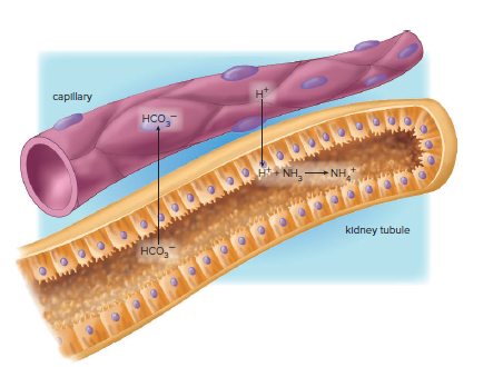
Kidneys regulate pH of blood through bicarbonate buffer system and regulation of breathing rate.
Kidneys excrete a wide range of acidic and basic substances.
Kidneys reabsorb bicarbonate ions and excrete hydrogen ions to maintain normal blood pH.
Urine is usually acidic, indicating excess hydrogen ions are produced and excreted.
Ammonia and phosphate provide means of buffering hydrogen ions in urine.
Regulation of Water-Salt Balance in Other Animals:
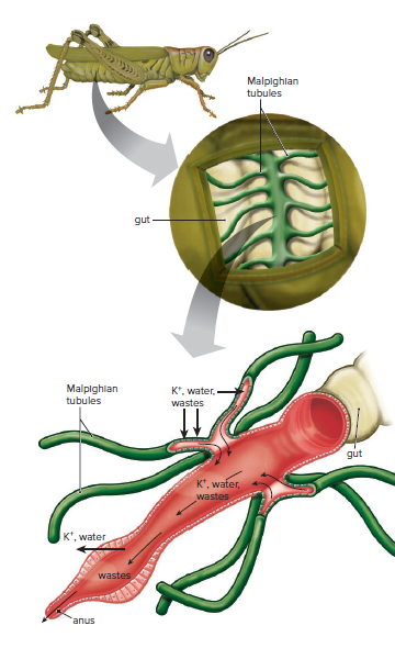
Insects have a unique excretory system consisting of Malpighian tubules attached to the gut.
Uric acid is the primary nitrogenous waste product of insects.
Uric acid diffuses from the hemolymph into the Malpighian tubules.
Water follows a salt gradient established by active transport of potassium (K+).
Water and useful substances are reabsorbed at the rectum.
Uric acid leaves the body at the anus.
Insects in dry environments reabsorb most of the water and excrete a dry, semisolid mass of uric acid.
Insects that live in water or eat large quantities of moist food reabsorb little water.
Most animals can regulate the blood levels of both water and salt.
Freshwater fishes take up salt in the digestive tract and gills and produce large amounts of dilute urine.
Saltwater fishes take in salt water by drinking, but then they pump ions out at the gills and produce only small amounts of concentrated urine.
Problems with Kidney Function
Illnesses such as diabetes, hypertension, and inherited conditions can cause progressive renal disease and renal failure.
Urinary tract infections, an enlarged prostate gland, pH imbalances, or excessive calcium intake can lead to kidney stones.
Kidney stones form in the renal pelvis and can block the renal pelvis or ureter, leading to back pressure that destroys nephrons.
The presence of albumin, white blood cells, or red blood cells in the urine is one of the first signs of nephron damage.
If more than two-thirds of the nephrons are inoperative, urea and other waste substances accumulate in the blood.
Retention of water and salts is of greater concern than nitrogenous waste in the blood.
Edema, fluid accumulation in the body tissues, may occur.
An imbalance in the ionic composition of body fluids can lead to loss of consciousness and even heart failure.
Hemodialysis and Kidney Transplants
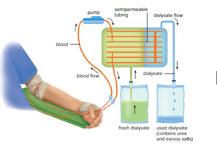
Patients with renal failure can undergo hemodialysis using an artificial kidney machine or continuous ambulatory peritoneal dialysis (CAPD).
Dialysis is the diffusion of dissolved molecules through a semipermeable membrane with pore sizes that allow only small molecules to pass through.
In an artificial kidney machine, the patient's blood is passed through a membranous tube in contact with a dialysis solution or dialysate.
Substances more concentrated in the blood diffuse into the dialysate, and substances more concentrated in the dialysate diffuse into the blood.
The dialysate is continuously replaced to maintain favorable concentration gradients.
In CAPD, the peritoneum is the dialysis membrane, and a fresh amount of dialysate is introduced directly into the abdominal cavity from a bag.
Waste and salt molecules pass from the blood vessels in the abdominal wall into the dialysate before the fluid is collected 4 or 8 hours later.
Patients with renal failure may undergo a transplant operation, receiving a functioning kidney from a donor.
Receiving a kidney from a close relative has the highest chance of success.
The current 1-year survival rate for a kidney transplant is 98%, and the 5-year survival rate is more than 91%.