

Neurobiology and Behavior
Neurulation and Embryogenesis
The development of a fully-formed organism from a fertilized egg is called embryogenesis
All tissues are derived from three initial germ layers (ectoderm, mesoderm, endoderm) formed via gastrulation
In chordates, a flexible notochord will develop during gastrulation and lead to the subsequent formation of a neural tube
The formation of a neural tube in embryonic chordates occurs via the process of neurulation
Cells located in the outer germ layer (ectoderm) differentiate to form a neural plate
The neural plate then bends dorsally, folding inwards to form a groove flanked by a neural crest
The infolded groove closes off and separates from the neural crest to form the neural tube
The neural tube will elongate as the embryo develops and form the central nervous system (brain and spinal cord)
The cells of the neural crest will differentiate to form the components of the peripheral nervous system
Xenopus are a genus of frog that possess robust embryos that can tolerate extensive manipulation
This makes them a suitable animal models for investigating the developmental stages of embryogenesis
During neurulation, the following embryonic tissues should be easily identifiable:
Three germ layers (outer = ectoderm ; middle = mesoderm ; inner = endoderm)
A hollow cavity called the archenteron (will develop into the digestive tract)
Notochord (flexible rod that stimulates neurulation)
Neural tube (developed from the infolding of the neural plate)
Closure of the neural tube does not occur simultaneously along the entire length of the embryo
The area where the brain forms is well advanced over the caudal (tail) region, where closure occurs more slowly
Spina bifida is a birth defect resulting in the incomplete closure of the neural tube (and associated vertebrae)
It is most commonly seen in the lumbar and sacral areas, as these are the regions where closure is slowest
The vertebral processes do not fuse, leaving the spinal cord nerves exposed and prone to damage
The severity of the condition can vary from mild to severe depending on the consequence of the incomplete closure
In cases of spina bifida occulta, the splits in the vertebrae are so small that the spinal cord does not protrude
In spina bifida cystica, a meningeal cyst forms (meningocele) which may include the spinal elements (myelomeningocele)
In the more severe cases, patients may typically suffer some degree of paralysis, as well as bowel and bladder dysfunction
Spina bifida is believed to be caused by a combination of genetic and environmental factors
The average worldwide incidence of the condition is ~1 in 1,000 births, however marked geographic variation occurs
Not having enough folate in the diet during pregnancy is believed to play a significant role in causing spina bifida
The neural tube contains multipotent neuronal stem cells which can differentiate to form the different types of nerve cells:
Neurons are specialised nerve cells that conduct messages – they can be sensory, motor or relay (interneurons)
Glial cells provide physical and nutritional support for the neurons – roughly 90% of nerve cells in the brain are glial cells
Neurons are produced by progenitor neuroblasts via a process known as neurogenesis
Most neurons survive for the lifetime of the individual and do not proliferate following embryogenesis (they are 'post-mitotic’)
Certain brain regions may be capable of adult neurogenesis, but most of the nervous system is incapable of regeneration
Immature neurons must migrate in order to adopt precise final positions that allow for the formation of neural circuitries
This migration process is critical for the development of brain and spinal architecture
Neural migration may occur via one of two distinct processes – glial guidance or somal translocation
Glial cells may provide a scaffolding network along which an immature neuron can be directed to its final location
Alternatively, the neuron may form an extension at the cell’s perimeter and then translocate its soma along this length
An immature neuron consists of a cell body (soma) containing a nucleus and cytoplasm
Axons and dendrites will grow from each immature neuron in response to chemical signals from surrounding cells
Some axons may be quite short (within the CNS) but others may extend to other parts of the body (within the PNS)
An axon has a growth cone at its tip that contains highly motile growth filaments called filipodia
Extension of these filipodia causes the expansion of the internal cytoskeleton within the growth cone – resulting in growth
The direction of this expansion is controlled by chemical stimuli released from surrounding cells
These cells may release chemoattractant signals (grow towards) or chemorepellant signals (grow away)
Using these molecular guidance signals, axon growth cones may navigate long distances to reach specific targets
A synapse is a junction at which a neuron transmits a signal to another cell (relay neuron or effector)
Most synapses transmit chemical signals, although electrical synapses also exist
A developing neuron will form multiple synapses, creating a vast array of permutable communication pathways
Within the CNS, a neuron may form a synapse with another axon, dendrite or cell body (soma)
Within the PNS, a neuron may form a synapse with a muscle fibre (neuromuscular) or gland (neuroglandular)
Some neurons may form a synapse with capillaries and secrete chemicals directly into the bloodstream (neurosecretory)
During embryonic and early post-natal development, neurons will form multiple synapses to maximise available connections
As an organism matures, some synapses are used more frequently and these connections are consequently strengthened
Other synapses are not used as often and these connections are weakened and do not persist
This strengthening and weakening of certain neural pathways is central to the concept of how organisms learn
Neural pruning involves the loss of unused neurons (by removing excess axons and eliminating their synaptic connections)
Infant and adult brains typically have the same total number of neurons (roughly 100 billion neurons in total)
However infant brains form vastly more synaptic connections (approximately twice the number found in adult brains)
The purpose of neural pruning seems to be to reinforce complex wiring patterns associated with learned behavior
Pruning is influenced by environmental factors and is mediated by the release of chemical signals from glial cells
Neuroplasticity describes the capacity for the nervous system to change and rewire its synaptic connections
Neuroplasticity enables individuals to reinforce certain connections (learning) or circumvent damaged regions
This adaptive response is achieved via two primary mechanisms – rerouting and sprouting
Rerouting involves creating re-establishing an existing nervous connection via an alternative neural pathway
Sprouting involves the growth of new axon or dendrite fibres to enable new neural connections to be formed
This reorganization of the architecture of the nervous system enables memory retention and learning
Strokes
A stroke is the sudden death of brain cells in a localized area due to inadequate blood flow
This results in the improper functioning of the brain, due to the loss of neural connections in the affected area
There are two main types of stroke – ischemic strokes and hemorrhagic strokes
Ischemic strokes result from a clot within the blood restricting oxygenation to an associated region of the brain
Hemorrhagic strokes result from a ruptured blood vessel causing bleeding within a section of the brain
Strokes symptoms may be temporary if the brain is able to reorganize its neural architecture to restore function
Following a stroke, healthy areas of the brain may adopt the functionality of damaged regions
This capacity for the restoration of normal function is made possible due to the neuroplasticity of the brain
Gastrulation
Gastrulation is an early phase of embryogenesis whereby a single-layered blastula differentiates into three germ layers
The organisation of cell layers occurs by different mechanisms in different types of animals
The end result in all cases is a trilaminar (three layered) mass of cells called a gastrula
Gastrulation precedes further cellular differentiation by processes such as neurulation
Gastrulation results in the production of three germ layers – ectoderm (outer), mesoderm (middle) and endoderm (inner)
The ectoderm will form the nervous system (via neurulation) and outer surfaces such as skin, pigment cells and hair cells
The mesoderm will form the majority of body organs, including muscle, blood vessels, kidney, heart and skeleton
The endoderm will form the respiratory and digestive tracts, as well as associated organs such as the liver and pancreas
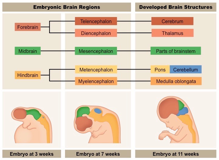
The Formation of The Human Brain
During embryonic development, the neural tube will enlarge and develop into different components of the nervous system:
The anterior part of the neural tube will expand to form the brain during cephalization (development of the head)
The remainder of the neural tube will develop into the spinal cord
Cells that comprised the neural crest will differentiate to form most of the peripheral nervous system
The embryonic brain will initially be composed of three primary structures – the forebrain, midbrain and hindbrain
These structures will eventually give rise to the identifiable components of the developed brain
Formation of the Human Brain
The human brain acts as an integration and coordination system for the control of body systems
It processes sensory information received from the body and relays motor responses to effector organ
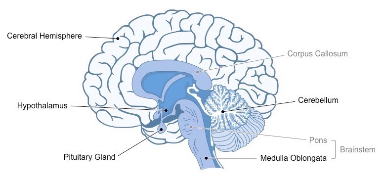
The human brain is organised into clearly identifiable sections that have specific roles
The major external structures include the cerebral cortex, cerebellum and brainstem
Internal structures include the hypothalamus, pituitary gland and corpus callosum
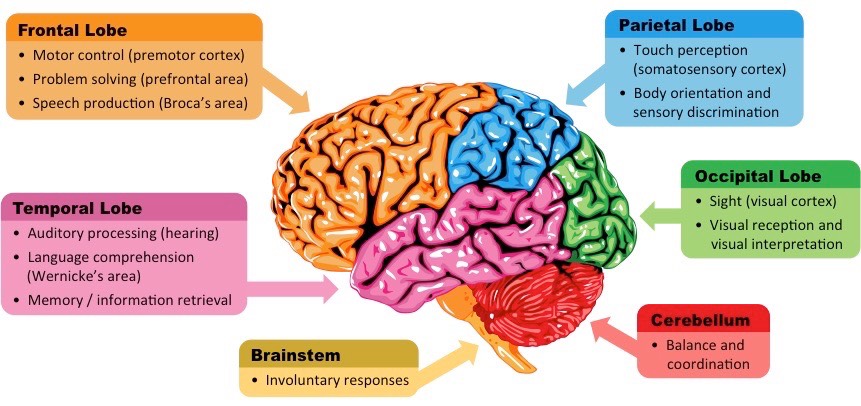
The External Structures
Cerebral Cortex
The cerebral cortex is an outer layer of tissue organised into two cerebral hemispheres and composed of four distinct lobes
The frontal lobe controls motor activity and tasks associated with the dopamine system (memory, attention, etc.)
The parietal lobe is responsible for touch sensation (tactility) as well as spatial navigation (proprioception)
The temporal lobe is involved in auditory processing and language comprehension
The occipital lobe is the visual processing centre of the brain and is responsible for sight perception
Cerebellum
The cerebellum appears as a separate structure at the base of the brain, underneath the cerebral hemispheres
It is responsible for coordinating unconscious motor functions – such as balance and movement coordination
Brainstem
The brainstem is the posterior part of the brain that connects to the spinal cord (which relays signals to and from the body)
The brainstem includes the pons, medulla oblongata (often referred to as the medulla) and the midbrain
The brainstem (via the medulla) controls automatic and involuntary activities (breathing, swallowing, heart rate, etc.)
External Structures of the Brain
Internal Structures
Hypothalamus
The hypothalamus is the region of the brain that functions as the interface with the pituitary gland
As such, the hypothalamus functions to maintain homeostasis via the coordination of the nervous and endocrine systems
The hypothalamus also produces some hormones directly, which are secreted via the posterior pituitary (neurohypophysis)
The pituitary gland is considered the ‘master’ gland – it produces hormones that regulate other glands and target organs
The anterior lobe is called the adenohypophysis and secretes hormones such as FSH, LH, growth hormone and prolactin
The posterior lobe is called the neurohypophysis and secretes hormones such as ADH and oxytocin
Corpus Callosum
The corpus callosum is a bundle of nerve fibres that connects the two cerebral hemispheres
It is the largest white matter structure in the brain, consisting of roughly 250 million axon projections
Damage to the corpus callosum can prevent information exchange between left and right hemispheres (split brain disorders)
Internal Structures of the Brain
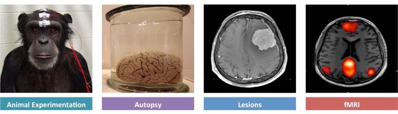
The role of a specific brain part can be identified by either stimulating or removing the region to assess its effect
Identification of brain roles can be made via the use of animal experiments, autopsy, lesions and fMRI
Animal Experiments
Animal experimentation can be used to identify function by stimulating regions with electrodes or removing via lobotomy
Because such methods are highly invasive and potentially damaging, animal models are frequently used
Experimentation on animals involves less ethical restrictions than human studies (although ethical standards do exist)
Animal studies are limited by the differences between animal and human brains, making valid comparisons difficult
Example: Animal studies using mice and rats have been used to develop drug treatments for diseases such as MS
Lesions
Lesions are abnormal areas of brain tissue which can indicate the effect of the loss of a brain area
Lesions can be identified via post-mortem analysis (autopsy) or via scans of the brain (CT scans or MRI)
The effects of lesions can be difficult to identify, as many functions may involve multiple brain areas
Additionally, the brain has the capacity to re-learn certain skills by re-routing instructions to other areas (plasticity)
Example: Split brain patients have been used to identify specific roles of the left and right cerebral hemisphere
Autopsy
An autopsy is a post-mortem examination of a corpse via dissection in order to evaluate causes of death
Comparisons can be made between the brains of healthy and diseased corpses to identify affected brain areas
Example: Cadavers who suffered from aphasia (language impairment) in life demonstrate damage to specific areas
fMRI
Functional magnetic resonance imaging (fMRI) records changes in blood flow within the brain to identify activated areas
Oxygenated haemoglobin responds differently to a magnetic field than deoxygenated haemoglobin
These differences in oxygenation can be represented visually and reflect differences in the level of brain activity
fMRI is non-invasive and can be used to identify multiple brain regions involved in complex, integrated brain activities
Example: fMRI studies have been used to diagnose ADHD and dyslexia, as well as monitor recovery from strokes
Methods for Identifying Brain Functions
While complex activities may require integration of multiple regions, some specific functions are localised to particular areas
Examples of brain areas with clearly defined functions include the visual cortex, Broca’s area and the nucleus accumbens
Visual Cortex
Located within the occipital lobe of the cerebrum and receives neural impulses from light-sensitive cells in the eyes
The visual cortex is the region of the brain responsible for visual perception (sight)
Broca’s Area
Located within the frontal lobe of the left cerebral hemisphere (not present in the right hemisphere)
Is responsible for speech production (if damaged, the individual cannot produce meaningful speech despite intending to)
Nucleus Accumbens
The nucleus accumbens is involved in the pleasure reward pathway and is found within each cerebral hemisphere
It secretes neurotransmitters responsible for feelings of pleasure (dopamine) and satiety (serotonin)
It communicates with other centres involved in the mechanisms of pleasure, such as the ventral tegmental area (VTA)
The cerebral cortex is the outer layer of neural tissue found in the cerebrum of humans and other mammals
It is composed of grey matter and is involved in complex actions, such as memory, perception, consciousness and thought
The cerebral cortex is much more highly developed in humans than other animals and forms a larger proportion of the brain
The cerebral cortex can be externally classified according to four topographical lobes – frontal, parietal, temporal, occipital
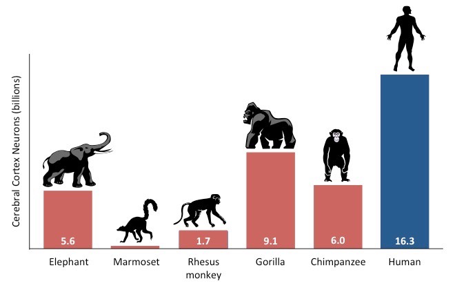
Comparison of Cerebral Cortex Density in Humans and Other Mammals
Link: The Human Brain in Numbers (Frontiers in Human Neuroscience)
Through evolution, the human cerebral cortex has been greatly enlarged in comparison to other brain structures
The disproportional enlargement of the cerebral cortex in humans is responsible for our capacity for cognitive thought
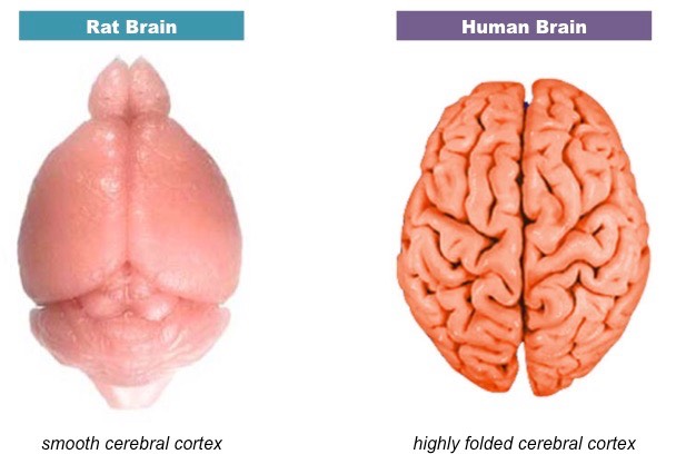
The increase in total area is mediated by extensive folding (gyrification) to form wrinkled peaks (gyrus) and troughs (sulcus)
This greatly increases surface area without increasing volume – allowing the brain to fit within the cranium
The extent of gyrification of the cerebral cortex is a reliable indicator of potential cognitive capacity
Primates and humans have a greater degree of folding compared to lower mammals (e.g. rats have a smooth cortex)
Brain Comparison – Human versus Rat (Not to Scale)
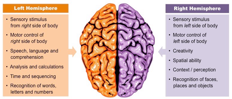
The cerebrum is organised into two hemispheres that are responsible for higher order functions and complex skills
These functions include memory, speech, cognitive thought, problem solving, attention and emotions
Not all complex tasks are equally represented by both cerebral hemispheres – some activities are localised to a single side
Speech production is coordinated by Broca’s area, which is situated in the left frontal lobe of the brain
Information can be passed between the two hemispheres by a bundle of myelinated nerve fibres embedded within the brain
These fibres form the corpus callosum to facilitate interhemispheric communication
Lateralization of Brain Function
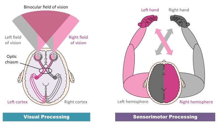
The left cerebral hemisphere is responsible for processing sensory information from the right side of the body (and vice versa)
Tactile sensation from the left side of the body is processed by the right side of the brain (at the somatosensory cortex)
Objects on the left side of the visual field in both eyes are processed on the right side of the visual cortex
The processing of information on the opposite side of the body is called contralateral processing (same side = ipsilateral)
Tactile information from the left side of the body is transferred to the right side in the spinal cord or brainstem
Visual information from the left visual field is transferred to the right cerebral hemisphere at the optic chiasma
The left cerebral hemisphere is also responsible for processing motor information for the right side of the body (and vice versa)
Muscular contractions are coordinated by the motor cortex (premotor cortex = preparation ; primary motor cortex = execution)
A consequence of this contralateral processing is that damage to one side of the brain affects the other side of the body
For instance, a stroke in the left hemisphere may cause paralysis to the right side of the body
Contralateral Processing
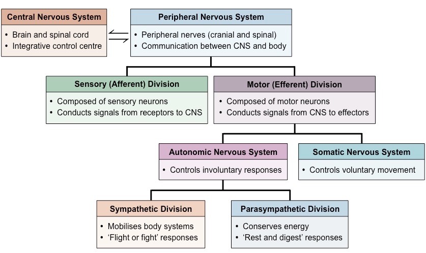
The human nervous system can be organised into several sub-divisions:
Firstly, the nervous system can be divided into the central nervous system (brain and spine) and peripheral nervous system
The peripheral nervous system (PNS) can be divided into the sensory (afferent) pathway or the motor (efferent) pathway
The motor pathway can be subdivided according to whether the response is voluntary (somatic) or involuntary (autonomic)
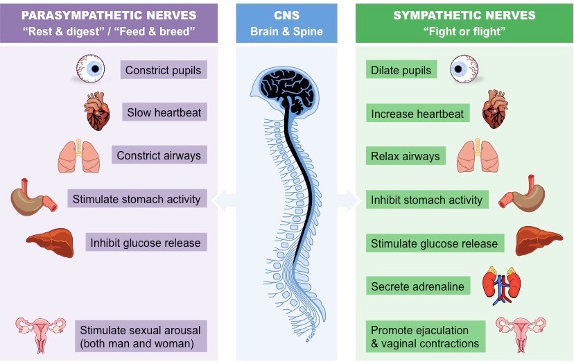
The autonomic nervous system controls involuntary processes in the body using centres located mostly within the brainstem
Sympathetic nerves release noradrenaline (adrenergic) to mobilise body systems (‘fight or flight’ responses)
Parasympathetic nerves release acetylcholine (cholinergic) to relax body systems and conserve energy (‘rest and digest’)
Divisions of the Nervous System
The medulla oblongata is a part of the brainstem responsible for coordinating many autonomic (involuntary) activities
This includes the regulation of body activities such as swallowing, breathing and heart rate
Sympathetic Responses (‘Fight or Flight’)
Decreases salivary release and blood flow to the gut in response to swallowing
Increases ventilation rate and dilates airways in response to a reduction in blood pH (caused by increased levels of CO2)
Increases heart rate by raising the normal sinus rhythm of the pacemaker of the heart
Parasympathetic Responses (‘Rest and Digest’ / ‘Feed and Breed’)
Increases salivary release and blood flow to the gut in response to swallowing
Lowers ventilation rate and constricts airways in response to an increase in blood pH (caused by lower levels of CO2)
Reduces heart rate (via vagus nerve) by lowering the normal sinus rhythm of the pacemaker of the heart
Autonomic Control of Body Systems
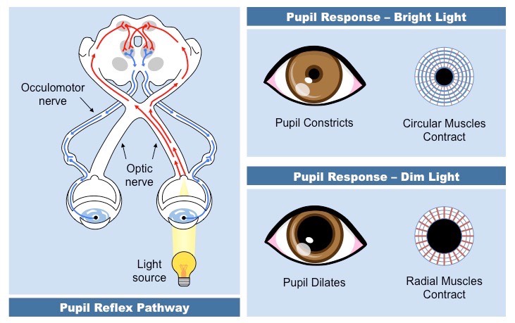
The Pupil Reflex
The pupil reflex is an involuntary response originating at the brainstem and under the control of the autonomic nervous system
It involves the resizing of the iris to regulate the amount of light that reaches the retina (excess light can damage the retina)
Pupils constrict in bright light (to prevent overstimulation of photoreceptors) and dilate in dim light (to maximise light exposure)
In bright light, parasympathetic nerves trigger circular muscles to contract and cause the pupils to constrict
In dim light, sympathetic nerves trigger radial muscles to contract and cause the pupils to dilate
Overview of the Pupil Reflex
Brain Death
Brain death is defined as the permanent absence of measurable activity in both the cerebrum and brainstem
The brainstem is responsible for involuntary autonomic responses and may function alone to maintain homeostasis
Hence, individuals with a non-functioning cerebrum but a functioning brainstem may be kept alive in a vegetative state
Brain death can be determined by medical professionals by testing the function of specific autonomic responses
The pupil reflex is one autonomic test used to assess brain death – brain dead individuals will not exhibit a pupil reflex
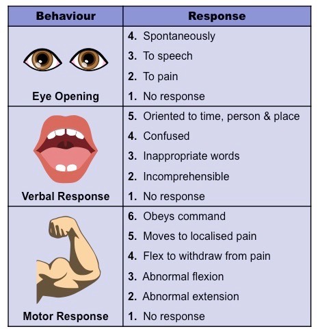
The Glasgow Coma Scale uses multiple tests to determine the neurological health of someone with suspected brain injury
Testing Levels of Consciousness
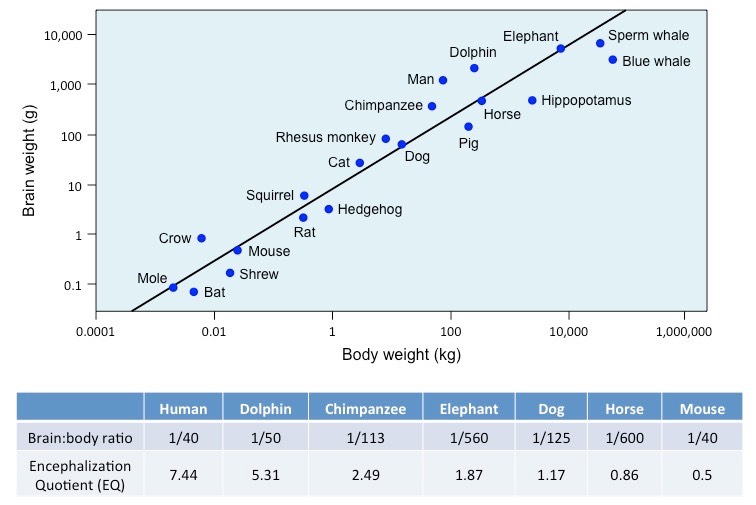
There is a positive correlation between body size and brain size in different animals – larger animals have larger brains
This correlation follows a linear pattern of progression but is not directly proportional
While an increase in body size results in an increase in brain size, the brain:body ratio decreases in larger animals
Body mass increases disproportionately to an increase in brain mass as most tasks only require a fixed brain capacity
While there is a correlation between body size and brain size, there is not a correlation between brain size and intelligence
Encephalization is defined as the amount of brain mass relative to an animal's body mass
Scientists have derived an encephalization quotient (EQ), which attempts to provide a rough estimate of potential cognition
The quotient is only applied to mammals – higher values are indicative of a higher predicted capacity for intelligence
Relationship between Brain Size and Body Size
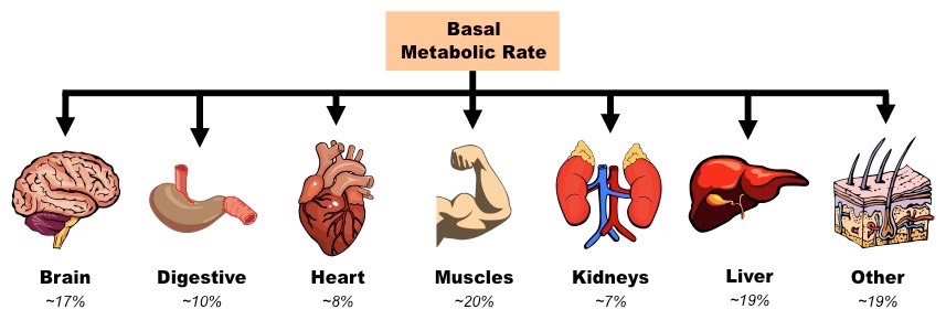
The human brain consumes ~20% of the body’s energy levels, despite making up only ~2% of the body’s mass
The brain’s rate of energy consumption varies little, regardless of the level of physical exertion by the body
The large amounts of energy required by the brain are used to sustain neurons and their processes
Energy is needed to maintain a resting potential when neurons are not firing (Na+/K+ pump uses ATP)
Energy is used to synthesise large numbers of neurotransmitters to facilitate neuronal communication
Metabolic Activity of Body Organs
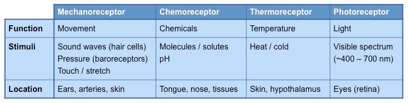
Sensitivity describes the ability of an organism to detect external and internal changes and respond accordingly
Receptors detect these changes as stimuli, and generate nerve impulses which are relayed to the brain and effector organs
There are different types of receptors that each recognise a different type of stimulus (temperature, light, etc.)
Main Types of Receptors in Humans
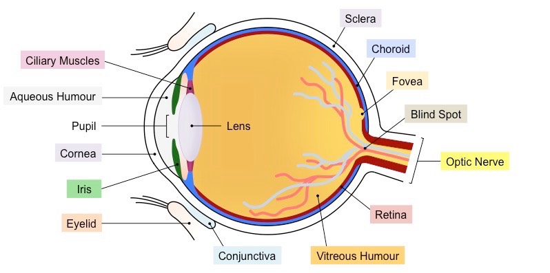
The human eye is the sensory organ responsible for vision (sight perception)
It consists of two fluid-filled cavities separated by a lens (anterior = aqueous humour, posterior = vitreous humour)
The lens is attached to ciliary muscles, which can contract or relax to change the focus of the lens
The amount of light that enters the eye via the pupil is controlled by the constriction and dilation of the iris
The exposed portion of the eye is coated by a transparent layer called the cornea, which is lubricated by conjunctiva
The internal surface of the eye is composed of three layers – the sclera (outer), choroid (middle) and retina (inner)
The region of the retina responsible for sharpest vision (i.e. focal point) is the fovea centralis (or fovea for short)
Nerve signals from the retina are sent via an optic nerve to the brain (no retina in this region creates a visual blind spot)
Diagram of the Human Eye
⇒ Click on the diagram to show / hide labels
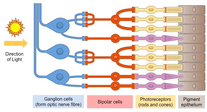
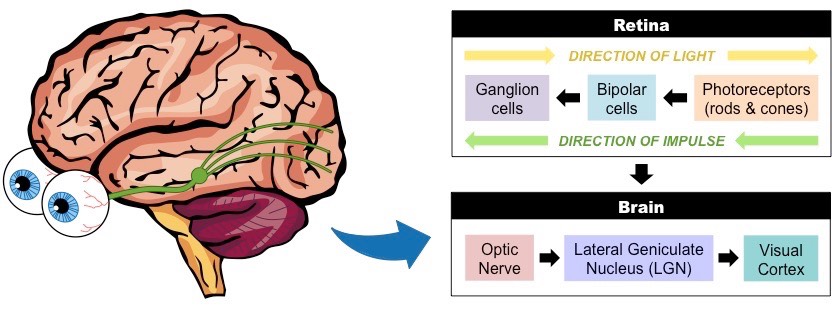
The retina is the light-sensitive layer of tissue that forms the innermost coat of the internal surface of the eye
Two types of photoreceptors (rods and cones) convert light stimuli into electrical nerve impulses
These nerve impulses are transmitted via bipolar cells to ganglion cells, whose fibres from the optic nerve tract
The photoreceptors line the rear of the retina (adjacent to the choroid), meaning light passes through the other cell layers
Diagram of the Human Retina
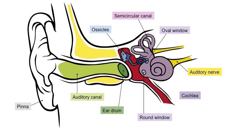
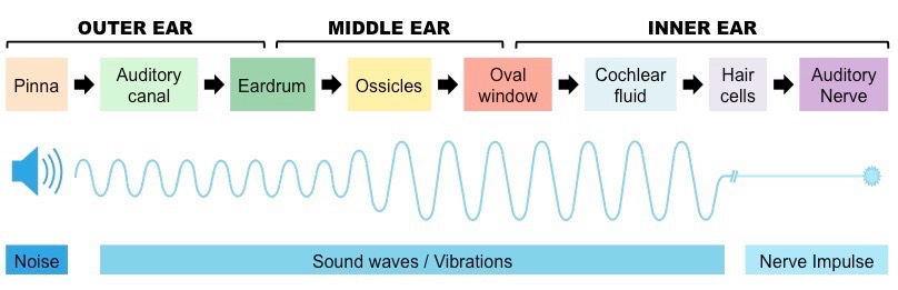
The human ear is the sensory organ responsible for hearing (sound perception)
The external part of the ear is called the pinna, whereas the internal part of the ear is divided into three sections
The outer ear contains the auditory canal, which channel sound waves to the tympanic membrane (or eardrum)
The middle ear contains three small bones called the ossicles, which transfer vibrations to the oval window
The inner ear consists of the cochlea and semicircular canals, as well as a round window which dissipates vibrations
The cochlear converts sound stimuli into electrical nerve impulses, which are transmitted via the auditory nerve to the brain
Diagram of the Human Ear
Photoreception is the mechanism of light detection (by the eyes) that leads to vision when interpreted by the brain
Light is absorbed by specialised photoreceptor cells in the retina, which convert the light stimulus into nerve impulses
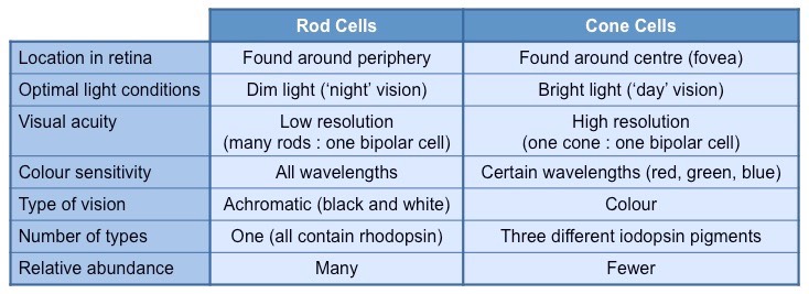
There are two different types of photoreceptors located within the retina – rod cells and cones cells
These cells differ in both their morphology (shape) and function
Types of Photoreceptors (Rods and Cones)
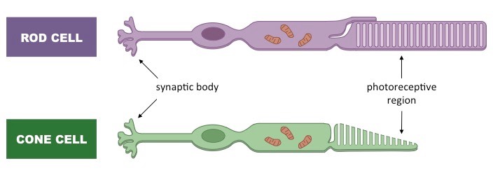
Rod Cells
Rod cells function better in low light conditions (twilight vision) – they become quickly bleached in bright light
Rod cells all contain the same pigment (rhodopsin) which absorbs a wide range of wavelengths
Rod cells cannot differentiate between different colours (monochromatic)
Rod cells are abundant at the periphery of the retina and hence are responsible for peripheral vision
Rod cells produce poorly resolved images as many rod cells synapse with a single bipolar neuron
Cone Cells
Cone cells function better in bright light conditions (daylight vision) – they require more photons of light to become activated
There are three different types of cone cells, each with a different pigment that absorbs a narrow range of wavelengths
Cone cells can therefore differentiate between different colours (red, blue and green)
Cone cells are abundant at the centre of the retina (within the fovea) and hence are involved in visual focusing
Cone cells produce well defined images as each cone cell synapses with a single bipolar neuron
Comparison of Rods and Cones
Photoreceptors (rods and cones) convert light stimuli into an electrical nerve impulse (action potential)
This neural information is relayed to the brain via bipolar cells and ganglion cells
Bipolar cells transmit the nerve impulses produced by the photoreceptors to ganglion cells
Many rod cells may synapse with a single bipolar cell, resulting in low resolution of sensory information (poor acuity)
Most cone cells only synapse with a single bipolar cell, resulting in high resolution of sensory information (high acuity)
Ganglion cells transmit nerve impulses to the brain via long axonal fibres that compose the optic nerve
Signals from ganglion cells may be sent to the visual cortex to form a composite representation of surroundings (i.e. sight)
Alternatively, signals may be sent to other brain regions to coordinate eye movements or maintain circadian rhythms
There are no photoreceptors present in the region of the retina where ganglion axon fibres feed into the optic nerve
This region is called the 'blind spot’ as visual information cannot be processed at this location
The brain interpolates details from the surrounding regions, such that individuals do not perceive a visual blind spot
Transmission of Visual Stimuli
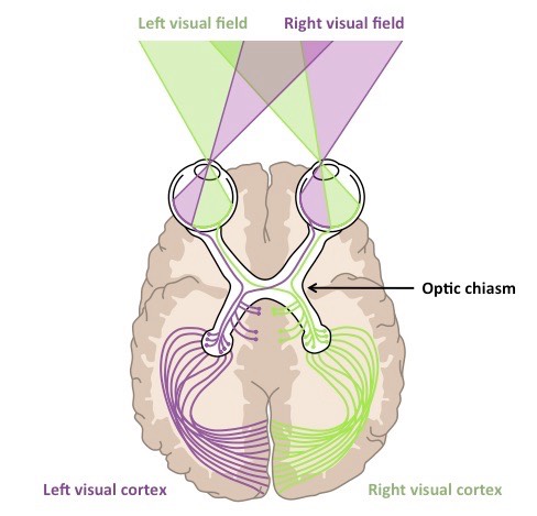
Contralateral processing is when a stimulus is processed on the opposite side to where it was detected
Information from the right half of the visual field is detected by the left half of the retina in both eyes and is processed by the left hemisphere (and vice versa for the left half of the visual field)
Information from each eye may swap at the optic chiasma, so that the right or left visual field is processed together
The optic nerves that swap sides are moving contralaterally, while those that stay on the same side remain ipsilateral
Impulses are conducted by the optic nerve to the thalamus, before being transmitted to the visual cortex (occipital lobe)
Thalamic structures (e.g. lateral geniculate nuclei) are involved in coordinating eye movements and circadian rhythms
Visual Processing
Sound travels as pressure waves in the air, which travel down the auditory canal and cause the eardrum to vibrate
The degree of vibration of the eardrum (tympanic membrane) will depend on the frequency and amplitude of the sound wave
The eardrum transfers the vibrations via the bones of the middle ear (the ossicles) to the oval window of the cochlea
The function of these bones is to amplify the vibrations from the eardrum (can increase magnification by ~ 20 times)
The vibration of the oval window causes fluid within the cochlea to be displaced – this displacement is detected by hair cells
Activation of these hair cells generates nerve impulses which are transmitted via the auditory nerve to the brain
Overview of Sound Perception
The middle ear is separated from the outer ear by the eardrum and the inner ear by the oval window
It is an air-filled chamber that houses three small bones (collectively called the ossicles)
The bones of the middle ear are individually called the malleus (hammer), incus (anvil) and stapes (stirrup)
The malleus is in contact with the eardrum and the stapes contacts the oval window (while the incus connects the two)
The function of the ossicles is to amplify the sound vibrations by acting like levers to reduce the force distribution
Sound travelling through air is mostly reflected when contacted by a liquid medium (due to the incompressibility of fluids)
The amplification of sound by the ossicles allows the vibrational pressure to pass to the cochlear fluid with very little loss
The oval window is smaller than the ear drum, which also assists in amplifying the sound energy
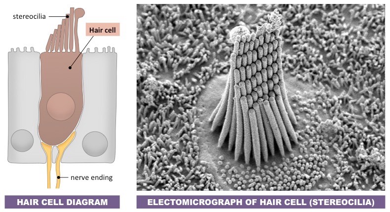
The cochlea is a fluid-filled spiral tube within the inner ear that converts sound vibrations into nerve impulses
Displacement of fluid by sound vibrations activates sensory hair cells within the spiral part of the cochlea (organ of Corti)
Hair cells are mechanoreceptors that possess tiny hair-like extensions called stereocilia
The cilia on hair cells vary in length and will each resonate to a different frequency of sound (i.e. specific wavelengths)
When the stereocilia are moved by the cochlear fluid, the hair cell will depolarise to generate a nerve impulse
The nerve impulse will be transmitted via the auditory nerve to the auditory centres of the brain
The kinetic movement of the cochlear fluid (and stereocilia motion) is dissipated by the vibration of the round window
Sensory Hair Cells
The vestibular system is a sensory system in the inner ear that is involved in balance and spatial orientation (proprioception)
Within the semicircular canals are gelatinous caps called cupula, which are embedded with numerous hair cells
When the head moves, the fluid in the semicircular canals (endolymph) follows the direction of movement (due to inertia)
This fluid movement exerts pressure on the hair cells embedded in the cupula, triggering nerve impulses
There are three semicircular canals at 90º angles to one another, allowing head movement to be detected in all three planes
The brain integrates information from the semicircular canals in each ear in order to identify head position and movement
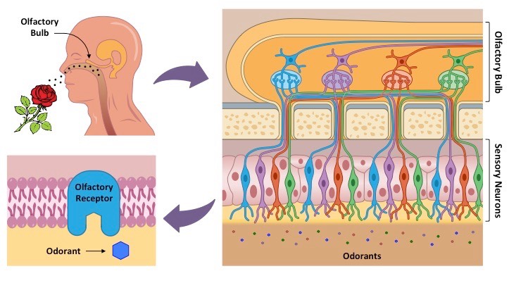
Olfaction is the ability to detect airborne chemicals (odorants) as scents or smells
At the back of the nasal cavity is a patch of tissue called the olfactory epithelium, which is embedded with chemoreceptors
The olfactory epithelium is lined with mucus, in which odorant molecules will dissolve before binding to the chemoreceptors
Binding of an odorant molecule will trigger a nerve impulse, which is transferred via the olfactory bulb to the brain
The combination of olfactory receptors activated determines the specific scent perceived by the brain
Olfactory Receptors
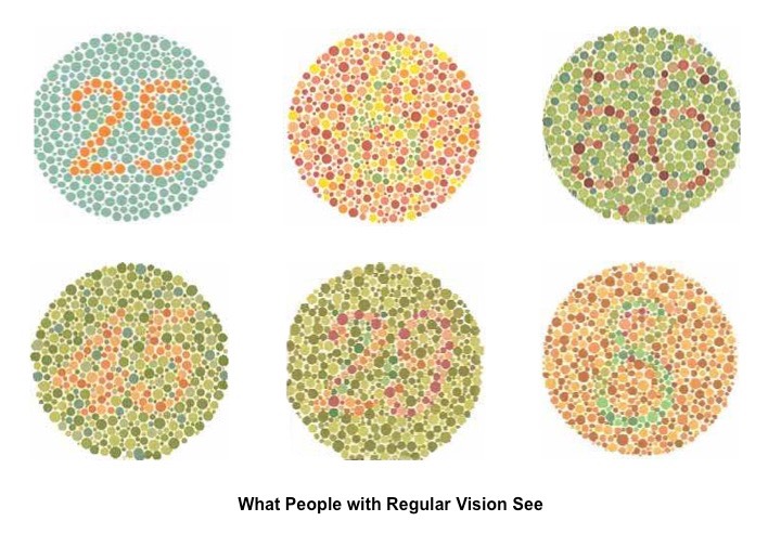
Red-green colour blindness is a genetic disorder whereby an individual fails to discriminate between red and green hues
There are three different types of cone cells, each of which absorbs different wavelengths (trichromatic: red, green, blue)
The genes responsible for producing red or green photoreceptors are located on the X chromosome (sex-linked)
If either of these genes are mutated, red and green wavelengths cannot be distinguished
As these genes are recessive and located on the X chromosome, red-green colour-blindness is more common in males
Red-green colour-blindness can be diagnosed using the Ishihara colour test
The Ishihara Color Test
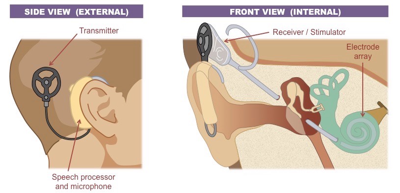
Cochlear implants may be used to stimulate the auditory centres of the brain in patients with non-functioning hair cells
Standard hearing aids are ineffective in deaf patients as they amplify sounds but do not bypass defective hearing structures
Cochlear implants consist of two parts – an external part (microphone / transmitter) and an internal part (receiver / stimulator)
The external components detect sounds, filter out extraneous frequencies and then transmit the signals to the internal parts
The internal components receive the transmissions and produce electrical signals via electrodes embedded in the cochlea
The electrical signals are then transferred via the auditory nerve to be processed by the brain
Cochlear Implants
A behaviour is typically defined as any observable action by a living organisms
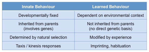
Behaviours can be categorised as either innate or learned
An innate behaviour is an instinctive response that is developmentally fixed – it is independent of environmental context
Innate behaviours have a genetic basis and are hence inherited from parents
Any instinctive response that improves survival and reproductive prospects will become more common by natural selection
Examples of innate behavioural responses seen in invertebrates include taxis and kinesis
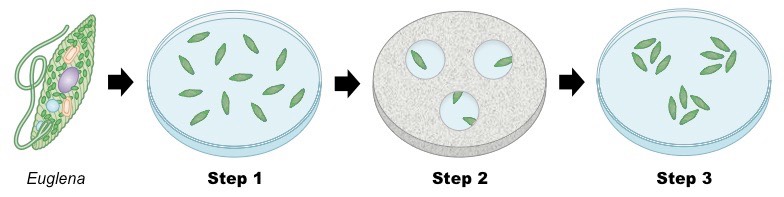
Taxis
Taxis is a change in movement in response to an environmental stimulus – either towards (positive) or away (negative)
Euglena is a photosynthetic microorganism that requires light as an energy source and hence displays positive phototaxis
Step 1: Place Euglena in a petri dish with appropriate environmental conditions for survival
Step 2: Cover the dish with aluminium foil, excluding a few small exposed sections
Observation: With a light source placed above the dish, the Euglena should migrate towards the exposed sections
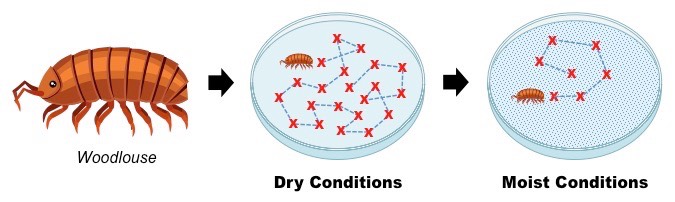
Kinesis
Kinesis is a change in the rate of activity in response to an environmental stimulus
Woodlice have gills for respiration and tend to prefer moist conditions (their gills may dry out in dry conditions)
Step 1: Place a woodlouse in a dry petri dish and mark its movements every 30 seconds
Step 2: Repeat this process for a second woodlouse placed in moist conditions (i.e. petri dish lined with wet paper towel)
Observation: The woodlouse in dry conditions should have a higher rate of movement (improve chances of finding moisture)
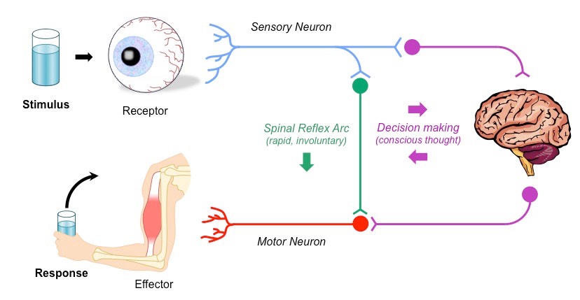
The basic pathway for a nerve impulse is described by the stimulus response model
A stimulus is a change in the environment (either external or internal) that is detected by a receptor
Receptors transform the stimuli into nerve impulses that are transmitted to the brain where decision-making occurs
When a response is selected, the signal is transmitted via neurons to effectors, promoting a change in the organism
Some responses may be involuntary and occur without conscious thought – these actions are called reflexes
Reflex actions do not involve the brain – instead sensory information is directly relayed to motor neurons within the spine
This results in a faster response, but one that does not involve conscious thought or deliberation
Stimulus–Response Pathway
Reflex actions are particularly beneficial in survival situations, when quick reactions are necessary to avoid permanent damage
Because reflex arcs don’t involve the brain (only the spine and possibly brainstem), reflex actions are more rapid
Reflex responses also include autonomic actions such as modifications to heart rate, breathing and pupil accommodation
A common example of a reflex action is the patellar reflex (‘knee jerk’ response) that occurs when the patellar tendon is tapped
The patellar reflex is a common test employed by doctors to determine the presence of spinal lesions
Example of a Reflex Arc
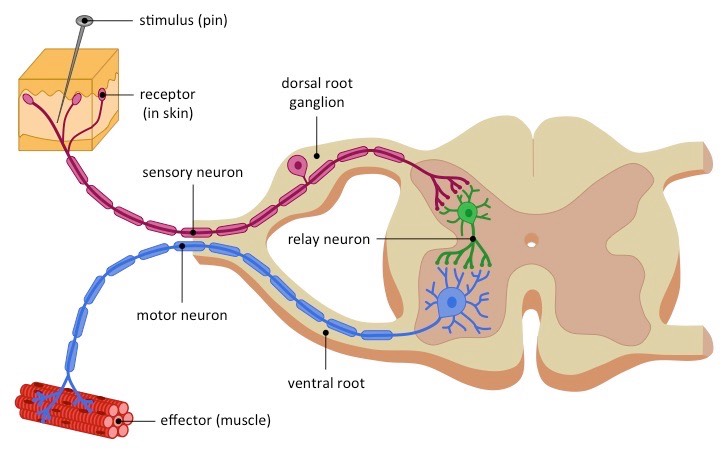
In a pain withdrawal reflex arc:
A pain stimulus is detected by a receptor (nocireceptor) and a nerve impulse is initiated in a sensory neutron
The sensory neuron enters the spinal cord via the dorsal root and synapses with a relay neuron in the grey matter
The relay neuron synapses with a motor neuron, which leaves the spinal cord via the ventral root
The motor neuron synapses with a muscle (effector), causing it to contract and remove the limb from the pain stimulus
Pain Withdrawal Reflex
Innate vs. Learned Behavior
Learned behaviour is not developmentally fixed and can be modified by experience
Learned behaviour shows significant variation as it is influenced by environmental context
Learning involves acquiring information from past experiences to adapt to new situations
The capacity to learn particular skills may be influenced by genes, but will not develop without appropriate experiences
Learning improves an organism’s survival prospects as they can modify their responses to changing environmental conditions
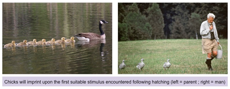
Imprinting is any kind of phase-sensitive learning that is rapid and independent of behavioural consequences
Imprinting occurs during a short critical period in which the organism adopts behavioural characteristics from a stimulus
Imprinted behaviour is not influenced by consequences – it does not require reinforcement to develop
Examples of imprinting include filial imprinting (bonding to a parent) and sexual imprinting (developing sexual preferences)
Filial imprinting was demonstrated by Konrad Lorenz, who imprinted baby geese to recognise him as a parental figure
Filial Imprinting
Conditioning is a process of behaviour modification whereby desired behaviours become associated with unrelated stimuli
This process can be achieved via either classical (reflex) conditioning or operant (instrumental) conditioning
Reflex conditioning involves placing a neutral signal before a reflex in order to create an association between the two
Reflex conditioning focuses on involuntary and autonomic behaviours
It involves associating a desired behaviour with a new stimulus
Operant conditioning involves applying reinforcement or punishment after a behaviour to increase or reduce its occurrence
Operant conditioning focuses on strengthening or weakening voluntary behaviours
It involves associating a particular behaviour with a specific consequence (either reward or punishment)
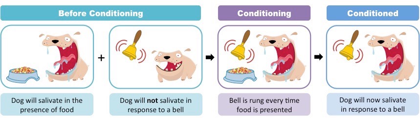
Reflex Conditioning
Reflex conditioning was first described by Ivan Pavlov, a Russian physiologist who experimented on dogs
Dogs normally salivate (unconditioned response) in anticipation of being fed (unconditioned stimulus)
Pavlov sounded a bell (neutral stimulus) prior to feeding a dog
After many repetitions, the dog came to associate the bell with food and began to salivate to the bell (conditioned response)
Pavlov described this as a conditioned reflex – the stimulus that prompted the response had been changed
Reflex Conditioning in Dogs
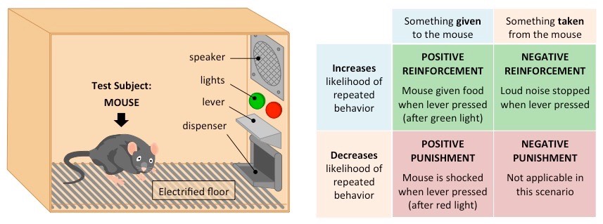
Operant Conditioning
Operant conditioning was first described by B. F. Skinner, an American psychologist who experimented on rats
Rats were placed in a controlled chamber (called a Skinner box) that contained a responsive lever
The pushing of the lever by the rat was accidental but resulted in several possible outcomes, including:
The delivery of food in response to light (desirable outcome = positive reinforcement)
The silencing of a loud noise from a speaker (desirable outcome = negative reinforcement)
The activation of an electrified floor if not pressed in response to light (negative outcome = punishment)
By trial and error, the mice learned to press the lever in response to the different environmental contexts
Operant Conditioning in Rats
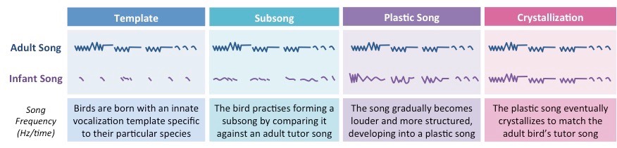
The development of birdsongs in fledglings is an example of an action that involves both innate and learned behaviours
Birds will use songs as a means of communication – either signalling courtship or establishing territorial boundaries
Most birds are born with a crude template song that is genetically inherited (innate behaviour)
The possession of an innate template prevents birds from adopting the songs of a different species of bird
Whilst young, fledglings learn to expand and refine their song by listening to, and mimicking, the adult version (motor learning)
Birds raised in isolation will lack the necessary song complexity that develops through social interaction
The time taken to develop a birdsong differs between species and songs, but once established, the final song is rarely altered
Stages in the Development of Birdsong
Learned behaviour is modified by experiences and thus requires memory to recall and process this information
If we could not remember past events, we couldn’t adapt our behaviour to new situations
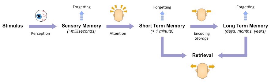
Memory is the faculty of the mind by which information is encoded, stored and retrieved
Encoding involves converting information into a form that can be stored (e.g. visual cues, sounds, semantics)
Accessing involves the retrieval of stored information to be actively used in cognitive processes
Information can be stored as a short term memory (short recall duration) or long term memory (indefinite recall period)
Short term memories can be converted to long term via the repetitive recall and consolidation of the information
Information that is not stored as a memory will be forgotten and will have to be re-learned
Many parts of the brain are involved in memory – including the prefrontal cortex and the hippocampus
Process of Memory Formation
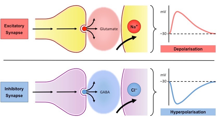
Presynaptic neurons release neurotransmitters that diffuse into the synapse and bind receptors on postsynaptic neurons
Some neurotransmitters generate excitatory post-synaptic potentials (EPSPs) by causing depolarisation (e.g. glutamate)
Some neurotransmitters generate inhibitory post-synaptic potentials (IPSPs) by causing hyperpolarisation (e.g. GABA)
If the combination of excitatory and inhibitory signals reaches a threshold limit, an action potential will be generated
Excitatory vs Inhibitory Neurotransmitters
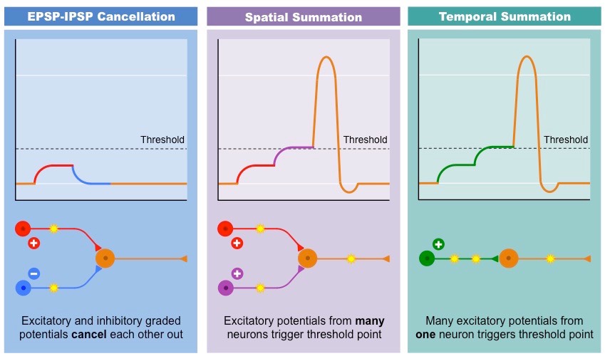
The combination of graded potentials (EPSPs and IPSPs) in the post-synaptic neuron is known as summation
Cancellation occurs when excitatory and inhibitory graded potentials cancel each other out (no threshold potential reached)
Spatial summation occurs when EPSPs are generated from multiple presynaptic neurons simultaneously to reach threshold
Temporal summation occurs when multiple EPSPs are generated from a single presynaptic neuron in quick succession
These summative effects determine which nerve pathways are activated and hence lead to alternate decision-making processes
Types of Summation
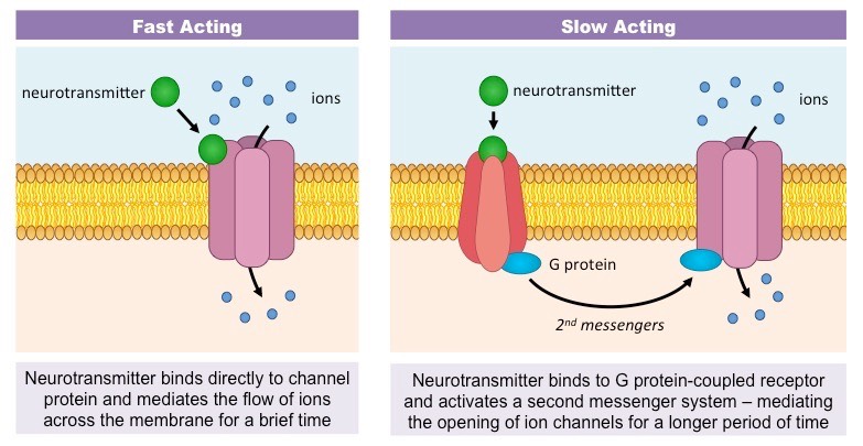
Neurotransmitters within the brain can be classified as either fast-acting or slow-acting according to their action
Fast-acting neurotransmitters bind directly to ligand-gated ion channels to initiate a rapid response (<1 millisecond)
Slow-acting neurotransmitters bind to G-protein coupled receptors to initiate a slower response (milliseconds – minute)
Slow-acting neurotransmitters trigger second messenger pathways within the post-synaptic cell, which allows for:
A longer, more sustained duration of action (i.e. ion channels remain open for longer to mediate greater depolarisation)
Long term alterations to cellular activity to improve synaptic transfer (i.e. increased expression of ion channels)
Slow-acting neurotransmitters are called neuromodulators because they can modulate the efficiency of synaptic transfer
Examples of fast-acting neurotransmitters include glutamate (excitatory) and GABA (inhibitory)
Examples of slow-acting neurotransmitters include dopamine, serotonin, acetylcholine and noradrenaline
By modulating the efficiency of synaptic transfer, slow-acting neurotransmitters can regulate fast synaptic transmission
Fast Acting versus Slow Acting Neurotransmission
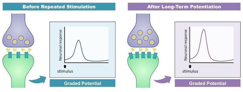
Slow-acting neurotransmitters can strengthen the neural pathways involved in learning and memory
By activating second messenger systems, they can trigger long-lasting changes to synaptic activity (long-term potentiation)
When a neuron is repetitively stimulated by slow-acting neurotransmitters, second messengers promote cellular changes:
There is an increase in dendritic receptors in the post-synaptic neutron (improving post-synaptic stimulation)
There is an increase in the production of neurotransmitters in the pre-synaptic cell
Neurons may undergo morphological changes to enlarge existing synaptic connections or form new synapses
The net effect of this long-term potentiation is that certain neural pathways become easier to stimulate
This makes certain memories easier to recall (i.e. forming long-term memories)
This makes certain actions easier to repeat (i.e. learning of a new skill or aptitude)
Long Term Potentiation
Psychoactive drugs affect the brain and personality by either increasing or decreasing postsynaptic transmissions
Drugs that increase neurotransmission levels are called stimulants and increase psychomotor arousal and alertness
Drugs that decrease neurotransmission levels are called depressants and slow down brain activities and relax muscles
Stimulant drugs mimic the stimulation provided by the sympathetic nervous system (i.e. 'fight or flight’ responses)
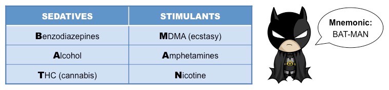
Examples of stimulants include caffeine, cocaine, amphetamines, ecstasy (MDMA) and nicotine
Depressants reduce stimulation of the central nervous system and may induce sleep (sedatives)
Examples of sedatives include benzodiazepines, barbiturates, alcohol and tetrahydrocannabinol (THC = cannabis)
Stimulants and Sedatives
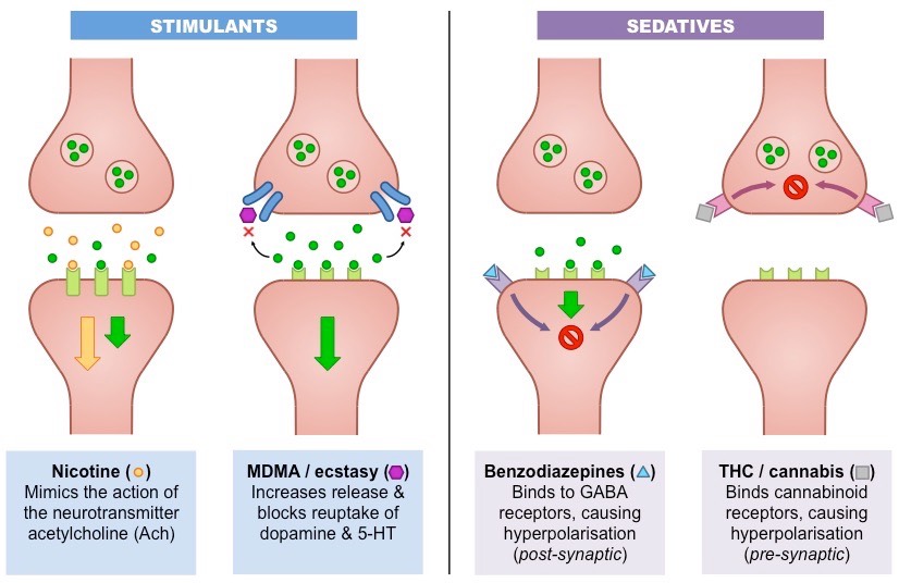
Stimulants
1. Nicotine
Nicotine stimulates the cholinergic pathways by mimicking the action of acetylcholine (binds Ach receptors)
Nicotine is not broken down by the enzyme acetylcholinesterase, resulting in overstimulation of Ach receptors
Nicotine raises dopamine levels in the brain (leading to addiction) and activates parasympathetic pathways (calming effect)
2. MDMA (ecstasy)
MDMA binds to reuptake pumps on presynaptic neurons and blocks the recycling of dopamine and serotonin (5-HT)
MDMA also enters the presynaptic neurons via the reuptake pumps and triggers the secretion of neurotransmitter
This increases levels of neurotransmitter in the synaptic cleft, prompting feelings of euphoria and heightened sensation
Sedatives
1. Benzodiazepine
Benzodiazepines bind to GABA receptors on the post-synaptic neuron and increase the efficiency of GABA action
GABA triggers the opening of chloride channels to cause hyperpolarisation – benzodiazepines enhance this effect
Benzodiazepines promote sleep-inducing and muscle relaxing responses by the body
2. Tetrahydrocannabinol (THC)
THC mimics the neurotransmitter anandamide by binding to cannabinoid receptors on presynaptic neurons
Anandamide (and THC) blocks the release of inhibitory neurotransmitters that prevent dopamine secretion
By preventing the inhibition of dopamine secretion, THC causes a sense of euphoria and emotional well-being
Mechanism of Drug Action
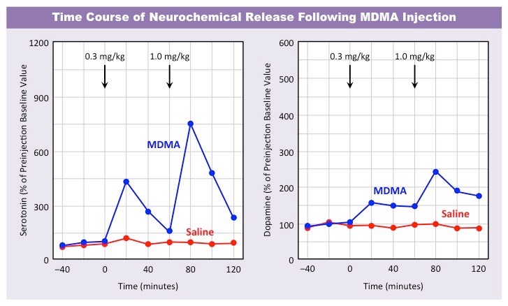
MDMA (ecstasy) is a recreational drug known to increase the activity of specific neurotransmitters – serotonin and dopamine
Serotonin (5-HT) is found in regions of the brain associated with sleep and emotion and is involved in regulating mood
Dopamine is involved in the brain’s reward pathway and plays an important role in regulating motivation and pleasure
MDMA binds to reuptake pumps and increases the release of neurotransmitter whilst slowing its rate of uptake
This causes an overstimulation of post-synaptic receptors until neurotransmitter reserves are depleted
Long-term usage of MDMA can cause adverse changes to brain architecure and result in cognitive impairment
Effect of MDMA on Serotonin and Dopamine Activity
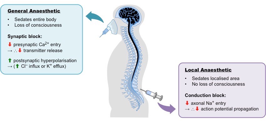
Anesthetics act on ion channels to block the conduction of sensory nerve signals to the central nervous system
This results in the loss of sensation (numbness) in the affected region, allowing for surgical interventions to occur
Anesthetics can be grouped into two classes – local anesthetics and general anesthetics
Local anesthetics only affect a localised region – usually by blocking axonal sodium influx (conduction block)
General anesthetics affect the whole body – this may involve blocking calcium influx to prevent neurotransmitter exocytosis
General versus Local Anesthesia
Different types of anesthetics will affect consciousness in different ways:
General anesthetics will induce a temporary loss of consciousness as they interfere with neural transmissions in the brain
Local anesthesia will not result in a loss of consciousness and only cause a reversible loss of sensation to the affected area
General anesthetics are typically inhaled (to affect the whole body), while local anaesthetics are injected into specific regions
General anesthetics are administered by trained specialists who monitor patient vitals for the duration of the procedure
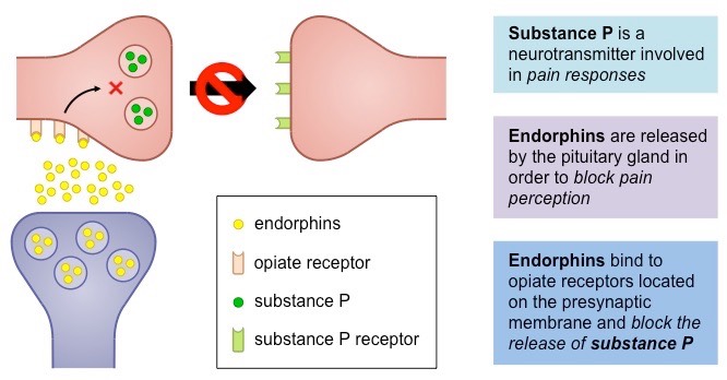
Endorphins are endogenous neuropeptides produced by the pituitary gland that functions as the body’s natural painkiller
Endorphins are typically released by the body during periods of stress, injury or physical exercise
Pain is perceived in body tissues when impulses are sent from pain receptors (nocireceptors) to sensory areas of the brain
Endorphins bind to opiate receptors on pre-synaptic neurons to block the transmission of pain signals
Endorphins differ from anesthetics in that they reduce pain perception but do not necessarily block all sensory perception
Endorphins can also promote feelings of euphoria (as they target opioid receptors)
Suppression of Pain Perception by Endorphins
An addiction is a dependence on a substance or an activity which results in its repeated and compulsive use
Stopping is very difficult and can cause severe mental and physical reactions (withdrawal symptoms)
Addictions can be affected by genetic factors, social factors and dopamine secretion
Genetic Predisposition
Particular addictions can run in families, suggesting a genetic predisposition (although social factors may contribute)
Specific genes might influence the rate of drug metabolism or intensity of drug effect (i.e. dopamine secretion)
Genetic factors may also contribute to personality types that are more inclined towards addictive behaviours
The genetic predisposition for a particular addiction may be determined by polygenic inheritance
Social Environment
Individuals raised in environments with prevalent substance abuse are at higher risk of addiction (peer pressure risks)
Individuals treated with neglect (child abuse) or suffering significant personal trauma are at a higher risk of addiction
Certain cultures have a higher incidence of addictions (may reflect demographic influences or marketing forces)
Low socioeconomic status (i.e. poverty) may increase the likelihood of addiction (poor education / lack of support networks)
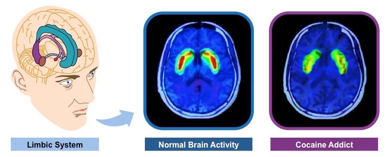
Dopamine Secretion
Dopamine is a neurotransmitter released within the limbic system in response to reward (activates pleasure pathways)
Certain drugs (e.g. cocaine, heroin) and particular activities (e.g. sex, gambling) enhance dopamine activity
Long-term substance abuse will lead to the down-regulation of dopamine receptors, requiring higher doses to achieve effect
Consequently, addicts must continue to repeat the addictive activity in order to achieve a diminishing level of reward
Effect of Drug Addiction on Dopamine Activity
Ethology is the scientific study of animal behaviour under natural conditions (i.e. observational not experimental)
As it is a biological perspective, behaviour is considered to be an evolutionary adaptive trait developed via natural selection
The modern field of ethology includes a number of well-known investigations into animal behaviour:
Migratory patterns in birds (such as blackcaps)
Reciprical altruism in animal species (such as vampire bats)
Breeding and courtship strategies in a number of different animals
Natural selection is a mechanism of evolution by which the frequency of inherited traits change as a result of external agents
Characteristics which promote survival and reproduction (i.e. beneficial alleles) become more prevalent in a population
Any behaviour that has a genetic basis (i.e. innate) and confers reproductive success will become more common
Learned behaviours may also evolve via natural selection if the capacity for learning has a genetic basis (e.g. language)
Natural selection will promote “optimal” behaviours for the given set of environmental conditions in which the organism lives
As these external conditions change, the frequency of certain behavioural responses will vary accordingly
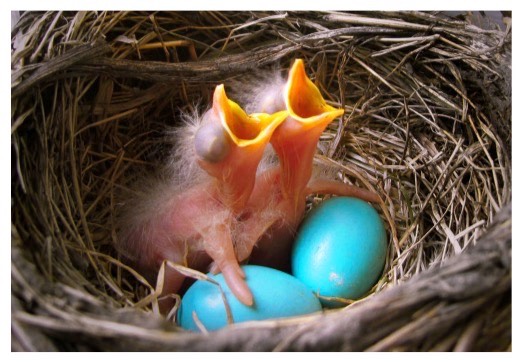
An example of the evolution of behaviour via natural selection can be demonstrated by the feeding habits of fledgling birds
Within a nest, baby birds (chicks) will gape and chirp as fledglings in order to be fed by their parents
The chicks that chirp louder and gape more obviously are more likely to receive parental attention and be fed more
These chicks are more likely to survive and pass their alleles for chirping and gaping on to their offspring
Over many generations, the frequency of excessively overt chirping and gaping behaviours has increased
Gaping Chicks
The blackcap (Sylvia atricapilla) exhibits behavioural variation in its seasonal migratory patterns
This behaviour is genetically predetermined and not learned (i.e. it is instinctive / innate)
Genetic Basis of Migratory Behaviours
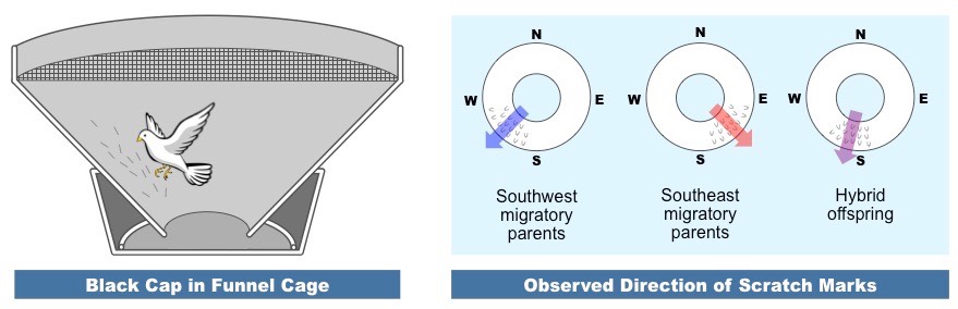
The migratory behaviours in blackcaps has been demonstrated to have a genetic basis via a number of experiments
Chicks raised in isolation will follow the migratory routes of their parents (hence it is an innate trait and not learned)
Hybrid chicks of parents with different migration routes will migrate in a direction between the two parental directions
This suggests heterozygote hybrids exhibit a combination of the migratory tendencies from each homozygous parent
Experiment Demonstrating the Genetic Basis of Migratory Behaviours
Natural Selection of Migratory Behaviours
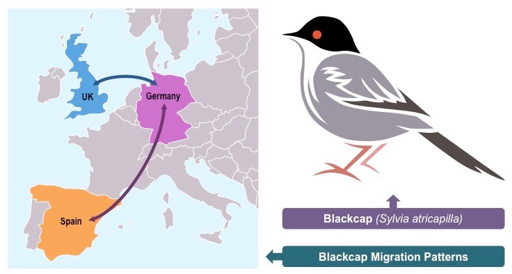
Blackcaps occupy summer breeding grounds in Germany, but migrate to different locations during the winter months
Historically, most birds migrated south to Spain in the winter, with a minority migrating west to the UK
Spain is further away but has generally had a more temperate winter climate than the UK, improving reproductive success
With an increase in global temperatures, the migratory patterns of blackcaps are changing due to natural selection
Blackcap populations in the UK are rising, as warmer temperatures are improving survival rates during the winter months
UK blackcaps are reproducing more, as the shorter migration allows them to select the best breeding territories in Germany
Migratory Patterns of European Blackcaps
Altruism is behaviour which benefits another individual at the cost of the performer
Ostensibly, it is in opposition to natural selection as it reduces the potential for the altruistic individual passing on their genes
However, it improves the chances of the other individual passing on genes into the same gene pool (i.e. inclusive fitness)
If the individuals are closely related, altruistic genes will persist in the gene pool and be naturally selected
Enhancing the reproductive success of relatives who share common genes is called kin selection
Organisms that live in social clusters will also promote the conservation of altruistic genes via reciprocal altruism
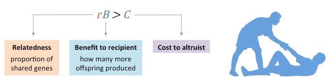
The occurrence of altruistic behaviours will be determined by three factors (known as Hamilton’s rule: rB > C)
The cost to the performer (C) should be small, while the benefit to a receiver (B) and degree of relatedness (r) should be large
Hamilton’s Rule
Blood Sharing Among Vampire Bats
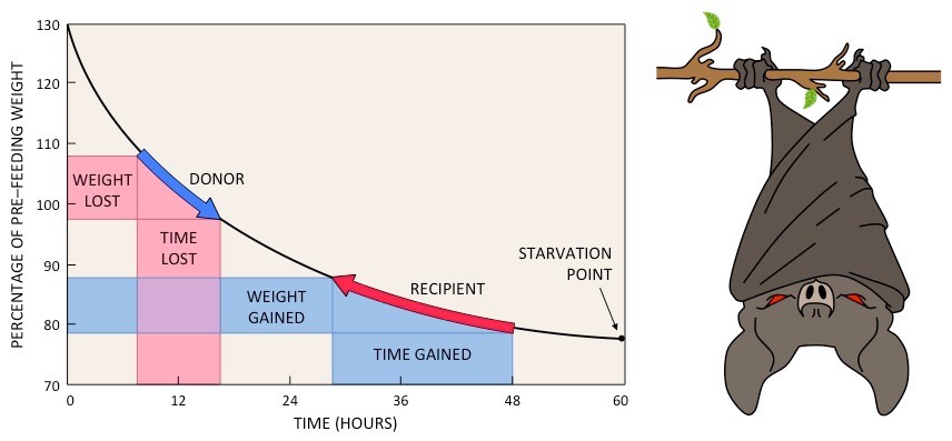
Vampire bats commonly regurgitate blood to share with unlucky roost mates who were unable to gain independent sustenance
Vampire bats cannot survive multiple successive days without food, however food can often be difficult to find
The small cost of sharing blood (lost time until starvation) is less than the benefit received (time gained)
Hence sharing blood improves the fitness of the entire brood (via reciprocal altruism), increasing the occurrence of altruism
Cost-Benefit Analysis of Blood Sharing
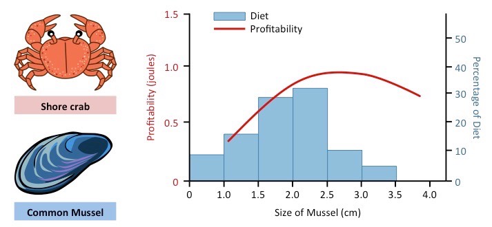
Foraging is the act of searching for (and potentially finding) food resources in nature
As availability and abundance of food resources vary, animals must adapt their foraging practices to account for any changes
Animals with optimal foraging strategies will have more available energy with which to survive and reproduce
According to the optimal foraging theory, animals will adopt strategies that:
Minimise the cost of foraging (i.e. the amount of energy used to capture and consume prey)
Maximise the benefits to the consumer (i.e. the amount of energy yielded by a particular food source)
Shore crabs demonstrate selectivity in the type of mussel foraged when the mussel population is abundant:
Crabs will ignore smaller mussels (as the energy yield is less than that obtained from larger mussels)
Crabs will also ignore larger mussels (difficult to crush, also risks potential damage to the crab’s claws)
Crabs will selectively identify and feed on mid-sized mussels (provided the mussel supply is in abundance)
Foraging Behaviour in Shore Crabs
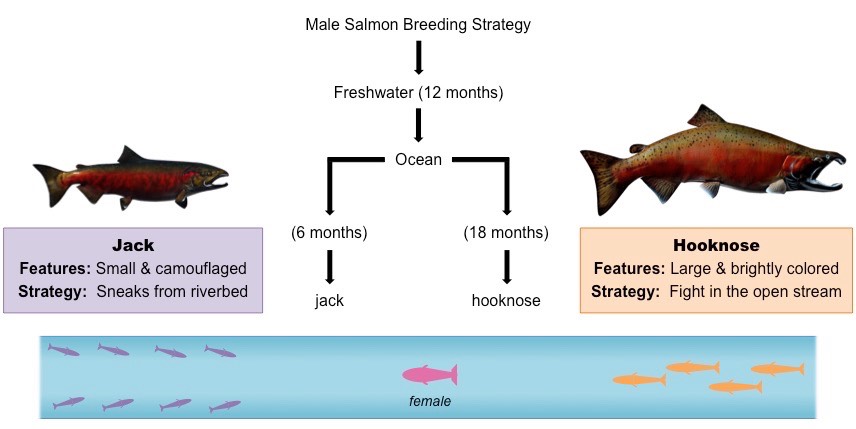
Breeding Strategies
Male coho salmon form two different breeding populations according to the strategy used for passing on genes:
All males initially undergo a development phase as juveniles in which they grow within freshwater rivers (~12 months)
Following that, the males migrate out to the ocean for a period of maturation, whereby they differentiate into two populations
Some of the male salmon develop into ‘jacks’, while other male salmon will develop into ‘hooknoses'
Jacks are smaller and well camouflaged – they only require ~ 6 months in the seawater to reach maturity
Hooknoses are larger and brightly coloured – they require ~ 18 months in the seawater to reach maturity
Jacks and hooknoses employ different breeding strategies in order to successfully reproduce with female coho salmon:
Jacks sneak out from behind rocks or recesses in the riverbed and attempt to stealthily mate with a female
Hooknoses swim within the open water and fight aggressively amongst one another for the opportunity to mate
Having two breeding pathways improves the rates of successful reproduction and also increases levels of genetic variation
Jacks have higher rates of survival (as they spend less time in seawater), but have more competition for reproduction
Hooknoses have lower rates of survival but consequently experience less direct competition for successful mating
Breeding Strategies in Coho Salmon Populations
Mate Selection
Courtship describes a set of behavioural patterns whereby potential mates inform each other of a readiness to reproduce
Courtship stimuli may be species-specific and will be performed differently by different individuals
Courtship stimuli are often competitive among males and form the basis of assessment by females
Courtship behaviour is especially pronounced in the different species of birds of paradise
Whereas females appear drab, males will have bright plumage and display fancy behaviours to demonstrate their virility
While these features make them a target for predators, they improve chances of attracting female attention (mate selection)
Any exaggerated trait that improves reproductive fitness will become more prominent in future generations (sexual selection)
Synchronised Oestrus
Female lions synchronise their sexual receptiveness (oestrus) to increase chances of survival and reproduction of offspring
Lionesses remain in the same pride their entire lives, living with genetic relatives (sisters, aunts, nieces)
Male lions leave their birth group at a young age and in order to reproduce must replace males in existing prides
Upon establishing dominance within a pride, a male lion will kill all cubs already present
The loss of cubs triggers an innate, synchronised response whereby all lionesses enter a period of oestrus
This synchronised oestrus is mediated by pheromone signals
There are many advantages to synchronising oestrus:
It increases the number of offspring the male lion can produce (risks of displacement are always present)
It allows for shared lactation and nursing of cubs within the pride (all female lions nurse indiscriminately)
It is easier for the lionesses to hunt and defend the pride if all cubs are of a comparable age
A Pride of Lions
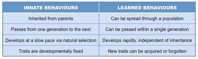
Learned behaviour describes the process of acquiring new knowledge or skills (which can be improved with practice)
Learned behaviour is dependent on environmental context and can disappear over time if the context is absent
Innate behaviour describes instinctive responses that are ingrained in an animal (it is encoded in the DNA)
It can only be modified by genetic change (mutation) which would take place over many generations
Properties of Innate versus Learned Behaviour
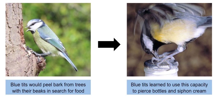
At the start of the 20th century, two bird species in the UK – blue tits and robins – would feed on cream from milk bottles
These bottles had no lids, allowing the birds easy access to the top layer of cream
Eventually, aluminium seals were placed over the tops of the milk bottles to close access to this food source
The blue tit population as a whole learnt how to penetrate the foil lids, whereas the robins as a population did not
This difference in learning development is attributed to the different social organisation of the two bird populations
Blue tits are flock birds and hence were more likely to observe and pass on newly acquired skills
Robins are territorial (solitary) birds and could not effectively propagate the information within the population
The ability to pierce the seals and siphon the cream has since been lost from the blue tit population
Milk is now capped with a plastic lid and is no longer delivered so the situational context is no longer present
Blue tits have a relatively short life span, so a learned behaviour can be removed from the population rapidly
Learned Behaviour in Blue Tits