


Chapter 12 - Coordination and Response : Human Eye
External Structures of an Eye:
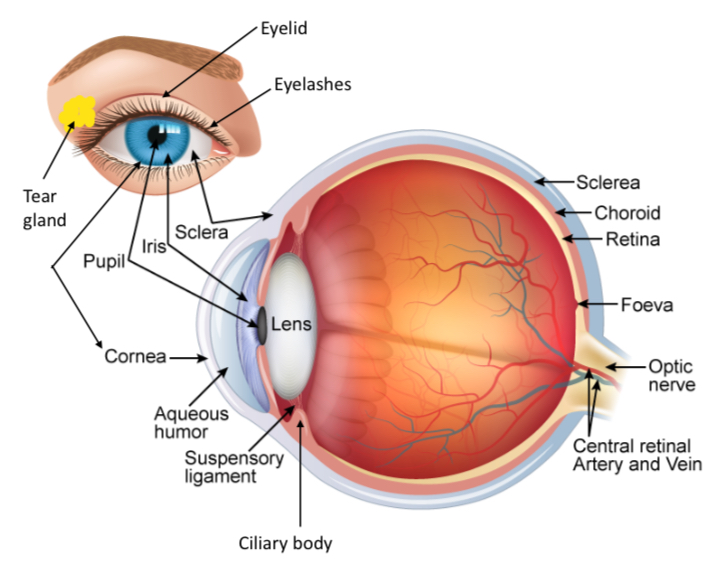
Tear gland:
Secretes tears which lubricate the eye, nourish the cornea and keeps it free from dust.
Eyelid:
Squinting is the partial closure of eyelids. This prevents excess light from entering the eye and damaging the light-sensitive tissues
Blinking spreads tears over the cornea and conjunctiva and wipes dust particles off the cornea
Eyelashes:
Shield eyes from dust particles.
Iris:
Contain radial and circular muscles that control the size of pupil
Pigment of iris gives the colour of eyes
Pupil:
Allows light to enter the eye
Sclera:
Tough white outer layer of connective tissue
Continuous with cornea
Conjunctiva:
Thin, transparent mucous membrane that covers the sclera
Secretes mucous to lubricate the eye
Cornea:
Transparent refractive layer covering the iris and pupil.
continuous with the sclera.
Internal Structure of Eye:
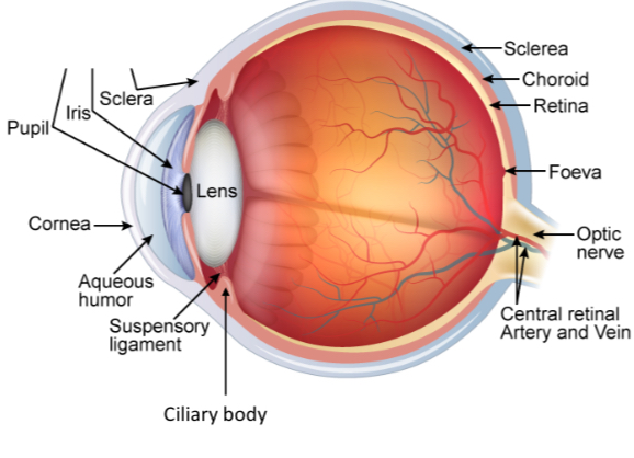
Choroid:
Middle layer of the eyeball, between the sclera and retina.
Contains blood vessels that supply oxygen and nutrients, and remove metabolic waste products.
Pigmented black to prevent an internal reflection of light.
Retina:
Innermost layer of the eyeball which contains photoreceptors, which are connected to the optic nerve.
Lens:
Transparent biconvex structure that refracts light onto the retina.
The lens can change its curvature to focus light onto retina
Fovea:
Also called yellow spot, a small depression in the retina where images are usually focused
The fovea contains the greatest concentration of cones, but no rods.
Ciliary body:
Contains ciliary muscles which control the curvature of the lens.
It is also responsible for producing aqueous humour.
Suspensory ligament:
Connects the ciliary body to the lens
Aqueous chamber:
The space between the lens and the cornea.
Transparent aqueous humour keeps the front of the eyeball firm and helps to refract light into the pupil.
Vitreous chamber:
The space behind lens
Transparent vitreous humour keeps the eyeball firm and helps to refract light onto the retina.
Optic nerve:
Transmits signal from the retina to the brain.
There are no photoreceptors in the area of the retina where the optic nerve leaves. This area is called the blind spot
Photoreceptors:
Photoreceptors in the retina consist of rods and cones. The photoreceptors are connected to the nerve endings from the optic nerve.
CONES:
Cones enable us to see colours in bright light.
There are three types of cones, red, blue, and green (RBG) that allow us to see a wide variety of colours by containing different pigment which absorbs light of different wavelengths.
Cones do not work well in dim light.
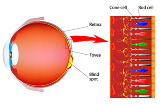
RODS:
Rods enable us to see in dim light, but only in black and white.
Rods are sensitive to light of low intensity as they contain pigment called visual purple. When the eye is exposed to bright light, all the visual purple is bleached.
Visual purple must be reformed for a person to see in the dark. Therefore, it takes awhile for one to see in dark after being in a bright environment as time is taken for visual purple to reform.
Formation of visual purple requires vitamin A.
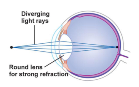
How we see
When light falls on an object, light rays are reflected from the object
Light rays are refracted through the cornea and the aqueous humour onto the lens
The lens causes further refraction and the rays are brought to a focus on the retina.
The image on the retina stimulates the photoreceptors, either the rods or the cones, depending on the intensity of the light.
Nerve impulses are produced and sent to the brain via optic nerve. The brain interprets the impulses and the person sees the object.

Accommodation:
A reflex action where the lens is adjusted so that clear images of objects at different distances are formed on the retina.
Focusing on a distant object:
Light rays reflected off distant objects are nearly parallel and enter the eyes
Ciliary muscles relax, causing suspensory ligaments to tighten
Suspensory ligaments become taut, pulling on the edge of the lens.
Lens becomes thinner and less convex, increasing its focal length, causing less refraction of the rays of light
Light rays from the distant object are sharply focused on the retina.

Focusing on a close object:
Light rays reflected off close objects are diverging and enter the eyes
Ciliary muscles contract, causing suspensory ligaments to become relax
Suspensory ligaments slacken, relaxing their pull on the lens.
The lens becomes thicker and more convex, decreasing its focal length, causing more refraction of the rays of light,
Light rays from the near object are sharply focused on the retina.
Pupil Reflex:
Reflex action (involuntary) where the pupil changes size in response to changes in light intensity.
In low light intensity, pupils dilate allow more light to enter the eye for better vision.
In high light intensity, pupils contract to restrict light to enter to prevent excessive light from damaging the retina.
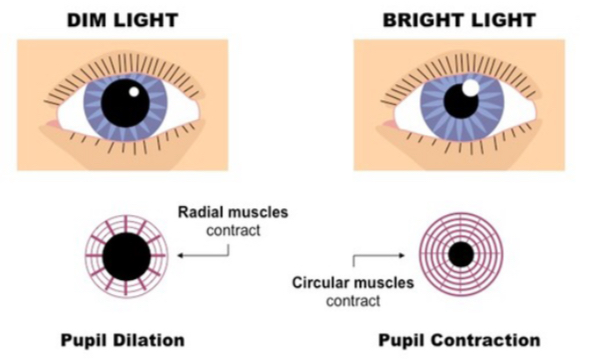
In dim light:
Radial muscles of the iris contract
Circular muscles of the iris relax
The pupil enlarges or dilates, increasing the amount of light entering the eye.
In bright light:
Circular muscles of the iris contract
Radial muscles of the iris relax
The pupil becomes smaller or constricts, reducing the amount of light entering the eye.