


Chap 3: Biological Bases of Behaviour
Source: Barron’s AP Psychology
Neuroanatomy
Neuroanatomy
The study of the parts and function of neurons
Neurons
Individual nerve cells
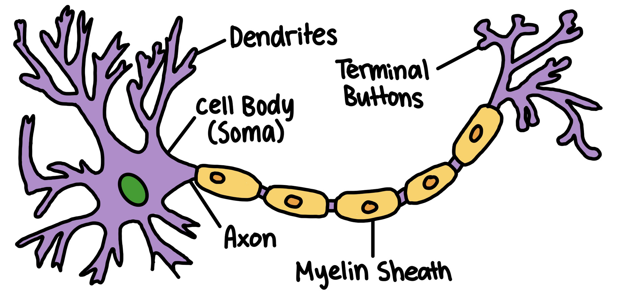
Dendrites
Root-like parts of the cell that stretch out from the cell body.
Grow to make synaptic connections with other neurons
Cell Body (Soma)
Contains the nucleus and other parts of the cell needed to sustain its life
Axon
Wirelike structure ending in the terminal buttons that extends from the cell body
Myelin Sheath
A fatty covering around the axon of some neurons that speeds neural impulses
Terminal Buttons
The branched end of the axon that also contain neurotransmitters
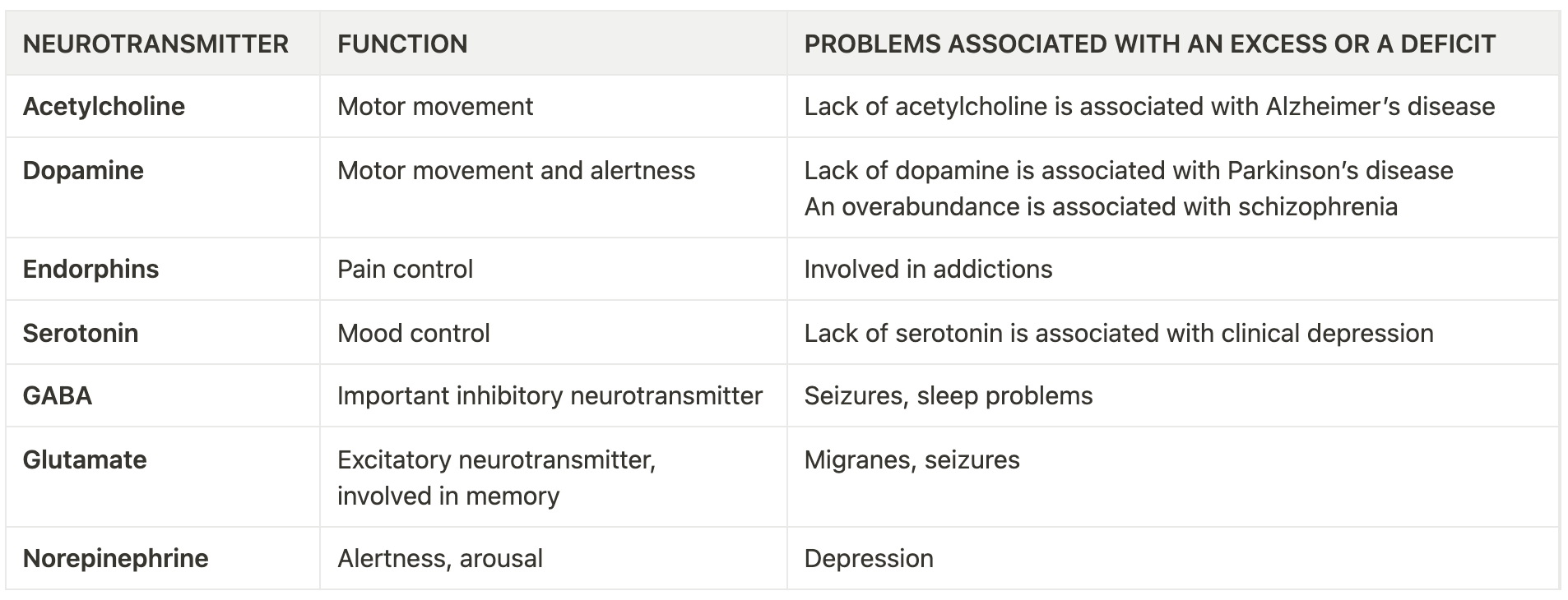
Neurotransmitters
Chemicals contained in terminal buttons that enable neurons to communicate
Fit into receptor sites on the dendrites of neurons like a key into a lock
Synapse
Space between the terminal buttons of one neuron and the dendrites of the next neuron
How a Neuron “Fires”
Background info: neuron membranes are selectively permeable; neurons start by having a slightly negative charge while + ions surround it
Begins when the terminal buttons of neuron A are stimulated and release neurotransmitters into the synapse
The neurotransmitters fit into neuron B’s receptor sites on the dendrites
If enough neurotransmitters are received (level is called absolute threshold), neuron B’s membrane becomes permeable and positive ions rush through the cell (action potential)
When the charge reaches the terminal buttons of neuron B, the buttons release their neurotransmitters into the synapse
Process begins again if enough neurotransmitters are received by the next cell to pass on the threshold
All-or-none principle
A neuron either fires completely or it does not fire.
Excitatory Neurotransmitters
Excite the next cell into firing
Inhibitory Neurotransmitters
Inhibit the next cell from firing
The Nervous System
Afferent Neurons (Sensory)
Take information from senses to brain
Interneurons
Once info reaches the brain or spinal cord, interneurons take the messages and send them elsewhere in the brain or onto efferent neurons
Efferent Neurons (Motor)
Take information from the brain to the rest of the body
Central Nervous System
Consists of brain + spinal cord (all nerves encased in bones)
Peripheral Nervous System
All nerves not encased in bone
Somatic Nervous System
Controls voluntary muscle movements
Autonomic Nervous System
Controls automatic functions of our body
Also control response to stress
Parasympathetic and sympathetic nervous systems
Sympathetic Nervous System
Mobilizes body to respond to stress
Alert system - accelerates some functions (e.g. heartbeat) but conserves resources needed for a quick response by slowing down other functions (e.g. digestion)
Parasympathetic Nervous System
Causes body to slow down AFTER a stress response (break pedal)
Reflexes → Reactions that occur the moment sensory impulses reach the spinal cord
The Brain
Ways of Studying the Brain
Accidents
By observing the brain damage and behaviour after an accident, researchers can determine the functions the damaged part played in behaviour.
Lesions
The removal or destruction of part of the brain
Observe behaviour afterwards to determine function of that part of the brain
Frontal Lobotomy (In the past, lesioning of frontal lobe was used to make the patients calm and relieve symptoms)
Electroencephalogram (EEG)
Detects brain waves
Examine what type of waves the brain produces during different stages of consciousness and use this information to generalize about brain function.
Computerized Axial Tomography Scan (CAT or CT)
Several X-ray cameras that rotate around the brain and combine all the pictures into a detailed 3D picture
Only show structure, not the functions or activity
Magnetic Resonance Imaging (MRI)
Uses magnetic fields to measure the density and location of brain material.
Only show structure, not functions or activity
Positron Emission Tomography Scan (PET)
Shows what areas of the brain are most active during certain tasks
Measures how much of a certain chemical parts of the brain are using
Functional MRI (fMRI)
Combines elements of MRI and PET scans
Show details of brain structures with information about blood flow in the brain
Brain Structure and Function
3 categories/sections: Hindbrain, Midbrain, Forebrain
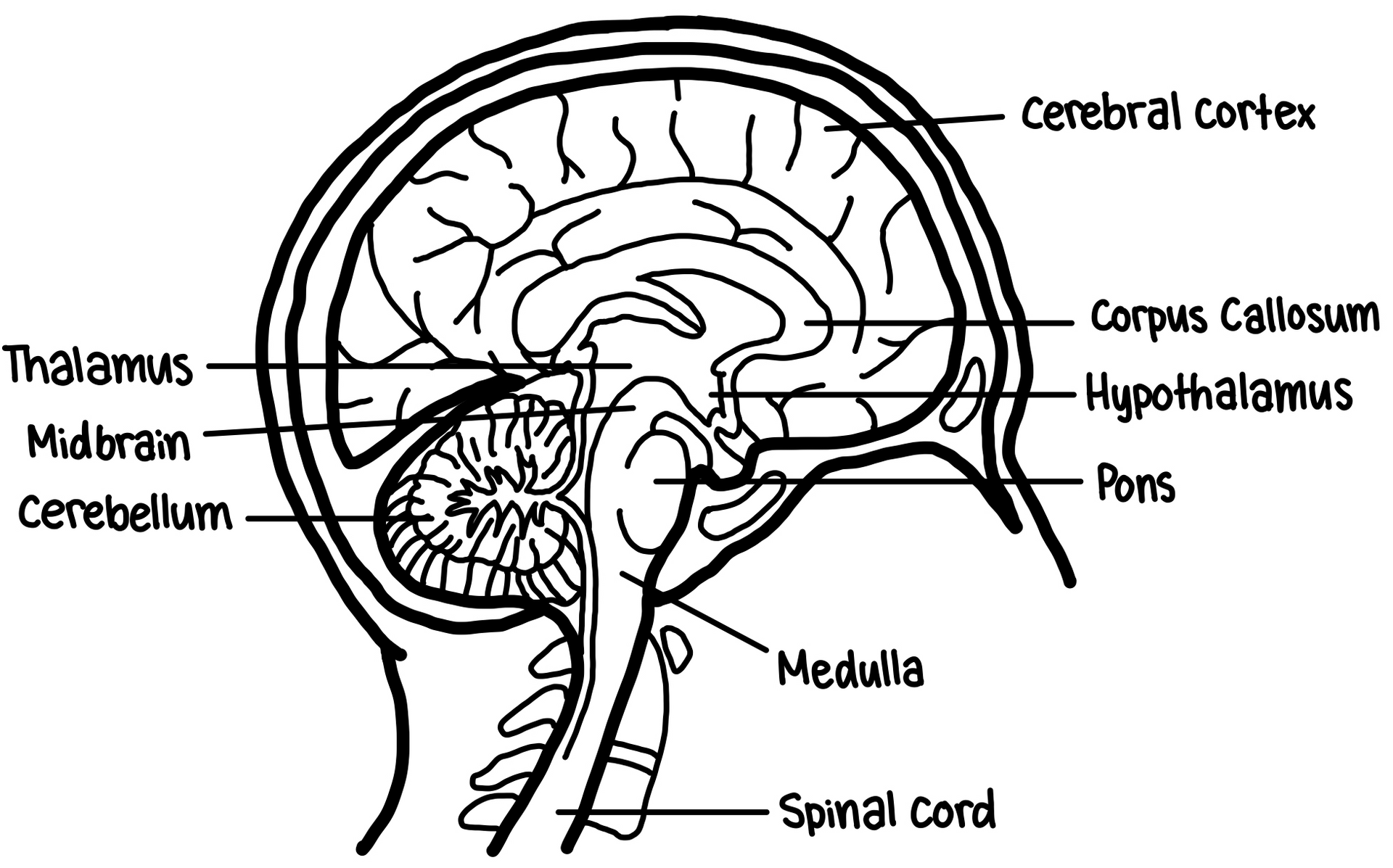
Hindbrain
Controls basic biological functions that keep us alive
Medulla
Control of blood pressure, heart rate, and breathing
Pons
Controls facial expressions
Cerebellum
Coordinates habitual muscle movements
Midbrain
Coordinates simple movements with sensory information
reticular formation
A netlike connection of cells throughout the midbrain that controls general body arousal and the ability to focus our attention
Forebrain
Controls thought and reason (what makes us human)
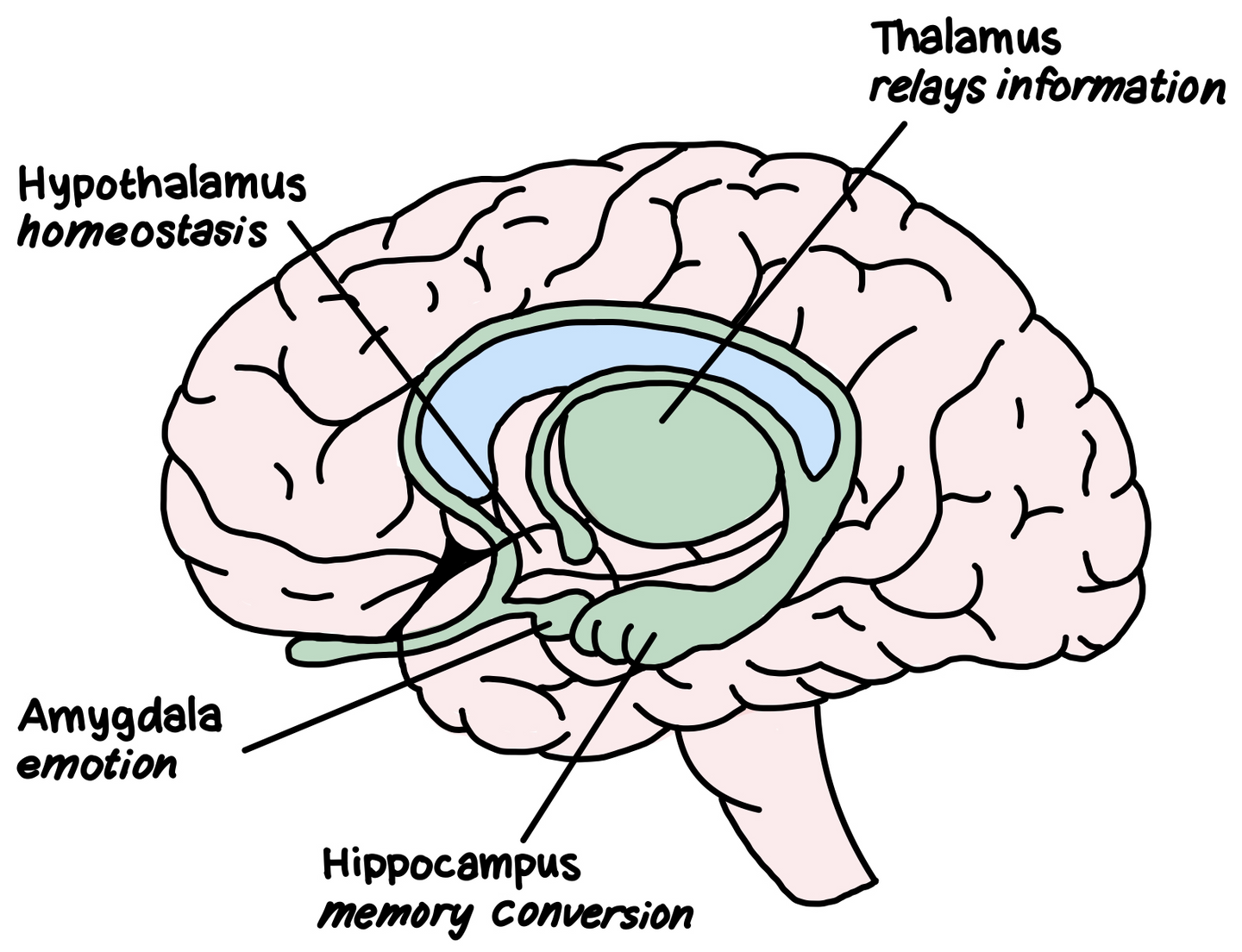
Thalamus
Receives the sensory signals coming up the spinal cord and sends them to the appropriate areas in the rest of the forebrain
Hypothalamus
Metabolic functions
e.g. body temperature, sexual arousal (libido), hunger, thirst, and the endocrine system
Amygdala
Vital to experiences of emotion
Hippocampus
Processes memory to be permanently stored in other areas of the cerebral cortex
Cerebral Cortex
Grey wrinkled surface of the brain (layer of densely packed neurons)
Overtime, the dendrites of the neurons grow and connect with other neurons to form the complex neural web
Fissures
Wrinkled surface of the cerebral cortex to increase surface area
Hemispheres
Theories: Left = logic and sequential tasks, Right = spatial and creative tasks
Contralateral control
Left hemisphere
Sensory and motor functions of RIGHT half of body
Right hemisphere
Sensory and motor functions of LEFT half of body
Brain Lateralization
Specialization of function in each hemisphere
Research is done by examining split-brain patients
Corpus callosum (nerve bundle that connects the two hemispheres) is cut to treat severe epilepsy
Operation pioneered by Roger Sperry and Michael Gazzaniga
Cannot orally report info only in the right hemisphere since spoken language is in the left hemisphere
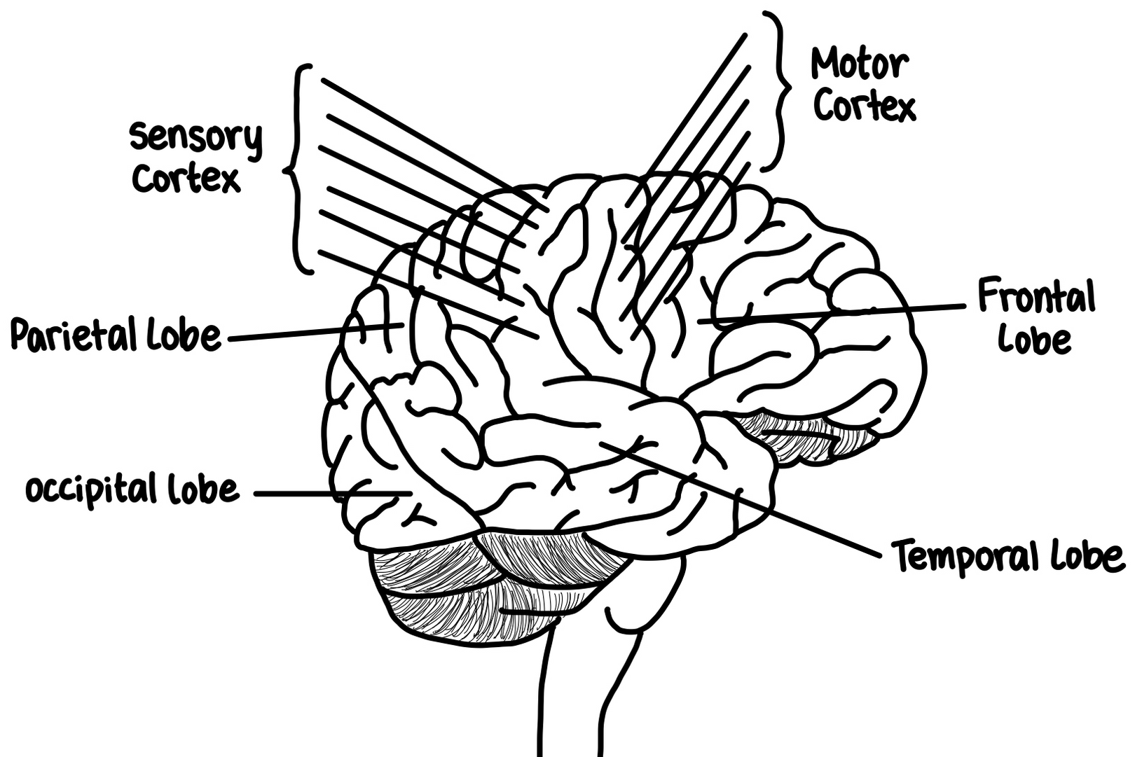
The cerebral cortex has eight (8) lobes (4 on each hemisphere - )
Association area
Any area of the cerebral cortex that is not associated with receiving sensory information or controlling muscle movements
Frontal Lobes
Large areas of the cerebral cortex located at the top front part of the brain behind the eyes
Anterior of frontal lobe is the prefrontal cortex
Critical role in thought directing process
Two special areas for language processing
Broca’s area (Paul Broca)
Frontal lobe; responsible for controlling muscles involved in producing speech
Wernicke’s Area (Carl Wernicke)
Temporal lobe; responsible for understanding of spoken and written language
Motor Cortex
Thin, vertical strip at the back of the frontal lobe
Sends signals to our muscles, controlling our voluntary movements
Top of the body is controlled my neurons at the bottom of this cortex, progressing down the body as you go up the cortex
Parietal Lobes
Located behind the frontal lobe but still on the top of the brain
Contain sensory cortex (somato-sensory cortex)
Thin vertical strip that receives incoming touch sensations from the rest of our body
Top of sensory cortex receives sensations from the bottom of the body and vice versa
Occipital Lobes
At the very back of our brain, farthest from our eyes
Interpret messages from our eyes in our visual cortex
Impulses from right half of each retina are processed in the visual cortex in the right occipital lobe
Impulses from left half of each retina are processed in the visual cortex in the left occipital lobe
Temporal Lobes
Process sound sensed by our ears (auditory cortex)
Sound received by either ear is processed in both auditory cortices
Damage to this area affects ability to interpret spoken language (Wernicke’s area)
Brain Plasticity
The ability of the nervous system to change its activity in response to intrinsic or extrinsic stimuli by reorganizing its structure, functions, or connections.
The Endocrine System
Endocrine System
A system of glands that secrete hormones that affect many different biological processes in our bodies
Controlled by hypothalamus
Adrenal Glands
Produce adrenaline
Signals body to prepare for fight or flight (autonomic nervous system - involuntary responses)
Ovaries and Testes
Produce sex hormones
Levels of estrogen and testosterone may explain gender differences (Developmental Psychology)
Genetics
Most traits are the results of nature and nurture
Monozygotic Twins
Identical twins - same genetic material
Thomas Bouchard studied twins raised in different families to see if traits were nature or nurture
Criticized because twins share the same physical characteristics, thus causing others to treat them in similar ways (effective psychological environment).
Chromosomal Abnormalities occur when chromosomes from the father (XY) and the mother (XX) fail to properly combine
Turner’s syndrome
Single X chromosome instead of a 23rd pair
Klinefelter’s syndrome
Extra X chromosome, thus XXY pattern
Down syndrome
Extra chromosome on 21st pair