


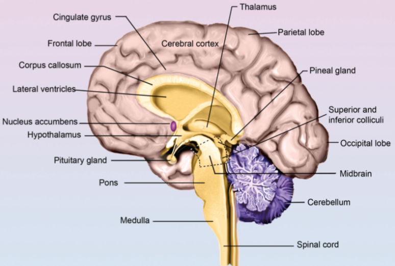
The Brain
Major Parts of the Brain
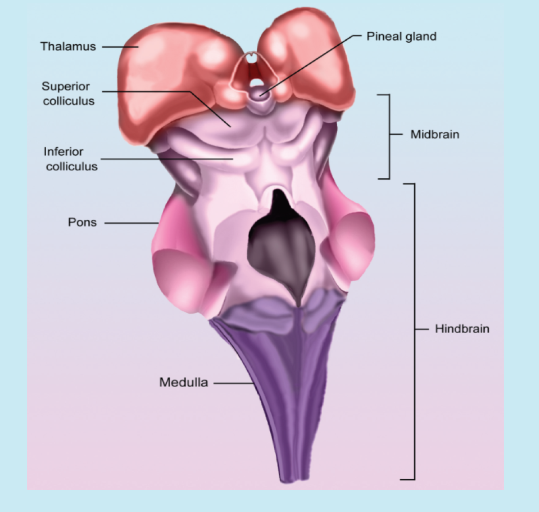
Hindbrain Includes:
Pons
Medulla
Cerebellum
Midbrain Includes:
Reticular Formation
Substantia Nigra: Movement
Colliculi
Superior: Visual reflexes
Inferior: Auditory reflexes
Forebrain Includes:
Basal Ganglia
Hypothalamus
Thalamus
Limbic system
Cerebral cortex
Pons
Consists of ascending and descending fibers.
Ascending fibers carry messages to higher parts of the brain.
Descending fibers are involved with sleep paralysis during REM.
Descending fibers inhibit motor neurons, causing sleep paralysis that occurs during the REM stage.
If the descending fibers were severed in a cat, it would result in the loss of sleep paralysis during the REM stage allowing cats to act out their dreams.
Medulla
Is located at the top of the spinal cord.
Controls vital processes, such as:
Respiration
Heart rate
Blood pressure
Vomiting
If the Medulla were to be destroyed, the results would be fatal.
Drugs which include alcohol can suppress the breathing centers in the medulla, which can result in death but can also trigger vomiting centers in the brain.
Cerebellum
The cerebellum coordinates motor behaviors, refining initiated movements by the motor cortex.
Alcohol has an effect on the cerebellum and motor skills which includes touching your nose with your finger when submitting to a sobriety test.
The cerebellum supports procedural memory, also known as motor programs, such as sports, bike riding, and piano playing.
Reticular Formation
A network of neurons that includes pathways extended from the lower parts of the brain to higher parts of the brain.
Is also known by the name reticular activating system.
Plays a key role in the arousal of higher parts of the brain as well as selective attention.
A theory is that problems such as selective attention, such as ADHD, are due to problems with reticular formation.
Substantia Nigra
Consists of fiber pathways extended from the substantial nigra in the midbrain to the basal ganglia in the forebrain.
The pathways use dopamine
Is involved with smooth, coordinated movements.
Parkinson's disease degenerates the pathways.
Colliculi
Superior Colliculus: This is important for visual reflexes, which include tracking, such as following a moving object with one’s eyes.
Inferior Colliculus: This is important for our auditory reflexes, such as orienting toward a sound.
Forebrain
Is the largest part of the brain.
Greatly expanded in humans.
Difficult to follow in humans vs. animals.
Consists of both the right and left hemispheres.
Its functions include:
Learning
Memory
Speaking
Language
Emotional responses
Experiencing
Sensations
Initiation of voluntary movements
Planning
Decision making
Basal Ganglia
Consists of a set of structures that includes:
Putamen
Globus
Pallidus
Caudate nucleus
Is involved in fine coordinated movements such as texting.
Is one of the areas of the brain responsible for allowing you to perform necessary movements.
Disorders that involve Basal Ganglia include:
Parkinson’s
OCD
Hypothalamus
Is responsible for many behaviors, which include:
Eating
Drinking
Sex
Sleep
Pleasure
Temperature
Part of the Endocrine system which regulates hormones released by the pituitary.
Associated with the four F’s, which include:
Feeding
Fighting
Fleeing
Fornicating
Controls the autonomic nervous system (ANS) Fighting and fleeing.
Thalamus
Is a sensory relay/relay station for sensory input.
Gathers information from all senses except olfactory.
Olfactory does not go through the thalamus.
Serves as a relay to the cortex.
Does some initial processing.
Limbic System
Is the accumulation of a number of structures.
In charge of emotional behaviors, which include:
Fear
Anger
Aggression
Is involved with learning and memory.
Composed of the Amygdala and Hippocampus.


Amygdala
Is in charge of evaluating the emotional significance of stimuli as well as facial expressions.
Is responsible for the fear response.
If you severed the Amygdala in a rat, it would then have no issue crawling all over a cat because it doesn’t have a fear response.
Hippocampus
Is critical in the formation of new memories and is responsible for transferring information from short-term memory to long-term memory.
The case of HM
Henry Molaison had his hippocampus removed in both hemispheres in order to control his seizures.
The surgery would help with controlling his seizures, but other consequences would arise, including the inability to form new memories.
Henry lost the ability to form any permanent memories and as a result, would talk to someone but later wouldn’t remember what was said.
Henry still had procedural memory; thus, his skills at golf would improve; however, he would have no memory of having gone golfing before.
Amygdala & Capgrad Syndrome
In Capgras Syndrome, a person thinks someone is an imposter.
Capgras Syndrome is caused by the damage being inflicted upon the connections from the temporal lobes to the amygdala.
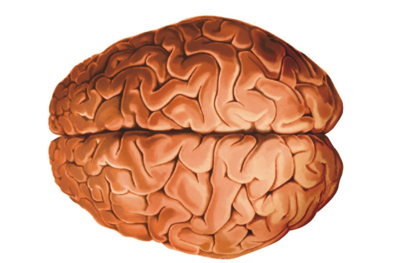
Cerebral Cortex
Is highly convoluted
Convolutions provide more surface area
A human brain compared to an animal’s brain shows that a human brain is more convoluted.
Convolutions
Gyrus (Singular).
Gyri (Plural): Bumps and hills.
Sulcus (Singular), Sulci (Plural): Indentations, Valleys, Grooves.
Fissures: Larger grooves are fissures.

Attention Deficit Hyperactivity Disorder (ADHD)
Characterized by the following:
Impulsiveness.
The inability to sustain attention.
Hyperactivity.
Neurotransmitter Anomalies include:
Reduced dopamine pathways activity.
Stimulants include:
Ritalin
Adderall
The Paradoxical Effect
Although it sounds counterintuitive to use simulants for ADHD, the stimulants increase dopamine and increase activity in the reticular formation.
Parkinson’s
Is caused by the death of cells that make up the dopaminergic tract between the substantial nigra and the basal ganglia.
Causes difficulties with movement.
Another side effect of Parkinson’s is Dementia which occurs in some people who have Parkinson’s.
Parkinson’s Disease Treatments
Levodopa (L-DOPA)
Takes the precursor for dopamine and gets converted in the brain into dopamine
Increases dopamine and thus can cause schizophrenic-like symptoms as well as other behavior changes.
Reduces symptoms but is not a cure for the progression of the disease.
Deep-brain stimulation
Electrodes are placed into the areas of the basal ganglia.
Surgical implantation of dopamine-producing cells (Cell replacement therapy)
MPTP and Parkinsons
A documentary titled The Case of the Frozen Addict.” was produced, which told the story of six young California drug users who got struck with the symptoms of Parkinson’s disease, which typically affects the elderly.
What had occurred was that the six young Californians had thought they had received synthetic heroin, but what they actually received was a neurotoxin (MPTP) which damaged cells in the substantial nigra and caused Parkinsonism.

Huntington’s Disease
A neurodegenerative disease.
Symptoms start with twitching muscles and then progress into involuntary movements.
Huntington’s disease also causes dementia as well as personality changes.
Degenerates neurons in the basal ganglia and then spreads to the vertebral cortex.
Begins between the ages of 30 and 35.
Strongly influenced by genetics and caused by one dominant gene.
Autosomal dominant: Get one bad gene from a parent, and you will have the disorder.
If a parent has the disorder, there is a 50% chance an offspring will inherit it.
Is fatal, and there is no known cure.
There is a test available; however, many wish not to take the test.
If a parent takes the test and has children, if tested early enough, they can make future decisions with the knowledge in mind.
Amnesia
A difficulty with memories caused by an event such as a stroke or accident which caused damage to the central nervous system.
Korsakoff Syndrome
Caused by chronic and excessive alcohol use.
The use of Alcohol leads to thiamine deficiency.
Anterograde Amnesia
Difficulty or inability to create new memories after an event.
Example: You answer the phone, go to your wife, and by the time you make it to the other side of the house, you forget who is on the phone.
Retrograde Amnesia
Difficulty or inability to remember events before an event.
Example: You can’t remember major events from the past that most would remember, such as the attack on 9/11.
Confabulation
Making up stories.
Korsakoff’s
Is progressive, irreversible brain deterioration.
Caused due to chronic alcoholism and Vitamin B1 (Thiamine) deficiency.
Damage to structures in the limbic system: mamillary bodies.
Symptoms include:
Anterograde amnesia
Retrograde amnesia
Confabulation
Hemispheres
There are two hemispheres in the brain.
Hemispheric lateralization:
The idea is that the left and right cerebral hemispheres take on different roles and cognitive specialties.
The left hemisphere: Is used more often for language and comprehension and speech production.
The right hemisphere: The reason for our visuospatial skills (Our ability to identify the relationship between objects in a 3D space), social communication, empathy, nonverbal cues, and emotional interpretation.
Responsible for our visual-spatial abilities.
Ability to detect an emotional tone of language.
Split Brain Research
Severs the corpus callosum.
The corpus callosum serves as a joint between the left and right hemispheres of the brain as two fibers.
You can sever the corpus callosum in order to control seizures.

Split Brain Surgery
By severing the corpus callosum, you create two separate brains, which cause the following effects:
Objects, when presented in the left visual field, go to the right hemisphere and cannot be identified verbally.
Objects, when presented to the right visual field, go to the left hemisphere and can be identified verbally.
The WADA Test
Tests for hemispheric lateralizations.
The majority of people’s language is in the left hemisphere; however, a small percentage of people have language in their right hemisphere.
Was used before open-brain surgery to test where an individual’s language was located in.
This was done by injecting barbiturate (sodium amytal) into one of the carotid arteries of an individual. Once the barbiturate made it to the brain, if the hemisphere had language, the individual wouldn’t be able to speak till the drug wore off.
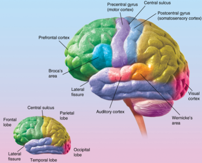
The Four Lobes
Frontal:
Involved with personality, emotions, and motor behaviors.
Parietal:
Involved with perception and sensory experiences.
Occipital:
Involved with visual processing.
Temporal:
Involved with hearing and speaking.


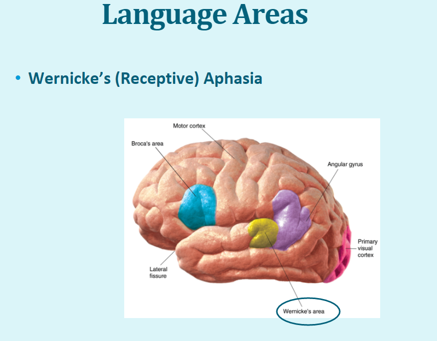
The Two Language Areas
Broca’s aphasia.
Wernicke’s aphasia.
The Temporal Lobe
The location of the primary auditory cortex.
The location of the Wernicke’s area: Important for language comprehension.
Wernicks Area
Located in the temporal lobe just above the auditory cortex.
Responsible for speech comprehension and creating understandable speech.
If the Wernicke’s area were damaged, you would have difficulties with speech comprehension causing receptive aphasia.
Receptive Aphasia
Issues with comprehension of spoken and written communication (A person can speak, but what they say makes no sense to you and is gibberish.)
Wernickes Aphasia
A type of language disorder that occurs after a stroke which causes poor comprehension, your speech is effortless, but the meaning is impaired.
The Frontal Lobe
The location for executive functions, planning, and impulse control.
Associated with Phineas Gage and Pre-frontal lobotomy.
Location of the motor cortex.
Location of the Broca’s Area: Expressive Aphasia.
The inability to speak in fluent sentences, but you can understand written and spoken words.
Executive Functions
Damage to the executive functions affects your understanding of abstractions, decision-making, personality, and other higher cognitive functioning.
Frontal Lobes: Phineas Gage
A massive 13-pound ride was accidentally driven through the front part of Phineas Gage’s frontal lobe; what resulted was similar to that of a frontal lobotomy; it caused Phineas to have emotional outbursts and problems in decision-making, something he had not experienced before the incident.
Broca’s Area
Located in the frontal lobe.
Is critical for language production.
Damage to the Broca’s Area results in expressive Aphasia.
Expressive Aphasia
Difficulty with speaking; issues orchestrating the necessary motor movements necessary to speak; you know what you want to say but can’t say it. You still have intact language comprehension and can still understand written and spoken words.
Broca’s Aphasia
A type of language disorder after a stroke with effortful speech, you can understand words, but finding the words is difficult.
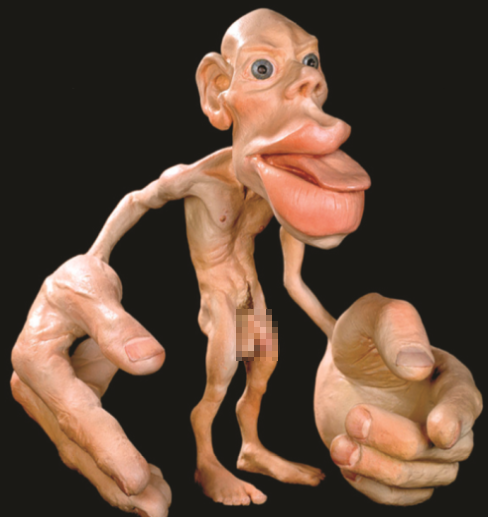
Characteristics of the Motor Cortex
Initiation of voluntary movements.
Characteristics:
Contralateral control.
The right side of the brain controls the left side of the body; the left brain controls the right side of the body.
If a person has a stroke in their right hemisphere, they will have paralysis on the left side of the body and so on.
Topographical representation.
Cortical areas are systematically arranged in a manner that is consistent with body part control.
Amount of cortical areas assigned to a body part.
The amount of cortical area assigned is related to the complexity of a movement, not how large the body part is.
The hands and mouth perform complex movements and have more cortical area assigned to them.
Homunculus means “little man” and shows this well.
Parietal Lobe
Home to the somatosensory cortex.
Brings information from the skin, muscles, etc.
You touch something, and that information gets sent to the somatosensory cortex.
Associated with unilateral neglect.
Characteristics of Somatosensory
Contralateral sensations:
The left side of the brain receives sensations from the right side of the body, and so on.
Topographical representation:
The cortical areas are systematically arranged in a way that is consistent with body parts that receive sensations from the areas previously shown.
The amount of cortical areas assigned is related to the sensitivity of the body part; examples include the mouth and hands; more cortex is assigned to those.
Neglect Syndrome
Caused by damage to the right parietal lobe due to injury or a stroke.
Also known as unilateral neglect and hemispatial.
Results in lack of recognition of the left visual field or left parts of the body.
Phantom Limb
When a person still feels sensation, including pain in a limb that has been removed.
Primary Visual Cortex
Receives electrical signals from receptors in the eyes and transforms signals into basic visual sensations.
Starts the processing of visual information.
More advanced visual recognition occurs in other areas of the brain; the recognition of the face of a friend or family is done in other areas of the brain.