


Chapter 8: The Appendicular Skeleton
The Pectoral Girdle
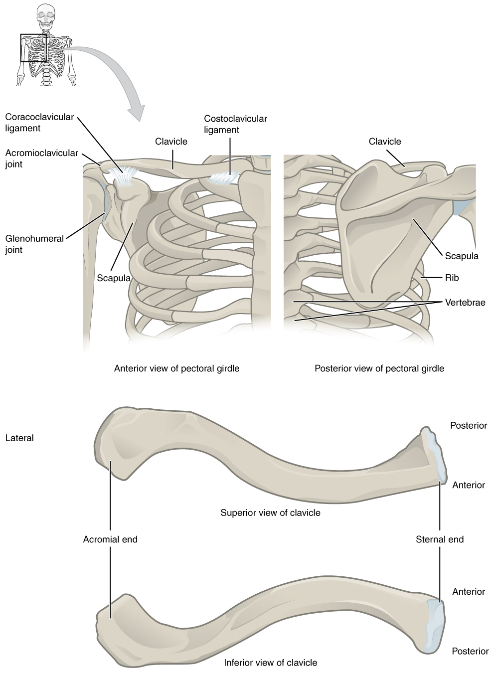
The scapula (shoulder blade) lies on the posterior aspect of the shoulder.
Supported by the clavicle, which also articulates with the humerus (arm bone) to form the shoulder joint.
The clavicle of each pectoral girdle is anchored to the axial skeleton by a single, highly mobile joint.
Clavicle
The clavicle is the only long bone that lies in a horizontal position in the body.
The clavicle has three regions: the medial end, the lateral end, and the shaft.
The medial end, known as the sternal end of the clavicle, has a triangular shape and articulates with the manubrium portion of the sternum.
The sternoclavicular joint, which is the only bony articulation between the pectoral girdle of the upper limb and the axial skeleton.
The sternoclavicular joint is indirectly supported by the costoclavicular ligament (costo- = “rib”), which spans the sternal end of the clavicle and the underlying first rib.
The lateral or acromial end of the clavicle articulates with the acromion of the scapula, the portion of the scapula that forms the bony tip of the shoulder.
Scapula
The scapula is also part of the pectoral girdle and thus plays an important role in anchoring the upper limb to the body.
The scapula is located on the posterior side of the shoulder.
The three margins or borders of the scapula, named for their positions within the body, are the superior border of the scapula, the medial border of the scapula, and the lateral border of the scapula.
The suprascapular notch is located lateral to the midpoint of the superior border.
The corners of the triangular scapula, at either end of the medial border, are the superior angle of the scapula, located between the medial and superior borders, and the inferior angle of the scapula, located between the medial and lateral borders.
The remaining corner of the scapula, between the superior and lateral borders, is the location of the glenoid cavity (glenoid fossa).
This shallow depression articulates with the humerus bone of the arm to form the glenohumeral joint (shoulder joint).
The small bony bumps located immediately above and below the glenoid cavity are the supraglenoid tubercle and the infraglenoid tubercle.
Toward the lateral end of the superior border, between the suprascapular notch and glenoid cavity, is the hook-like coracoid process (coracoid = “shaped like a crow’s beak”).
The spine of the scapula is a long and prominent ridge that runs across its upper portion.
Extending laterally from the spine is a flattened and expanded region called the acromion or acromial process.
The acromion forms the bony tip of the superior shoulder region and articulates with the lateral end of the clavicle, forming the acromioclavicular joint.
The scapula has three depressions, each of which is called a fossa (plural = fossae).
Superior to the spine is the narrow supraspinous fossa, and inferior to the spine is the broad infraspinous fossa.
The anterior (deep) surface of the scapula forms the broad subscapular fossa.
The primary support for the acromioclavicular joint comes from a very strong ligament called the coracoclavicular ligament.
Bones of the Upper Limb
The upper limb is divided into three regions:
These consist of the arm, located between the shoulder and elbow joints; the forearm, which is between the elbow and wrist joints; and the hand, which is located distal to the wrist.
The humerus is the single bone of the upper arm, and the ulna (medially) and the radius (laterally) are the paired bones of the forearm.
The base of the hand contains eight bones, each called a carpal bone, and the palm of the hand is formed by five bones, each called a metacarpal bone.
The fingers and thumb contain a total of 14 bones, each of which is a phalanx bone of the hand.
Humerus
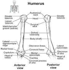
The margin of the smooth area of the head is the anatomical neck of the humerus.
Located on the lateral side of the proximal humerus is an expanded bony area called the greater tubercle.
The smaller lesser tubercle of the humerus is found on the anterior aspect of the humerus.
Passing between the greater and lesser tubercles is the narrow intertubercular groove (sulcus), which is also known as the bicipital groove because it provides passage for a tendon of the biceps brachii muscle.
At its proximal end is the head of the humerus.
The surgical neck is located at the base of the expanded, proximal end of the humerus, where it joins the narrow shaft of the humerus.
The deltoid tuberosity is a roughened, V-shaped region located on the lateral side in the middle of the humerus shaft.
The prominent bony projection on the medial side is the medial epicondyle of the humerus.
The much smaller lateral epicondyle of the humerus is found on the lateral side of the distal humerus.
The roughened ridge of bone above the lateral epicondyle is the lateral supracondylar ridge.
The distal end of the humerus has two articulation areas, which join the ulna and radius bones of the forearm to form the elbow joint.
The more medial of these areas is the trochlea, a spindle- or pulley-shaped region (trochlea = “pulley”), which articulates with the ulna bone.
Immediately lateral to the trochlea is the capitulum (“small head”), a knob-like structure located on the anterior surface of the distal humerus.
Superior to the trochlea is the coronoid fossa, which receives the coronoid process of the ulna, and above the capitulum is the radial fossa, which receives the head of the radius when the elbow is flexed.
Similarly, the posterior humerus has the olecranon fossa, a larger depression that receives the olecranon process of the ulna when the forearm is fully extended.
Ulna
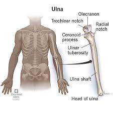
The ulna is the medial bone of the forearm. It runs parallel to the radius, which is the lateral bone of the forearm.
The proximal end of the ulna resembles a crescent wrench with its large, C-shaped trochlear notch.
The inferior margin of the trochlear notch is formed by a prominent lip of bone called the coronoid process of the ulna.
Just below this on the anterior ulna is a roughened area called the ulnar tuberosity.
To the lateral side and slightly inferior to the trochlear notch is a small, smooth area called the radial notch of the ulna.
This area is the site of articulation between the proximal radius and the ulna, forming the proximal radioulnar joint.
The posterior and superior portions of the proximal ulna make up the olecranon process, which forms the bony tip of the elbow.
More distal is the shaft of the ulna. The lateral side of the shaft forms a ridge called the interosseous border of the ulna.
This is the line of attachment for the interosseous membrane of the forearm, a sheet of dense connective tissue that unites the ulna and radius bones.
The small, rounded area that forms the distal end is the head of the ulna.
Projecting from the posterior side of the ulnar head is the styloid process of the ulna, a short bony projection.
Radius
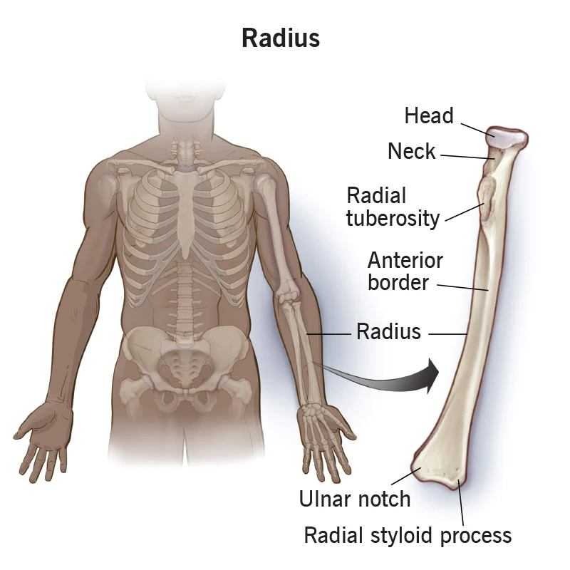
The head of the radius is a disc-shaped structure that forms the proximal end.
The neck of the radius is the narrowed region immediately below the expanded head. Inferior to this point on the medial side is the radial tuberosity, an oval-shaped, bony protuberance that serves as a muscle attachment point.
The shaft of the radius is slightly curved and has a small ridge along its medial side.
This ridge forms the interosseous border of the radius, which, like the similar border of the ulna, is the line of attachment for the interosseous membrane that unites the two forearm bones.
The distal end of the radius has a smooth surface for articulation with two carpal bones to form the radiocarpal joint or wrist joint.
On the medial side of the distal radius is the ulnar notch of the radius.
This shallow depression articulates with the head of the ulna, which together form the distal radioulnar joint.
The lateral end of the radius has a pointed projection called the styloid process of the radius.
Carpal Bones
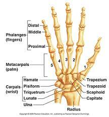
The carpal bones are arranged in two rows, forming a proximal row of four carpal bones and a distal row of four carpal bones.
The bones in the proximal row, running from the lateral (thumb) side to the medial side, are the scaphoid (“boat-shaped”), lunate (“moon-shaped”), triquetrum (“three-cornered”), and pisiform (“pea-shaped”) bones.
The distal bones (lateral to medial) are the trapezium (“table”), trapezoid (“resembles a table”), capitate (“head-shaped”), and hamate (“hooked bone”) bones.
The hamate bone is characterized by a prominent bony extension on its anterior side called the **hook of the hamate bone**.
The proximal and distal rows of carpal bones articulate with each other to form the midcarpal joint.
A strong ligament called the flexor retinaculum spans the top of this U-shaped area to maintain this grouping of the carpal bones.
The carpal bones and the flexor retinaculum form a passageway called the carpal tunnel, with the carpal bones forming the walls and floor, and the flexor retinaculum forming the roof of this space.
Metacarpal Bones
The proximal end of each metacarpal bone articulates with one of the distal carpal bones. Each of these articulations is a carpometacarpal joint.
The expanded distal end of each metacarpal bone articulates at the metacarpophalangeal joint with the proximal phalanx bone of the thumb or one of the fingers.
Phalanx Bones
The fingers and thumb contain 14 bones, each of which is called a phalanx bone (plural = phalanges), named after the ancient Greek phalanx (a rectangular block of soldiers).
The thumb ( pollex) is digit number 1 and has two phalanges, a proximal phalanx, and a distal phalanx bone.
An interphalangeal joint is one of the articulations between adjacent phalanges of the digits.
The Pelvic Girdle and Pelvis
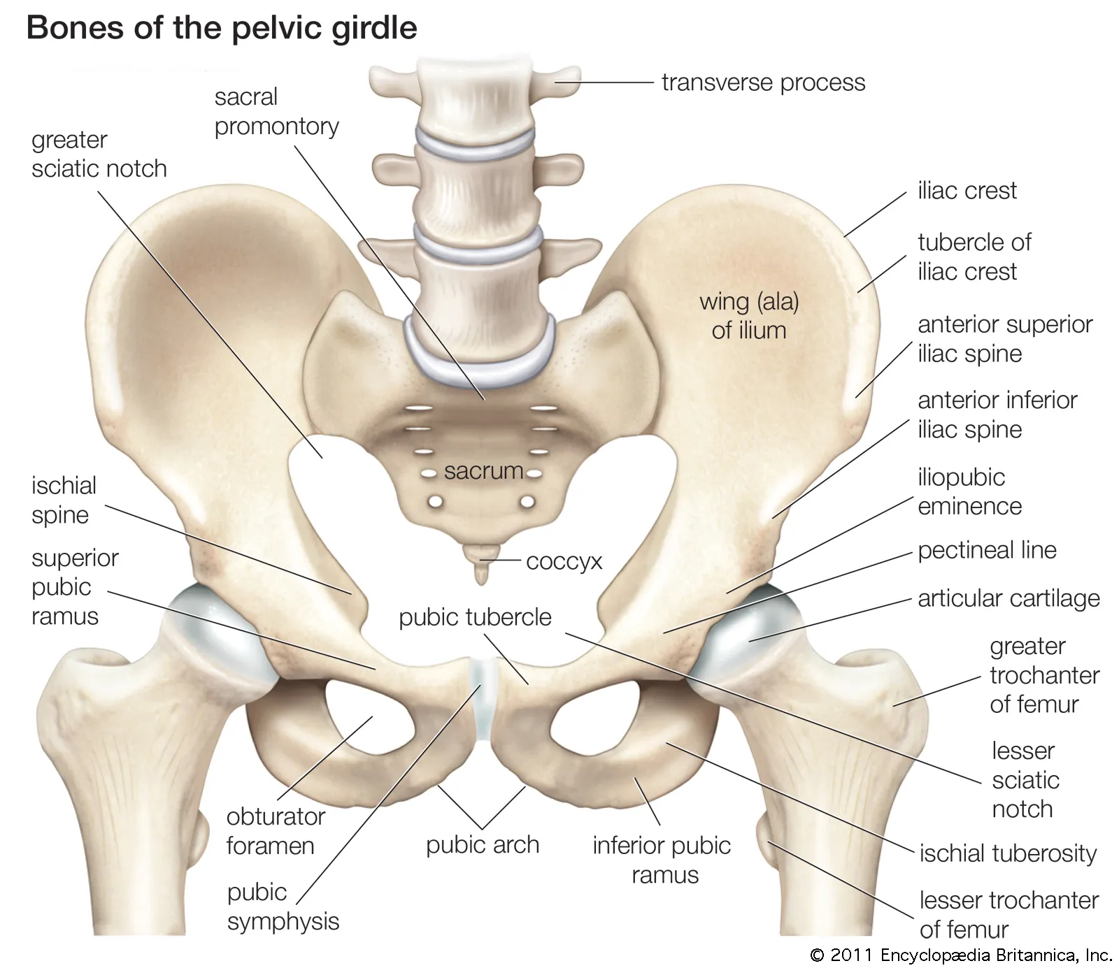
The pelvic girdle (hip girdle) is formed by a single bone, the hip bone or coxal bone (coxal = “hip”), which serves as the attachment point for each lower limb.
The bony pelvis is the entire structure formed by the two hip bones, the sacrum, and, attached inferiorly to the sacrum, the coccyx.
Hip Bone
The hip bone, or coxal bone, forms the pelvic girdle portion of the pelvis.
The ilium is the fan-like, superior region that forms the largest part of the hip bone.
It is firmly united to the sacrum at the largely immobile sacroiliac joint.
The ischium forms the posteroinferior region of each hip bone. It supports the body when sitting.
The pubis forms the anterior portion of the hip bone.
The pubis curves medially, where it joins to the pubis of the opposite hip bone at a specialized joint called the pubic symphysis.
Ilium
This curved, superior margin of the ilium is the iliac crest.
The rounded, anterior termination of the iliac crest is the anterior superior iliac spine.
Inferior to the anterior superior iliac spine is a rounded protuberance called the anterior inferior iliac spine.
Posteriorly, the iliac crest curves downward to terminate as the posterior superior iliac spine.
More inferiorly is the posterior inferior iliac spine.
This is located at the inferior end of a large, roughened area called the auricular surface of the ilium.
The shallow depression located on the anteromedial (internal) surface of the upper ilium is called the iliac fossa.
The inferior margin of this space is formed by the arcuate line of the ilium, the ridge formed by the pronounced change in curvature between the upper and lower portions of the ilium.
The large, inverted U-shaped indentation located on the posterior margin of the lower ilium is called the greater sciatic notch.
Ischium
The ischium forms the posterolateral portion of the hip bone.
The large, roughened area of the inferior ischium is the ischial tuberosity.
Projecting superiorly and anteriorly from the ischial tuberosity is a narrow segment of bone called the ischial ramus.
The slightly curved posterior margin of the ischium above the ischial tuberosity is the lesser sciatic notch.
The bony projection separating the lesser sciatic notch and greater sciatic notch is the ischial spine.
Pubis
The pubis forms the anterior portion of the hip bone.
The enlarged medial portion of the pubis is the pubic body.
Located superiorly on the pubic body is a small bump called the pubic tubercle.
The superior pubic ramus is the segment of bone that passes laterally from the pubic body to join the ilium.
The narrow ridge running along the superior margin of the superior pubic ramus is the pectineal line of the pubis.
Extending downward and laterally from the body is the inferior pubic ramus.
The pubic arch is the bony structure formed by the pubic symphysis, and the bodies and inferior pubic rami of the adjacent pubic bones.
The inverted V-shape formed as the ischiopubic rami from both sides come together at the pubic symphysis is called the subpubic angle.
Pelvis
The pelvis consists of four bones: the right and left hip bones, the sacrum, and the coccyx.
The three areas of each hip bone: the ilium, pubis, and ischium, converge centrally to form a deep, cup-shaped cavity called the acetabulum.
The large opening in the anteroinferior hip bone between the ischium and pubis is the obturator foramen.
The anterior sacroiliac ligament on the anterior side of the joint and the posterior sacroiliac ligament on the posterior side.
The sacrospinous ligament runs from the sacrum to the ischial spine, and the sacrotuberous ligament runs from the sacrum to the ischial tuberosity.
The superior opening is the greater sciatic foramen.
This large opening is formed by the greater sciatic notch of the hip bone, the sacrum, and the sacrospinous
The smaller, more inferior lesser sciatic foramen is formed by the lesser sciatic notch of the hip bone, together with the sacrospinous and sacrotuberous ligaments.
The broad, superior region, defined laterally by the large, fan-like portion of the upper hip bone, is called the greater pelvis (greater pelvic cavity; false pelvis).
The narrow, rounded space of the lesser pelvis (lesser pelvic cavity; true pelvis) contains the bladder and other pelvic organs, and thus is also known as the true pelvis.
The pelvic brim (also known as the pelvic inlet) forms the superior margin of the lesser pelvis, separating it from the greater pelvis.
The inferior limit of the lesser pelvic cavity is called the pelvic outlet.
Overview of Differences between the Female and Male Pelvis
Female pelvis | Male pelvis | |
|---|---|---|
Pelvic weight | Bones of the pelvis are lighter and thinner | Bones of the pelvis are thicker and heavier |
Pelvic inlet shape | Pelvic inlet has a round or oval shape | Pelvic inlet is heart-shaped |
Lesser pelvic cavity shape | Lesser pelvic cavity is shorter and wider | Lesser pelvic cavity is longer and narrower |
Subpubic angle | Subpubic angle is greater than 80 degrees | Subpubic angle is less than 70 degrees |
Pelvic outlet shape | Pelvic outlet is rounded and larger | Pelvic outlet is smaller |
Bones of the Lower Limb
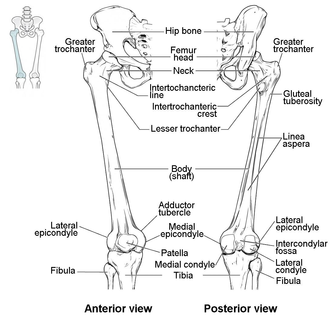
The thigh is that portion of the lower limb located between the hip joint and knee joint. The leg is specifically the region between the knee joint and the ankle joint. Distal to the ankle is the foot.
The femur is the single bone of the thigh.
The patella is the kneecap and articulates with the distal femur.
The tibia is the larger, weight-bearing bone located on the medial side of the leg, and the fibula is the thin bone of the lateral leg.
The posterior portion of the foot is formed by a group of seven bones, each of which is known as a tarsal bone, whereas the mid-foot contains five elongated bones, each of which is a metatarsal bone.
The toes contain 14 small bones, each of which is a phalanx bone of the foot.
Femur
The femur, or thigh bone, is the single bone of the thigh region.
The rounded, proximal end is the head of the femur, which articulates with the acetabulum of the hip bone to form the hip joint.
The fovea capitis is a minor indentation on the medial side of the femoral head that serves as the site of attachment for the ligament of the head of the femur.
The narrowed region below the head is the neck of the femur.
The greater trochanter is the large, upward, bony projection located above the base of the neck.
The lesser trochanter is a small, bony prominence that lies on the medial aspect of the femur, just below the neck.
Running between the greater and lesser trochanters on the anterior side of the femur is the roughened intertrochanteric line.
The trochanters are also connected on the posterior side of the femur by the larger intertrochanteric crest.
The elongated shaft of the femur has a slight anterior bowing or curvature.
The posterior shaft has the gluteal tuberosity, a roughened area extending inferiorly from the greater trochanter.
The gluteal tuberosity becomes continuous with the linea aspera (“rough line”).
On the lateral side, the smooth portion that covers the distal and posterior aspects of the lateral expansion is the lateral condyle of the femur.
The roughened area on the outer, lateral side of the condyle is the lateral epicondyle of the femur.
The smooth region of the distal and posterior medial femur is the medial condyle of the femur, and the irregular outer, medial side of this is the medial epicondyle of the femur.
The adductor tubercle is a small bump located at the superior margin of the medial epicondyle.
supporting ligaments of the knee.
The medial and lateral condyles are separated by a deep depression called the intercondylar fossa.
Anteriorly, the smooth surfaces of the condyles join together to form a wide groove called the patellar surface, which provides for articulation with the patella bone.
Patella: The patella (kneecap) is largest sesamoid bone of the body.
Tibia
The tibia (shin bone) is the medial bone of the leg and is larger than the fibula, with which it is paired.
The two sides of this expansion form the medial condyle of the tibia and the lateral condyle of the tibia.
These areas articulate with the medial and lateral condyles of the femur to form the knee joint.
Between the articulating surfaces of the tibial condyles is the intercondylar eminence, an irregular, elevated area that serves as the inferior attachment point for two supporting ligaments of the knee.
The tibial tuberosity is an elevated area on the anterior side of the tibia, near its proximal end.
The shaft of the tibia becomes triangular in shape.
MH this triangle forms the anterior border of the tibia, which begins at the tibial tuberosity and runs inferiorly along the length of the tibia.
A small ridge running down the lateral side of the tibial shaft is the interosseous border of the tibia.
This is for the attachment of the interosseous membrane of the leg, the sheet of dense connective tissue that unites the tibia and fibula bones.
The large expansion found on the medial side of the distal tibia is the medial malleolus (“little hammer”).
On the lateral side of the distal tibia is a wide groove called the fibular notch.
This area articulates with the distal end of the fibula, forming the distal tibiofibular joint.
Fibula
The fibula is the slender bone located on the lateral side of the leg.
The head of the fibula is the small, knob-like, proximal end of the fibula.
It articulates with the inferior aspect of the lateral tibial condyle, forming the proximal tibiofibular joint.
The thin shaft of the fibula has the interosseous border of the fibula, a narrow ridge running down its medial side for the attachment of the interosseous membrane that spans the fibula and tibia.
The distal end of the fibula forms the lateral malleolus, which forms the easily palpated bony bump on the lateral side of the ankle.
Tarsal Bones
The most superior bone is the talus. This has a relatively square-shaped, upper surface that articulates with the tibia and fibula to form the ankle joint.
The talus articulates with the calcaneus (heel bone), the largest bone of the foot, which forms the heel.
The medial calcaneus has a prominent bony extension called the sustentaculum tali (“support for the talus”) that supports the medial side of the talus bone.
The cuboid bone articulates with the anterior end of the calcaneus bone.
The talus bone articulates anteriorly with the navicular bone, which in turn articulates anteriorly with the three cuneiform (“wedge-shaped”) bones.
These bones are the medial cuneiform, the intermediate cuneiform, and the lateral cuneiform.
Metatarsal Bones
The base of the metatarsal bone is the proximal end of each metatarsal bone.
The expanded distal end of each metatarsal is the head of the metatarsal bone.
Each metatarsal bone articulates with the proximal phalanx of a toe to form a metatarsophalangeal joint.
Phalanges
The toes contain a total of 14 phalanx bones (phalanges), arranged in a similar manner as the phalanges of the fingers.
The toes are numbered 1–5, starting with the big toe ( hallux).
Limb Growth
Each upper and lower limb initially develops as a small bulge called a limb bud, which appears on the lateral side of the early embryo.
The ectoderm at the end of the limb bud thickens to form a narrow crest called the apical ectodermal ridge.