


Module 18: Vision
18-1: What is the energy that we see as visible light, and how does the eye transform light energy into neural messages?
Our eyes receive light energy and transduce (transform) it into neural messages our brain can process.
The Stimulus Input: Light Energy
Visible light is a very small section of the electromagnetic spectrum (figure 18.1)

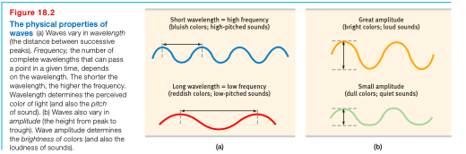
Wavelength: distance from the peak of one wave to the next peak
Wavelength determines the lights hue
Hue: dimension of color that is determined by the wavelength of light
Ex: the colors we know, blue, green, etc,.
Intensity: the amount of energy in light or sound waves, which we perceive as brightness or loudness, determined by the wave’s amplitude (height)
The Eye
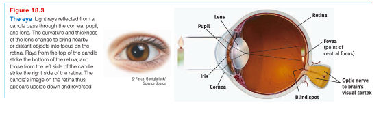
(1) Light enters the eye through the cornea, which protects the eye and bends light to provide focus. (2) Then the light passes through the pupil, a small adjustable opening in the center of the eye. (3) Surrounding the pupil is the iris, a colored muscle that dilates or constricts in response to light intensity or inner emotions. (4) Behind the pupil is a lens that focuses incoming light rays into an image on the retina, a multilayered tissue on the eyeball’s sensitive inner surface. (5) The Lens changes shape to focus the rays in a process accommodation
Pupil: adjustable opening in the center of the eye through which light enters
Iris: ring of muscle tissue that forms the colored portion of the eye around the pupil and controls the size of the pupil opening
Lens: transparent structure behind the pupil that changes shape to help focus images on the retina
Retina: light-sensitive inner surface of the eye, containing receptor rods and cones plus layers of neurons that begin the processing of visual information
Accommodation: process by which the eye’s lens changes shape to focus near or far objects on the retina
The Retina
(1) Light-energy particles enter your eye and make their way through the retina’s outer layer of cells to its buried receptor cells, the rods and cones (figure 18.4). (2) There, light-energy will trigger chemical changes that would spark neural signals, activating nearby bipolar cells. (3) The bipolar cells in turn would activate the ganglion cells, whose axons twine together like the strands of a rope to form the optic nerve. (4) The nerve will carry the information to your brain, where your thalamus stands ready to distribute the information.

Rods: retinal receptors that detect black, white, and gray; necessary for peripheral and twilight vision, when cones don’t respond
Cones: retinal receptor cells that are concentrated near the center of the retina and that function in daylight or in well-lit conditions. These give fine detail and give rise to color sensations
Optic nerve: nerve that carries neural impulses from the eye to the brain
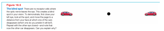
Blind spot: point at which the optic nerve leaves the eye, creating a “blind” spot because no receptor cells are located there (figure 18.5)
There are many differences with rods and cones, mainly where they’re found and in what they do (table 18.1)
Cones cluster in and around the fovea. Each cone transmits to a single bipolar cells that helps relay the cone’s individual messages to the visual cortex, which devotes and large area to input the fovea.
Also enable you to perceive color. In dim light they become ineffectual, so you see no colors
Fovea: central focus point in the retina, around which the eye’s cones cluster (figure 18.3)
Rods are the opposite. They share bipolar cells with other rods, sending combined messages
Enables black-and-white vision
Remain sensitive in dim light
Cones: detail and color
Rods: Faint light

Visual Information Processing
18-2: How do the eye and the brain process visual information?
Any given retinal area relays its information to a corresponding location in the visual cortex, in the occipital lobe at the back of your brain (figure 18.6)
Same sensitivity that enables retinal cells to fire messages can lead them to misfire
Ex: Turn your eyes to the left, close them, and then gently rub the right side of your eyelid with your fingertip. You should see the patch of light to the left, moving as your finger movies.
This happens because your retinal cells are so responsive that even pressure triggers them, but your brain interprets their firing as light . It interprets the light as coming from the left–the normal direction of light that activates the right side of the retina

Feature detection
David Hubel and Torsten Wiesel received a nobel prize for their work on feature detectors.
Feature detectors: nerve cells in the brain that respond to specific features of the stimulus, such as shape, angle, or movement
In the visual cortex, feature detectors respond to specific features of the visual stimulus
Supercell clusters in other critical brain areas respond to more complex patterns
Parallel Processing
Parallel processing: processing of many aspects of a problem simultaneously; the brain’s natural mode of information processing for many functions, including vision. Contrast with the step-by-step (serial) processing of most computers and of conscious problem solving
To analyze a visual scene, the brain divides it into subdimensions–motion, form, depth, color–and works on each aspect simultaneously. Other neural teams integrate the results, comparing them with stored information and enabling perceptions (figure 18.9)

Color Vision
18-3 What theories help us understand color vision?
If no one sees a tomato, is it red?
No. (1) the tomato is everything but red, because it reflects the long wavelengths of red. (2) the tomato’s color is our mental construction, color, like all aspects of vision, resided not in the object but in the theater of our brain, as evidenced by our dreaming in color
Young-Helmholtz trichromatic (three-color) theory: theory that the retina contains 3 different color receptors–one most sensitive to red, one to green, on to blue–which, when stimulated in combination, can produce the perception of any color
Ex: there are no receptors sensitive to yellow, we see yellow when mixing red and green light, which stipulates both red and green sensitive cones
Most people who are “color-blind” are actually not. They just lack functioning red and/or green sensitive cones. Their vision is either monochromatic or dichromatic
How come people blind to green and red can still see yellow? This theory leaves some parts of the color vision mystery unsolved
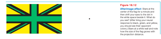
Opponent-process theory: theory that opposing retinal processes (red-green, yellow-blue, white-black) enable color vision
Ex: some cells are stimulated by green and inhibited by red; others are stimulated by red and inhibited by green
These two theories, and the research supporting them, show that color processing occurs in two stages