


Nervous System Part 1-1
Parts of the Nervous system
Central nervous system (CNS).
Brain & Spinal cord.
Peripheral nervous system.
Somatic: Skeletal & Sensory.
Autonomic: Sympathetic & Parasympathetic
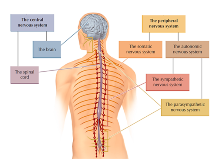
The Autonomic Nervous System
Parts of the nervous system include:
Part A | Central nervous system (CNS):
Brain & Spinal cord.
Part B | Peripheral nervous system:
Somatic: Skeletal & Sensory.
Autonomic: Sympathetic & Parasympathetic.
The sequence for activation of the fight-flight response
Sympathetic portion:
Gets ready for an emergency.
Becomes ready to be attacked.
Triggers multiple physiological responses.
Parasympathetic portion:
Returns the body to a relaxed state.
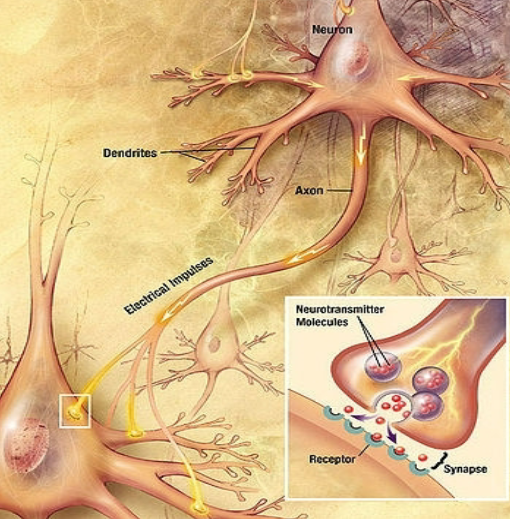
The Structure and Function of Neurons
The function of the neuron:
Neurons receive and transmit signals/information
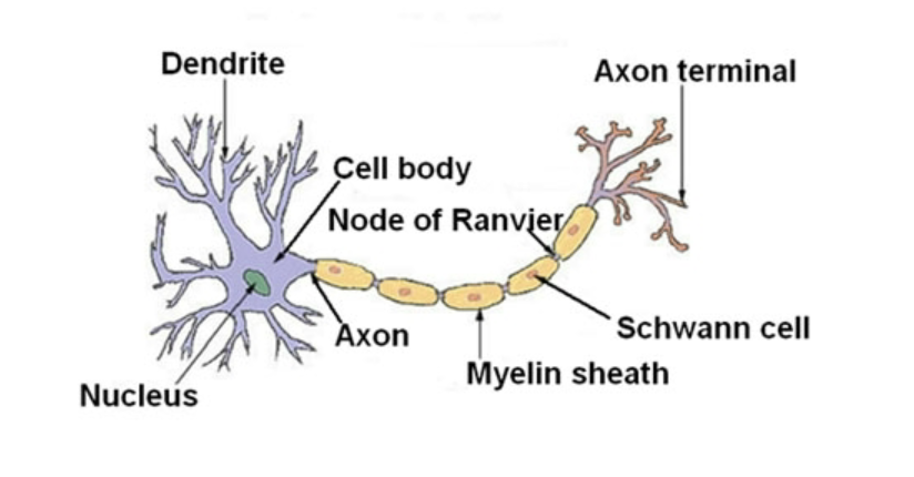
The Structure of the Neuron:
The neuron is composed of two branches that extend from the call body; those branches include the following.
Axon
Dendrites
Axons
Axons are part of the neuron that carries information away from the cell body and toward other cells.
There is only one axon per cell, and they are typically long, but it is not unusual for them to be extremely long.
There isn’t as much branching compared to dendrites.
Axons are also myelinated.
Dendrites
Dendrites are part of the neuron that receives information and brings the received data to the cell body.
Dendrites are typically short.
Typically have numerous branches as well as dendrite spines which allow for more surface area to receive data from other neurons.
Has no myelination.
Factors That Affect the Speed of Action Potential
The diameter of an axon:
Neurons that are larger in diameter fire faster.
Myelin sheath/myelination:
The Myelin sheath increases firing.
Myelin sheath:
A layer of fat cells that encases and insulates many axons.
Speeds up nerve impulses.
The interaction between the axon and the myelination/myelin sheath:
Nodes of Ranvier
Gaps of space between the myelin (bare portions.)
Saltatory Conduction
The action potential sequence jumps from node to node and speeds up AP.
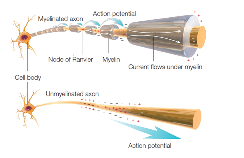
Due to the myelination of neurons, action potentials may only occur at the nodes of Ranvier.
Depolarization cannot occur in myelinated regions.
Depolarization can only occur at the nodes of Ranvier.
Saltatory conduction
Is when action potential jumps from node to node traveling down an axon.
It serves as a means of increasing the rate of propagation of an action potential.
It increases the speed of impulse transmission.
Conserves energy for the axon as depolarization only occurs at the node.
This causes 100x less movement of ions and allows the re-establishment of Na+ and K+ concentration across the membranes.
Multiple Sclerosis: Myelin Sheath Degenerates
The degeneration of the myelin sheath.
Causes weakness of limbs and difficulty walking. Standing, fatigue as well as tingling pain.
Classified as an autoimmune disease.
It is more common to occur in women than in men.
Reflex Arc Neurons
Sensory/Afferent:
Bring sensory information into the spinal cord.
Motor/Efferent:
Carry motor commands away from the spinal cord.
Interneuron/Association:
Neurons between the sensory and motor neurons.
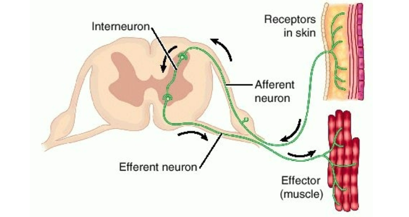
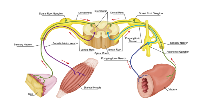
Example of the reflex arc in action:
Sensory information is carried into the spinal cord via sensory neurons.
The sensory information then travels across the interneurons to the motor neurons.
The motor neurons carry the sensory information out to pull a hand away.
Reflex Arc:
An involuntary response that occurs at the level of the spinal cord
A reflex is pulling a hand away without the need of your brain, like when touching something scolding hot.
Electrochemical activity terms:
Graded potential (Dendrites)
Graded:
The more intense the stimulation, the more electrochemical activity (more Na+) moves in.
Decremental:
As the electrochemical activity moves down the dendrite, less Na+ moves in (decreases).
Action Potential & Activity in the Axon
Action Potential (Axon) Nerve Impulse
All-or-None Effect:
Once the electrochemical activity reaches the threshold at the axon hillock, the axon fires with the same intensity.
Non-decremental:
The amount of electrochemical activity (Na+ moving in) does not decrease as it moves down the axon.

The Synapse
The Synapse
A gap/space between the neurons.
A functional contact between neurons.
Most neurons do not touch each other.
Neurotransmitters carry a message across a gap.
Parts of the Synapse
Pre-synaptic neuron
A neuron, before the synapse sends a message(s).
Post-synaptic neuron
Neurons after the synapse receive a message(s).
Telodendria/axonal branching/terminal branches
The branching at the end of the axon.
Synaptic knob/ Synaptic button/ End bulb/ Terminal bouton
The enlarged area at the end of the branching.
Pre-synaptic membrane
Membrane before the synapse.
Post-synaptic membrane
The membrane after the synapse (Has receptor sites.)
Synaptic vesicles
Fluid-filled structures (Hold and release transmitters .)
Steps at the Synapse
Action potential propagates down the axon (domino effect.)
Vesicles merge with the presynaptic membrane.
Released neurotransmitters into the cleft (vesicles don’t move across.
Transmitters travel; across the cleft & plug into the receptor sites.
Excite or inhibit the next neuron.
Inactivation & Deactivation of transmitters
There are two ways to deactivate a transmitter after it has plugged into receptor sites on the postsynaptic membrane and excited or inhibited the next neuron, which is:
Reuptake into the presynaptic knob.
Enzymes.
Antidepressants & Reuptake
Depression can occur for a multitude of reasons; one reason is too little serotonin; treatment for this is SSTIs or antidepressants.
Antidepressants selectively block the reuptake of serotonin, forcing it to stay out in the cleft longer.
Neurotransmitters
A chemical substance transmits information across the synaptic gap to the next neuron.
Stored in the synaptic vesicles.
Neurotransmitters
Dopamine
Pleasure transmitter: all drugs that are pleasurable increase DA in addition to other transmitters
Cocaine & Amphetamines: directly increase DA.
Schizophrenia: too much DA.
Antipsychotic drugs: block DA.
Parkinson’s: too little DA, Treatment: L-Dopa.
L-Dopa:precursor to DA. Gets converted to DA.
Tourette’s: imbalance in DA.
Serotonin
Involved in many disorders, including depression.
SSRIs: Increase SE.
Sleep.
Tryptophan is a precursor to SE: Efficacy of
tryptophan as sleep aid gets overstated.
Endorphins
Endogenous opiates/opioids: similar to heroin,
morphine, opium, etc.Pleasure transmitter: Such as a runner’s high.
Analgesic: pain relief.
Acetylcholine (ACh)
Memory: Alzheimer’s Acetylcholine pathways
are degenerate.The transmitter between the motor neurons & muscle.
Drugs that block or interfere with ACh can cause paralysis and even death.
Examples: Curare, Botulism
Botox is botulism: medical & cosmetic uses.
Norepinephrine
Similar to adrenaline.
Sympathetic nervous system.
Connected with mood disorders/depression.
Treatment: Antidepressants; some increase NE.
Gaba
Main inhibitory transmitter.
Depressant drugs such as tranquilizers,
barbiturates, and alcohol increase GABA.Withdrawal from these drugs can cause seizures.