


Cell Biology (IB)
Cell Biology:
1.1 Introduction to Cells
Cell theory:
The cell theory was proposed by Mathias Schleiden, Theodore Schwaan, and Rudolf Virchow.
All living organisms are composed of cells and its products.
The cell is the basic structural, and functional unit of all living organisms.
All cells arise from pre-existing cells that is they cannot be created from non-living material.
Functions of life:
A cell carries out many different functions of life which includes:
Metabolism:
It is the sum of all the chemical reactions in the body, including both anabolic (building) and catabolic (breakdown) processes.
Catabolism: These are the energy-releasing processes that break down complex molecules into simpler ones.
Anabolism: These are the energy-requiring processes that bild complex molecules from simpler ones.
It is very essential for maintaining various life functions.
It also controls and regulates the rate of chemical reactions in the body.
Response:
It is a fundamental aspect of human behaviour.
It is basically an action, behaviour, or answer that follows a particular stimulus or situation.
It can be voluntary, involuntary, or depending on whether they are under conscious control.
Homeostasis:
Homeostasis is the body's natural ability to maintain internal stability and balance.
Homeostasis is a fundamental biological process that ensures the body's internal environment remains stable despite external changes.
It involves the regulation of various physiological variables, including temperature, blood pressure, blood sugar levels, and pH, among others.
Homeostasis is essential for proper bodily function.
When it fails, it can lead to health issues or even life-threatening conditions.
Homeostasis helps the body adapt to changing external conditions, such as temperature fluctuations, exercise, or dietary changes.
Growth:
Growth is one of the essential characteristics of living organisms.
It is a fundamental biological process.
It can occur at the cellular, tissue, organ, and organismal levels.
Types of growth:
Primary Growth:
Typically seen in plants and involves the lengthening of stems and roots.
Driven by cell division in the apical meristems.
Secondary Growth:
Also in plants, it involves the thickening of stems and roots.
Occurs due to the activity of lateral meristems, like the vascular cambium.
Limited Growth:
This type of growth is seen in animals where the growth happens upto a certain age.
Unlimited Growth:
This type of growth is seen in plants where the growth happens life time.
Reproduction:
It is the biological process by which new individuals of the same species are produced.
The primary purpose of reproduction is to ensure the survival and continuation of species.
Two types of reproduction:
Sexual: It involves the fusion of gametes from two parents.
Asexual: It involves the production of offspring without the fusion of gametes.
Excretion:
It is the process of eliminating waste products and excess substances from the body.
Nutrition:
It refers to the process by which organisms obtain and utilize the nutrients and substances necessary for their growth.
Surface and Volume ratio:
As, a cell grows in size its volume increases much more rapidly than its surface area.
The surface area-to-volume ratio decreases as cell increases in size.
The surface allows the entry of oxygen. So, large cells cannot get as much oxygen as they would need to support themselves.
Smaller single-celled organisms have a high surface area to volume ratio.
The higher the surface area to volume ratio, the more effective the diffusion process can be.
Magnification:
Magnification is the process of enlarging an object or image to make it appear larger than its actual size.
Commonly used in optics and microscopy to study fine details.
Calculation of magnification:
Magnification = Image size (with ruler) ÷ Actual size (according to scale bar)
Calculation of actual size:
Actual Size = Image size (with ruler) ÷ Magnification
Emergent Properties:
These are the characteristics or behaviours that arise in complex systems but are not evident in their individual components.
Due to this multicellular organisms are capable of completing functions that unicellular organisms could not undertake.
Multicellular organisms are living beings composed of multiple cells that work together to perform various functions.
Cell → Tissue → Organ → Organ system → Organism
Cell Differentiation:
The development and maintenance of multicellular organisms,
It refers to the transformation of unspecialized or stem cells into specialized cell types with distinct functions and characteristics.
This process is critical for the growth, repair, and functioning of various tissues and organs in an organism.
Gene Packaging:
It is a fundamental process in molecular biology that involves the organization and compaction of genetic material within the nucleus of eukaryotic cells.
Packaging includes:
Euchromatin:
Active genes are usually packaged as euchromatin.
Transcriptionally active.
Heterochromatin:
Inactive genes are usually packaged as heterochromatin.
Transcriptionally inactive.
Stem cells:
It is a type of unspecialized, undifferentiated cell that has the unique ability to develop into various specialised cell types.
Charactized by:
Self-renewal: The ability to create more stem cells.
Pluripotency/ Multipotency: The ability to differentiate into specific cell types.
Types of stem cells:
Stem cells come in various types, each with its own unique characteristics and differentiation potential. Here are the main types of stem cells:
Embryonic Stem Cells (ESCs):
Derived from the inner cell mass of early-stage embryos.
Pluripotent, meaning they can differentiate into almost any cell type in the body.
Used in research and regenerative medicine due to their high differentiation potential.
Adult (Somatic) Stem Cells:
Found in various tissues and organs throughout the body.
Multipotent, meaning they can differentiate into a limited range of cell types specific to the tissue or organ where they are located.
Responsible for tissue maintenance and repair.
Examples include hematopoietic stem cells (found in bone marrow) and mesenchymal stem cells (found in various tissues).
Induced Pluripotent Stem Cells (iPSCs):
Generated by reprogramming adult somatic cells, such as skin cells or blood cells, into a pluripotent state.
Similar to embryonic stem cells in terms of pluripotency.
Offer the advantage of patient-specific stem cells for personalized medicine, disease modeling, and drug testing.
Fetal Stem Cells:
Derived from the tissues of developing fetuses, typically obtained during prenatal testing or elective abortion procedures.
Show a degree of multipotency, with potential applications in regenerative medicine and research.
Perinatal Stem Cells:
Isolated from various tissues associated with childbirth, such as the placenta, amniotic fluid, and umbilical cord.
Can differentiate into a range of cell types and are considered a valuable source of stem cells for research and therapy.
Cancer Stem Cells:
Found within tumors and possess stem cell-like properties.
Thought to play a role in cancer growth, metastasis, and resistance to treatments.
Stem Cell Therapy:
Stem cell therapy, also known as regenerative medicine that utilizes stem cells to treat various diseases, injuries, and medical conditions.
It is based on the remarkable ability of stem cells to sef-renew and differentiate into different cell types.
Stem cell therapy has shown promise in treating a variety of medical conditions and diseases.
Examples of stem cell therapy:
Bone Marrow Transplantation:
Hematopoietic stem cells from the bone marrow have been used for decades to treat blood-related disorders, such as leukemia, lymphoma, and aplastic anemia.
They can re-establish the patient's blood and immune system.
Cardiovascular Diseases:
Stem cells, such as mesenchymal stem cells, have been investigated for their potential to repair damaged heart tissue in conditions like myocardial infarction (heart attack) and heart failure.
Neurodegenerative Diseases:
Stem cell therapy is being explored for conditions like Parkinson's disease, Alzheimer's disease, and amyotrophic lateral sclerosis (ALS) to replace damaged or lost neurons.
Spinal Cord Injuries:
Researchers are investigating stem cell therapy as a means to regenerate damaged spinal cord tissue and restore function in individuals with spinal cord injuries.
Orthopedic Injuries:
Mesenchymal stem cells have been used in the treatment of orthopedic injuries, such as osteoarthritis and sports-related injuries, to promote cartilage and bone regeneration.
Autoimmune Diseases:
Mesenchymal stem cells have shown potential in modulating immune responses and have been explored as a treatment for autoimmune diseases like multiple sclerosis and systemic lupus erythematosus (SLE).
Type 1 Diabetes:
Efforts are underway to develop insulin-producing beta cells from stem cells for transplantation to treat type 1 diabetes.
Eye Diseases:
Stem cell therapy is being researched for eye conditions like age-related macular degeneration (AMD) and retinal degenerative diseases to restore vision.
Burn Injuries:
Stem cell therapy, particularly with skin stem cells, has been used to promote skin regeneration in burn victims.
Cancer Treatment:
Hematopoietic stem cell transplantation is a critical component of treatment for certain cancers, such as leukaemia and lymphoma, allowing for high-dose chemotherapy and radiation therapy.
Autoimmune Diseases:
Stem cell therapy is under investigation for autoimmune diseases, like rheumatoid arthritis and Crohn's disease, to regulate the immune response and reduce inflammation.
Cell Theory History:
Stem The cell theory is a fundamental concept in biology that describes the basic structural and functional unit of life. Its development is a significant milestone in the history of biology. Here's a brief overview in bullet points:
17th Century: Early microscopes allowed scientists to observe cells. In 1665, Robert Hooke coined the term "cell" when he examined cork and saw small, box-like structures under his microscope.
Late 17th Century: Anton van Leeuwenhoek, a Dutch scientist, used a more advanced microscope to observe single-celled microorganisms, including bacteria and protists.
19th Century: The cell theory was formulated as follows:
Matthias Schleiden:
In 1838, Schleiden proposed that all plants are composed of cells.
Theodor Schwann:
In 1839, Schwann extended the theory by suggesting that all animals are also made up of cells.
Rudolf Virchow:
In 1855, Virchow added the concept that cells arise from pre-existing cells, completing the cell theory.
Cell Theory Postulates: The cell theory comprises three main principles:
All living organisms are composed of one or more cells.
The cell is the basic structural and functional unit of life.
All cells arise from pre-existing cells through cell division.
Microscopes:
Microscopes are essential scientific instruments that enable the observation of objects and structures at a microscopic level, revealing details that are not visible to the naked eye.
Types of Microscopes: There are several types of microscopes, each designed for specific applications. Common types include:
Optical Microscopes: These use visible light to magnify and observe specimens, including bright-field, dark-field, phase-contrast, and fluorescence microscopes.
Electron Microscopes: These use focused beams of electrons to achieve higher magnification and resolution, including transmission electron microscopes (TEM) and scanning electron microscopes (SEM).
Confocal Microscopes: These use lasers and pinhole apertures to create sharp, three-dimensional images, often used in biological and medical research.
Magnification and Resolution:
Microscopes allow for both magnification and improved resolution.
Magnification increases the apparent size of an object.
Resolution refers to the ability to distinguish between closely spaced objects.
Higher magnification and resolution are essential for detailed observations.
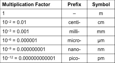
Cell Scale:
Cells are measured according to this metric:
1.2 Ultrastructure of Cells:
Organisms are divided based on cell structure:
Prokaryotes:
Primitive; Small; Simple.
No distinct nuclear compartment.
No membrane-bound organelles.
Live in variable ecological habitats.
The prokaryotes are divided into two groups:
Bacteria/ Eubacteria
Archaebacteria
Bacteria (the primitive organisms):
Bacteria are the sole members of Kingdom Monera.
Eubacteria are true bacteria characterized by the presence of a cell wall. Eg: Blue-green algae (Cyanobacteria)
Bacteria are classified based on shape into four types:
Cocci (spherical)
Bacilli (rod-shaped)
Spirillum (spiral like)
Vibrio (comma shaped)
Archaebacteria:
They are special and survive in some of the harshest habitats.
Extreme salty (halophiles), marshy areas (methanogens), and hot springs (thermoacidophiles)
Eukaryotes:
Advanced; Complex
Distinct nuclear compartment.
Membrane-bound organelles are present.
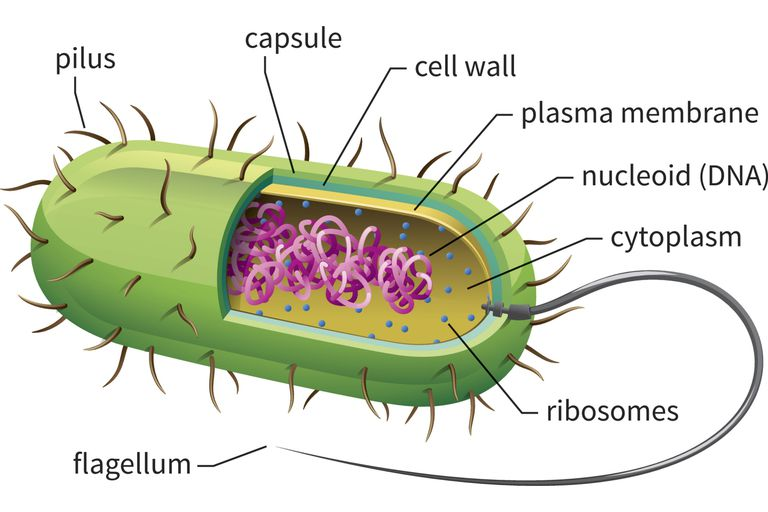
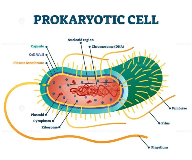
Structure and function of prokaryotic cell:
Flagella: A slender whip like structure used for locomotion
Pili and fimbrae: Attachment to substrate
Cell wall: Structural support and protection against damage
Cell membrane: Provide protection and allow movement of substance from in and out of cells.
Ribosome: Protein synthesis
Naked DNA (Nucleoid): Store genetic information and passed to daughter cell.
Structure some time present in prokaryotic cell:
Plasmid: Extra chromosomal DNA. Contain antibiotic resistance gene.
Used as vector for genetic engineering.
Capsule: Protect cell from chemical and dry environment
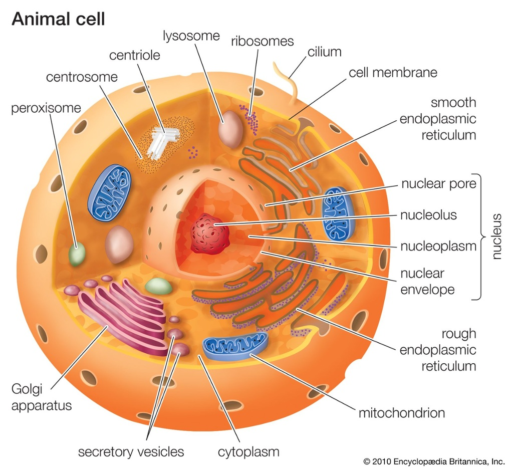
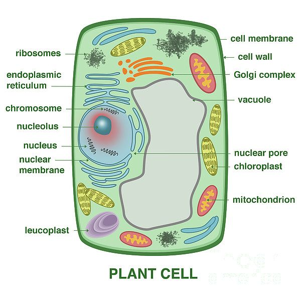
Structure and function of eukaryotic cell:
Cell membrane: Protect the cell from its surroundings and controls the exchange of substances with the outside.
Nucleus: It stores the cell's hereditary material, or DNA, and it coordinates the cell's activities, which include growth, intermediary metabolism, protein synthesis, and reproduction.
Cytoplasm: Contents of the cell located between the plasma membrane and the nuclear membrane.
Comprises a liquid medium or cytosol, comprising water and soluble substances, where the present other organelles.
Mitochondria: Called as power house of the cells. Responsible for breakdown of sugar molecules for releasing ATP (the energy currency of cells).
It also regulate cellular metabolism.
Endoplasmic reticulum: Structural frame work.
Production and processing of protein (RER)
Synthesis of carbohydrates and lipid (SER).
Ribosome: Ribosomes are a cell structure that makes protein.
Protein is needed for many cell functions such as repairing damage or directing chemical processes.
Ribosomes can be found floating within the cytoplasm or attached to the endoplasmic reticulum.
Golgi apparatus: A number of proteins synthesized by ribosomes on the endoplasmic reticulum are modified in the cisternae of the golgi apparatus before they are released from its trans face.
Golgi apparatus is the important site of formation of glycoproteins and glycolipids.
Lysosomes: Contain lytic enzyme. Site for intracellular digestion and destruction of certain organelles at the time of development.
Also called as suicidal sac.
Cytoskeleton: Mechanical support, motility, maintenance of the shape of the cell.
Centrosome and Centriole: The centrioles form the basal body of cilia, flagella, and spindle fibres that give rise to spindle apparatus during cell division in animal cells.
Chloroplast: Helps in photosynthesis. (only plant cell)
Cell wall: Maintain cell shape and avoid cell bursting (only plant cell).
Differences between plant and animal cell:
Plant Cell:
Plant cells have cell wall.
They contain chloroplast.
They donot have centriole.
Vacuole is large and present in centre of the cell.
Nucleus is present in the side of the plant cell.
Animal Cell:
Animal cells don’t hve a cell wall.
They don’t have chloroplasts.
Centriole is present in them.
Vacuole is small.
Nucleus is present in the centre of the animal cell.
Differences between prokaryotic and eukaryotic cell:
Prokaryotic cell:
Primitive cell
Generally small in size
Nucleus is absent
Chromosome is single, and circular.
Plasmid is present
Nucleolus is absent.
Membrane bound cell organelles are absent.
Pilli, and fimbrae helps in adhesion.
Eukaryotic cell:
True nucleus
Size is generally large
Nucleus is present
No plasmid
Nucleolus is present
Membrane bound organelles are present
Cillia and flagella helps in motility
1.3 Membrane Structure:
Cell Membrane:
Cell Membranes are the outermost layer in animals whereas in plants the second most layer after the cell wall.
It is the universal structure and structural cell membrane of prokaryotes that are similar to eukaryotes.
It encloses the boundaries and all the cellular interactions between cytosol and the external environment.
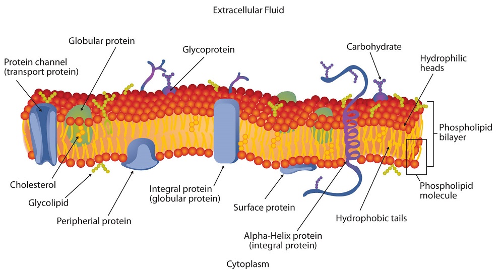
Cell Membranes are dynamic, fluid structures comprising lipid bilayers and protein molecules embedded in them.
They are held together by non-covalent interactions.
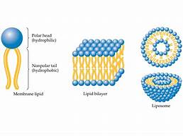
Lipid Bilayer:
The lipid bilayer comprises phosphoglycerides, sphingolipids, and sterols in the cell membrane.
All lipid molecules have a hydrophilic head and two hydrophobic tails. This makes nature to be amphillic.
Lipids are the main component of the cell membrane because it forms the continuous structural frame of the cell membrane.
The phospholipid layer provides fluidity to the plasma membrane because phospholipids are rich in unsaturated fatty acids.
The main phospholipids in most animal cell membranes are phosphoglycerides.
Glycolipids and cholesterol are also part of the lipid bilayer.
The reason behind the formation of the bilayers is their shape and amphiphilic nature.
The lipid bilayer is a two-dimensional fluid.
Liposomes (spherical vesicles) provide fluidity to the lipid bilayer.
When the lipid molecules in the plasma membrane of living cells segregate in their specialized domains called lipid rafts.
The excess lipids often get stored as fat droplets and these are often termed to be adipocytes.
As the lipid bilayer is known to be asymmetrical which is functionally very important when it comes to converting extracellular signals into intracellular ones.
Membrane Proteins:
Membrane proteins are responsible for performing most of the membrane’s specific tasks.
“A typical plasma membrane is somewhere in between, with protein accounting for about half of its mass”.
Membrane proteins are amphiphilic having both hydrophobic and hydrophilic regions.
Many of the membrane proteins also extend through the lipid bilayer called transmembrane proteins.
Other membrane proteins are located entirely in the cytosol.
There are membrane-associated proteins that do not extend into the hydrophobic interior of the lipid bilayer at all. These sorts of proteins are often referred to as peripheral membrane proteins.
Only transmembrane proteins can function on both sides of the bilayer or transport the molecules across it.
There are two types of protein that are present in the plasma membrane.
Carbohydrates:
Oligostructures of the glycolipids and glycoproteins on the outer surface of the plasma membrane are involved in cell to cell recognization mechanism.
1.4 Membrane Transport:
Transportation of molecules across lipid membrane:
The lipid layer restricts the passage of most polar molecules.
This barrier often allows the balance in concentrations of solutes in the cytosol and the external environment.
To regulate and transport materials across the membrane. It uses specialized membrane proteins.
Cells often transfer larger molecules (macromolecules), that take place with a different mechanism.
Principles of Membrane Transport:
The main classes of membrane proteins include:
Transporters: These undergo sequential conformational changes to transport the small molecules.
Channels: It forms narrow pores allowing passive transmembrane movement.
Transport through channels occurs much faster than transport mediated by transporters.
Protein-Free Lipid bilayers are impermeable to ions. The diffusion rate depends on the size of the molecule.
Transportation across a gradient:
Passive transport: All channels and many transporters allow solute to cross the membrane only passively.
Active transport: Here, it requires a certain form of energy to catalyze and
actively help the solute cross the membrane.Endocytosis: (bulk transport)
Pinocytosis: ingestion of liquid material.
Phagocytosis: ingestion of solid complex materials.
Exocytosis: egestion of waste materials from cells through the plasma membrane.
Due to the concentration gradient and the potential difference across the membrane, there exists the membrane’s potential difference which also influences the transport.
Membrane Structure:
Cell membranes are composed of a phospholipid bilayer with embedded proteins.
The hydrophobic interior of the membrane acts as a barrier to the movement of polar molecules.
Selective Permeability:
Membranes are selectively permeable, allowing certain substances to pass while restricting others.
Permeability is determined by factors such as molecular size, charge, and lipid solubility.
Passive Transport:
Passive transport occurs without the input of energy.
Diffusion is the primary mechanism of passive transport, allowing molecules to move from an area of high concentration to an area of low concentration.
Simple diffusion involves the movement of small, non-polar molecules directly through the lipid bilayer.
Facilitated diffusion relies on carrier proteins or channel proteins to facilitate the movement of specific molecules across the membrane.
Active Transport:
Active transport requires the expenditure of energy (usually ATP) to move molecules against their concentration gradient.
Primary active transport involves the direct use of ATP to pump molecules across the membrane, often against a concentration gradient.
Secondary active transport utilizes the energy stored in an ion gradient to transport molecules against their gradient.
Endocytosis and Exocytosis:
Endocytosis is the process by which cells engulf substances from the extracellular environment by forming vesicles.
Exocytosis is the reverse process, where vesicles fuse with the membrane, releasing their contents into the extracellular space.
Membrane Potential:
Membrane potential refers to the voltage difference across the cell membrane, usually maintained by ion concentration gradients.
It is crucial for various cellular functions, such as nerve impulse transmission and muscle contraction.
Transporters:
Transporters are integral membrane proteins that facilitate the movement of specific molecules across the membrane.
They can operate through active or passive mechanisms, depending on the energy requirement and directionality of transport.
Specialized Transport Processes:
Some specialized transport processes include symport, antiport, and uniport.
Symport involves the transport of two different molecules in the same direction.
Antiport involves the transport of two different molecules in opposite directions.
Uniport refers to the transport of a single molecule or ion.
Channels and Electrical Properties of Membranes
Ion Channels
Ion channels are specialized membrane proteins that allow the selective passage of ions across the cell membrane.
They play a crucial role in maintaining the electrical properties of membranes.
Types of Ion Channels:
Voltage-gated ion channels: These channels open or close in response to changes in the membrane potential.
Ligand-gated ion channels: These channels open or close in response to the binding of specific molecules (ligands) to the channel.
Mechanically-gated ion channels: These channels open or close in response to mechanical stimuli such as pressure or stretch.
Leak channels: These channels are always open, allowing a small and constant flow of ions across the membrane.
Ion Selectivity:
Ion channels exhibit selectivity for specific ions based on their size, charge, and hydration.
For example, potassium channels are highly selective for potassium ions, while sodium channels preferentially allow the passage of sodium ions.
Conductance and Permeability:
Conductance refers to the ability of an ion channel to allow the flow of ions.
Permeability is the measure of the ease with which ions can pass through a channel.
Channels with high conductance and permeability facilitate the movement of ions more effectively.
Resting Membrane Potential:
Resting membrane potential is the electrical potential difference across the cell membrane when the cell is at rest.
In most cells, the resting membrane potential is around -70 millivolts (mV).
It is primarily determined by the differential distribution of ions (such as potassium, sodium, and chloride) across the membrane.
Action Potentials:
Action potentials are rapid and transient changes in the membrane potential that allow for long-range communication in excitable cells such as neurons and muscle cells.
They are initiated by a depolarization event that reaches a threshold, leading to the opening of voltage-gated ion channels.
Depolarization and Repolarization:
Depolarization refers to a change in the membrane potential towards a more positive value, typically caused by the influx of positively charged ions.
Repolarization is the process of returning the membrane potential back to its resting state after depolarization.
This is achieved by the efflux of positively charged ions or the influx of negatively charged ions.
Refractory Period:
After an action potential, there is a refractory period during which the membrane is temporarily unresponsive to further depolarization stimuli.
The refractory period ensures the proper propagation of action potentials and prevents overlapping signals.
Saltatory Conduction:
In myelinated neurons, action potentials jump from one node of Ranvier to the next, a process known as saltatory conduction.
This increases the speed and efficiency of electrical signal transmission along the axon.
Synaptic Transmission:
Synaptic transmission involves the release of neurotransmitters from the presynaptic neuron, which then bind to receptors on the postsynaptic neuron.
This binding can result in the opening or closing of ion channels, leading to changes in the postsynaptic membrane potential.
1.5 The Origin of Cells:
Abiogenesis:
It is also known as spontaneous generation.
It is the scientific theory that proposes the natural, non-biological origin of life from simple organic compounds.
It suggests that life could emerge from inanimate matter under specific conditions, without the need for pre-existing life.
Historical Context:
Abiogenesis was a prevailing belief for centuries, with the notion that life could spontaneously arise from decaying matter or non-living substances.
This idea was challenged by the work of Louis Pasteur in the mid-19th century, who demonstrated through experiments that life only arises from pre-existing life (biogenesis).
Oparin-Haldane Hypothesis:
The modern scientific concept of abiogenesis gained prominence in the early 20th century with the work of Alexander Oparin and J.B.S. Haldane.
They proposed that life could have originated on Earth from simple organic molecules in a reducing atmosphere rich in methane, ammonia, and water vapor.
Miller-Urey Experiment:
In 1953, Stanley Miller and Harold Urey conducted a famous laboratory experiment that simulated the conditions proposed by Oparin and Haldane.
They produced amino acids, the building blocks of proteins, from simple inorganic compounds, providing experimental support for the idea that the building blocks of life could form abiotically.
RNA World Hypothesis:
Another hypothesis related to abiogenesis is the "RNA world."
It suggests that self-replicating ribonucleic acid (RNA) molecules could have preceded DNA as the genetic material.
RNA can both store genetic information and catalyze chemical reactions, making it a potential candidate for the first self-replicating molecule.
Hydrothermal Vent Hypothesis:
Some researchers propose that life could have originated in the high-temperature, high-pressure environment near hydrothermal vents on the ocean floor.
These conditions may have provided the necessary energy and chemical gradients for life to emerge.
Biogenesis:
It states that living organisms can only arise from pre-existing living orgranisms.
Louis Pasteur’s experiments provided strong evidence for biogenesis.
The acceptance of biogenesis was a significant factor in the development of cell theory.
It remains as a very fundamental concept in both modern biology and microbiology.
1.6 Cell Division:
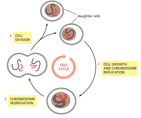
Overview of the Cell Cycle:
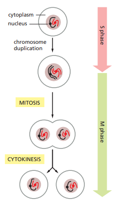
The cell cycle is a highly regulated process that ensures proper cell growth, DNA replication, and cell division, leading to the production of two daughter cells with identical genetic material.
The cell cycle is the series of events that occur in a cell leading to its division and the production of two daughter cells.
It consists of distinct phases, including interphase and mitotic phase.
Interphase
Interphase is the longest phase of the cell cycle, where the cell grows, carries out its normal functions, and replicates its DNA.
Interphase is further divided into three stages: G1 (Gap 1), S (Synthesis), and G2 (Gap 2).
G1 phase is characterized by cell growth, protein synthesis, and preparation for DNA replication.
S phase involves DNA replication, where the genetic material is duplicated, resulting in two identical copies of the genome.
G2 phase is a period of growth and preparation for cell division, during which the cell checks the accuracy of DNA replication and repairs any errors.
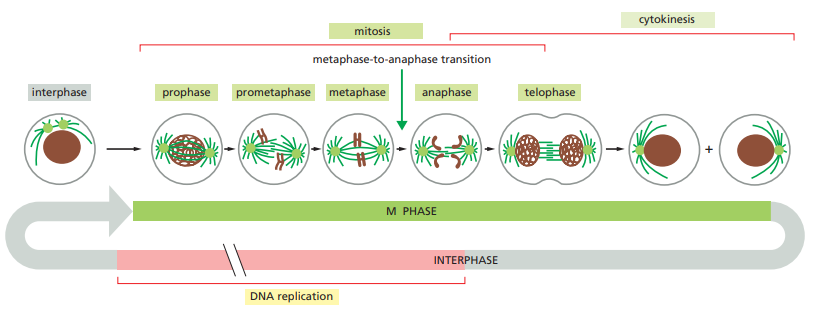
Mitotic Phase
The mitotic phase (M phase) is the shortest phase and involves the actual division of the cell into two daughter cells.
The M phase is subdivided into four stages: prophase, metaphase, anaphase, and telophase.
During prophase, the chromatin condenses into visible chromosomes, the nuclear envelope breaks down, and the mitotic spindle begins to form.
In metaphase, the chromosomes align at the equator of the cell, facilitated by microtubules attached to the kinetochores.
Anaphase is characterized by the separation of sister chromatids, which are pulled to opposite poles of the cell by the shortening of microtubules.
Telophase marks the end of mitosis, where the nuclear envelope reforms around the separated chromosomes, and the cell prepares for cytokinesis.
Cytokinesis
Cytokinesis is the final stage of the cell cycle, where the cytoplasm divides, and two daughter cells are formed.
Cytokinesis differs in animal and plant cells, with animal cells undergoing cell cleavage through the formation of a contractile ring, while plant cells form a cell plate to divide the cytoplasm.
The completion of cytokinesis marks the end of one cell cycle and the beginning of the next, resulting in the formation of two genetically identical daughter cells.
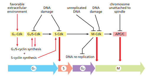
Cell-Cycle Control System
The cell-cycle control system is a complex network of molecular checkpoints and regulatory mechanisms that ensure the orderly progression of the cell cycle.
It functions to monitor and regulate the timing and coordination of cell-cycle events, including DNA replication, chromosome segregation, and cell division.
The cell-cycle control system consists of various proteins, enzymes, and signaling pathways that interact to regulate the cell cycle.
Key components of the control system include cyclins, cyclin-dependent kinases (CDKs), and checkpoints.
Cyclins are proteins that fluctuate in concentration during the cell cycle and bind to specific CDKs to activate them.
CDKs are enzymes that regulate the progression of the cell cycle by phosphorylating target proteins involved in cell-cycle events.
Checkpoints are critical control points that monitor the integrity of DNA, ensure proper replication and repair, and verify accurate chromosome alignment.
Checkpoints include the G1 checkpoint (restriction point), G2 checkpoint, and spindle assembly checkpoint (SAC) during mitosis.
At each checkpoint, the cell-cycle control system assesses the conditions and signals from the environment and other cellular processes before deciding whether to proceed or halt the cell cycle.
If the cell-cycle control system detects DNA damage, replication errors, or other abnormalities, it can activate DNA repair mechanisms or induce cell-cycle arrest to allow time for repair or prevent the transmission of damaged DNA.
Failure of the cell-cycle control system can lead to uncontrolled cell growth, genomic instability, and the development of cancer.
Abnormalities or mutations in cell-cycle control genes, such as tumor suppressor genes (e.g., p53) or oncogenes (e.g., cyclins, CDKs), can disrupt the normal functioning of the control system.
Studying the cell-cycle control system provides insights into the mechanisms underlying cell division, DNA replication, and cell-cycle regulation, with implications for understanding diseases like cancer and potential targets for therapeutic interventions.
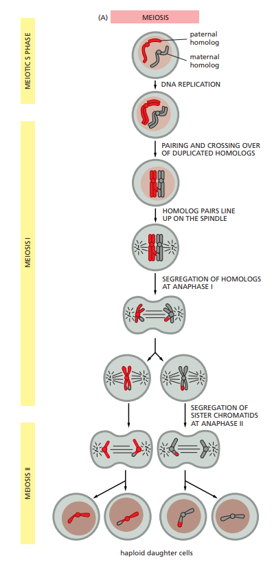
Meiosis
Meiosis is a specialized form of cell division that occurs in sexually reproducing organisms.
It involves the division of a diploid (2n) cell into four haploid (n) daughter cells, resulting in the production of gametes (sperm and eggs).
Meiosis consists of two successive divisions, known as meiosis I and meiosis II.
Meiosis I is the reduction division, where homologous chromosomes pair up and exchange genetic material through a process called crossing over.
Crossing over leads to genetic recombination and promotes genetic diversity among offspring.
During meiosis I, homologous chromosomes separate, resulting in the formation of two haploid cells with duplicated chromosomes (consisting of sister chromatids).
Meiosis II is similar to mitosis, where the sister chromatids of each chromosome separate, resulting in the formation of four haploid daughter cells.
The four daughter cells produced at the end of meiosis are genetically distinct due to the independent assortment of chromosomes during meiosis I and the random segregation of sister chromatids during meiosis II.
Meiosis I
Meiosis I is the first division of meiosis and consists of several distinct steps, including prophase I, metaphase I, anaphase I, and telophase I.
Prophase I is the longest phase of meiosis I and can be further divided into five sub-stages: leptotene, zygotene, pachytene, diplotene, and diakinesis.
During leptotene, the chromosomes condense, becoming visible as thread-like structures, and the nuclear envelope starts to break down.
In zygotene, homologous chromosomes pair up and form structures called synaptonemal complexes. This pairing is called synapsis.
Pachytene is characterized by the crossing over of genetic material between non-sister chromatids of homologous chromosomes.
This genetic exchange increases genetic diversity.
Diplotene is marked by the separation of homologous chromosomes but with the physical connection points known as chiasmata still present.
This is when the synaptonemal complexes dissolve partially.
During diakinesis, the chromosomes further condense, and the nuclear envelope completely disintegrates. The spindle apparatus starts to form.
Metaphase I is the stage where homologous chromosome pairs line up along the equator of the cell called the metaphase plate.
The orientation of the chromosomes is random, which contributes to genetic diversity through a process called independent assortment.
Anaphase I follows metaphase I, and it involves the separation of homologous chromosomes, with one member of each pair moving towards opposite poles of the cell.
Telophase I marks the end of meiosis I, where the chromosomes arrive at the poles and start decondensing.
Nuclear envelopes may form around each group of chromosomes, and cytokinesis typically occurs, resulting in the formation of two haploid daughter cells.
Meiosis II
Meiosis II is the second division of meiosis and follows meiosis I. It is similar to a mitotic division, consisting of four stages: prophase II, metaphase II, anaphase II, and telophase II.
Prophase II initiates with the condensation of the chromosomes, and the nuclear envelope disintegrates.
The centrosomes, containing the centrioles, duplicate and move towards opposite poles of the cell, forming the spindle apparatus.
Metaphase II is the stage where individual chromosomes line up along the equator of the cell, called the metaphase plate.
Unlike meiosis I, the chromosomes align individually in metaphase II, without homologous pairs.
Anaphase II follows metaphase II and is characterized by the separation of sister chromatids.
The connections between the sister chromatids break, and each chromatid is pulled towards opposite poles of the cell.
Telophase II marks the end of meiosis II, where the chromosomes arrive at the poles and start decondensing.
Nuclear envelopes may form around each set of chromosomes, and the spindle apparatus disintegrates.
Cytokinesis typically occurs, resulting in the division of the cytoplasm, and the formation of four haploid daughter cells.
Each daughter cell produced at the end of meiosis II contains a single set of chromosomes, making them genetically distinct from one another and the parent cell.
Meiosis II completes the process of meiosis, resulting in the production of four haploid daughter cells from the original diploid (2n) parent cell.
Meiotic phases