Chapter 4: Sources of Biological Evidence
4.1: Bodily Fluids
Bodily fluids and their stains are useful biological evidence for forensic, serological, and DNA analysis and may be useful in solving crimes.
The most common bodily fluids in the forensic analysis are blood, seminal fluid, and saliva.
Blood Evidence: One of the most common types of biological evidence that is found at crime scenes.
Plasma: The fluid portion of blood; a subcompartment of extracellular fluid, which is the bodily fluid outside cells.
The cellular portion of the blood consists of:
Erythrocytes
Leucocytes
Platelets
Because mature human erythrocytes and platelets do not have nuclei, they are not useful sources of nuclear DNA.
The nuclear DNA in blood samples is primarily isolated from leucocytes, which are nucleated.
Menstrual blood can be analyzed to investigate the possibility of the occurrence of sexual assault.
Transcellular fluids: These fluids are considered to be a subcompartment of the extracellular fluid that is contained within epithelial-lined extracellular spaces.
Extracellular Nucleic Acids
Blood plasma and other various bodily fluids usually contain small amounts of nucleic acids known as extracellular nucleic acids.
Circulating Nucleic Acids: The nucleic acids circulating in plasma
Cell-Free Nucleic Acids: The nucleic acids that are found in other body fluids, such as saliva and urine.
Extracellular Vesicles (EVs): The potential sources of extracellular nucleic acids.
EVs are endogenous vesicular structures, containing proteins and nucleic acids that are secreted by most eukaryotic cells.
Exosomes are one potential source of extracellular nucleic acids.
Exosomes are derived from multivesicular bodies (MVBs), which are intracellular organelles of the endocytic pathway.
Microvesicles: Shed from the plasma membrane and thus carry along the membrane and cytosolic materials including nucleic acids.
Apoptotic bodies: These are a special type of microvesicle that is formed in apoptotic cells.
4.2: Cells
Cell Surface Markers
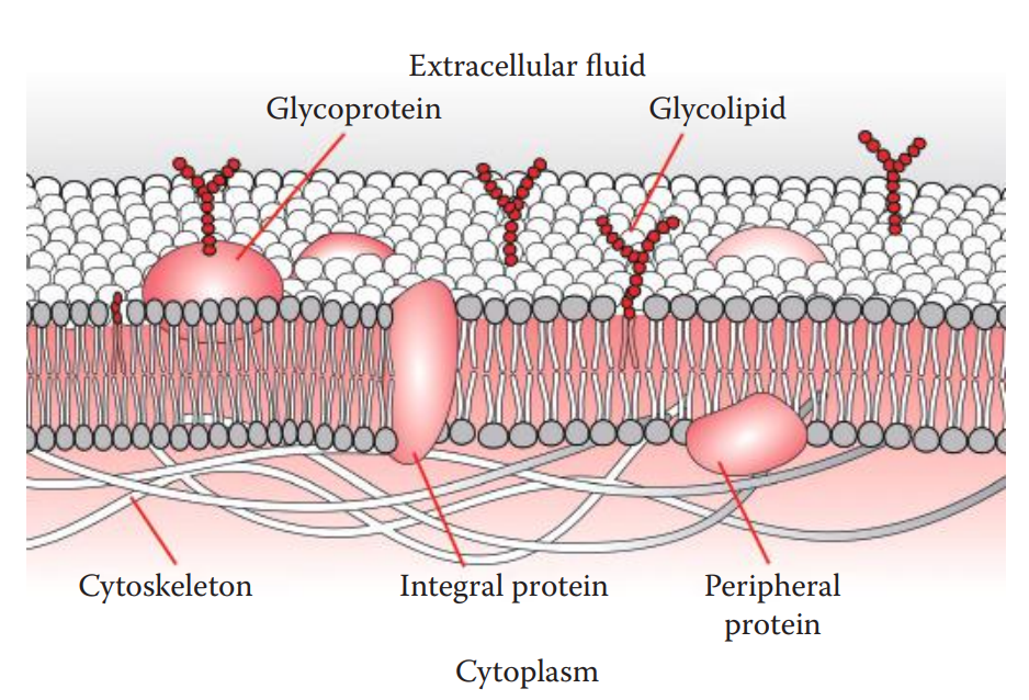
All cells have membranes, also known as plasma membranes, which constitute their outer boundaries.
Cell Membranes: A phospholipid bilayer containing lipids, proteins, and carbohydrates.
Membrane proteins: Act enzymes, receptors, or ion channels.
Nucleated Cells
Nucleus: It is surrounded by a nuclear envelope and contains chromosomes and a nucleolus.
Nucleolus: A dense, non-membrane-bound structure due to its high RNA content.
Its function is to transcribe ribosomal RNA and form ribosomes.
There are two types of cells in the body:
Sex cells: Sperms and oocytes.
Somatic cells: All other types of cells.
Spermatozoa and ova, which are formed by germ cells, are called gametes.
In humans, each gamete is haploid, containing 22 autosomes plus one sex chromosome.
In ova, the sex chromosome is always an X, while in spermatozoa it may be an X or a Y.
Most somatic cells are diploid.
It means that they have two copies of each autosome plus two sex chromosomes, XX for females or XY for males.
This results in a total of 46 chromosomes per diploid cell.
Homologous Chromosomes: The two chromosomes of a pair in a diploid cell.
Some differentiated cells such as red blood cells and platelets have no nuclei and are designated nulliploid.
Polyploid: Cells that have more than two sets of chromosomes as a result of DNA replication without cell division.
The nuclear chromosomes of humans consist of complexes of DNA, histone proteins, and nonhistone chromosomal proteins.
Each chromosome consists of one linear, double-stranded DNA molecule.
The large amounts of DNA present in the human chromosome are compacted by their association with histones into nucleosomes and even further compacted by higher levels of folding of the nucleosomes into chromatin fibers.
Each chromosome contains a large number of looped domains of chromatin fibers attached to a protein scaffold.
During the metaphase of mitosis and meiosis of the cell cycle, chromatin is the most condensed.
Euchromatin regions: These are areas of chromosomes that undergo normal chromosome condensation and decondensation during the cell cycle.
Heterochromatin: Comprises the chromosomal regions that usually remain condensed throughout the cell cycle.
Centromeres: These are the DNA sequences that are found near the points of attachment of mitotic or meiotic spindle fibers.
Telomeres: The ends of the chromosome; they help stabilize the chromosome and play a role in the replication of DNA in the chromosome.
Cytogenetic Mapping: Each chromosome arm is divided into regions based on the cytogenic bands.
The cytogenetic bands are labeled p1, p2, p3, q1, q2, q3, and so on, counting from the centromere out toward the telomeres.
Chromosomes can be identified on the basis of the size and the positions of the centromeres and cytogenetic banding patterns.
Karyotype: The chromosome constitution.
Karyogram: Includes a total number of chromosomes and the sex chromosome composition.
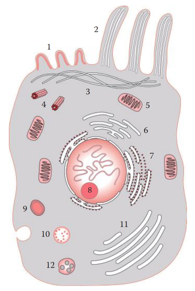
Mitochondria and Other Organelles
The cytoplasm contains the cytosol fluid in which organelles are suspended.
Multiple copies of mitochondria are located within the cytoplasm.
Mitochondria are surrounded by phospholipid membranes that separate them from the cytosol.
The mitochondria are responsible for energy production through aerobic metabolism by producing molecules containing high-energy bonds, such as adenosine triphosphate (ATP).
Two types of endoplasmic reticulum (ER) can exist within the cytoplasm:
Smooth ER: It is involved in lipid synthesis.
Rough ER: It contains ribosomes on its outer surface and forms transport vesicles.
Golgi apparatus: It is responsible for the production of secretory vesicles and new membrane components, and also for the packaging of lysosomes.
Lysosomes: Vesicles containing digestive enzymes for the degradation of injured cells).
Peroxisomes: Carry enzymes that neutralize potentially harmful free radicals.
Cytosol
Messenger RNAs
The genetic code is read as an array of triplet codes, a sequence of three bases that specifies the identity of a single amino acid.
As gene expression is activated, transcription occurs in which precursor mRNA is produced from a DNA template. After transcription, the pre-mRNA is capped, polyadenylated, and spliced to form matured mRNA. Only the matured mRNA is transported from the nucleus to the cytoplasm.
Tissue-specific mRNA can be potentially used for the identification of biological evidence.
The proteins are synthesized in a process known as translation, in which amino acids are assembled based on the codons derived from the triplet code of the DNA contained in the sequence of the mRNA strand.
MicroRNAs
MicroRNAs (miRNAs) are short RNA molecules that are 21–23 nucleotides in length.
In eukaryotic organisms, miRNAs function as negative regulators of gene expression.
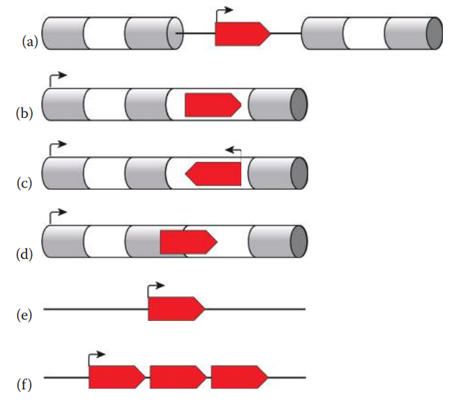
Three Types of MicroRNAs:
Intergenic miRNA genes are distinct transcriptional units that are found in genomic regions.
Intronic miRNAs reside within the introns of protein-coding and noncoding genes.
Exonic miRNAs are rare in eukaryotic genomes and reside in genomic regions that overlap with an exon and an intron of a pseudogene, which is a noncoding gene or is no longer transcribed.
A monocistronic miRNA has a single transcriptional unit with its own promoter.
In polycistronic miRNAs, several miRNAs reside as a cluster of transcriptional units with a shared promoter.
The biogenesis of miRNAs begins in the nucleus.
miRNAs are transcribed by RNA polymerase II.
Nascent transcripts, referred to as primary transcripts (pri-miRNAs), can be several hundred to thousands of nucleotides in length.

4.3: Tissues
Skin
Biology of Skin
Fingerprints: Are ridge skin impressions that are usually collected at crime scenes, and shed skin tissue is a source of DNA for human identification.
The skin of the dorsal area of the body is usually thicker than that of the ventral area of the body.
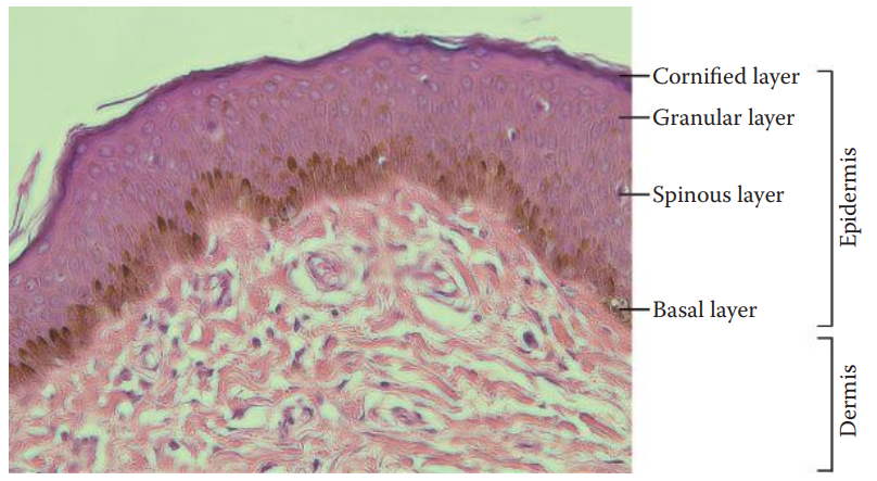
Epidermis: The outer layer of the skin. It also contains melanocytes that produce the skin pigment melanin.
The epidermis renews continually through the proliferation and differentiation of keratinocytes.
Basal Layer: Contains newly formed keratinocytes that are proliferative.
Epidermal differentiation begins with the migration of the keratinocytes from the basal layer toward the outer layer of the skin.
Once the migrating keratinocytes reach the spinous and granular layers, the keratinocytes become nonproliferating and partially differentiated.
As the cells reach the cornified layer, these cells are filled with keratin filaments and are differentiated into corneocytes, which are dead, and terminally differentiated keratinocytes.
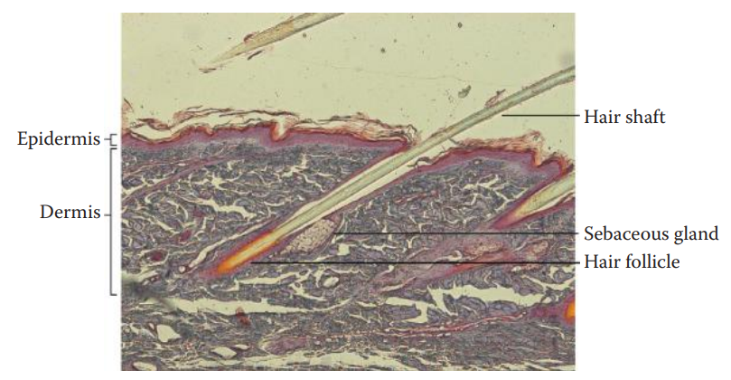
Dermis: The middle layer of the skin.
It is filled with fibrous collagen proteins secreted by fibroblasts and contains hair follicles and sweat glands.
It contains blood, lymph vessels, and nerves.
Subcutaneous layer: The deepest layer of the skin.
It consists of collagen networks and adipose tissue to prevent loss of heat.
Skin as Source of DNA Evidence
Touched Evidence: Evidence from skin contact; which can be collected and used for forensic DNA analysis. It usually contains minute quantities of nuclear DNA.
Transfer DNA: The DNA recovered from touched evidence.
Hair
Hairs, including the scalp and pubic hairs, frequently constitute biological evidence that is found at crime scenes, and their identification can be of great forensic importance.
The development of the polymerase chain reaction (PCR) amplification technique made it possible to analyze very small quantities of DNA in hair, and the use of hair as evidence of identification has become more significant.
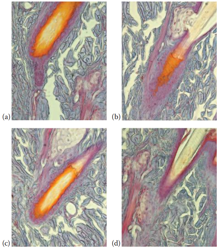
Biology of Hair
The human hair shaft is a keratinized cylindrical structure.
Medulla: The center or core of the hair.
Cortex: It surrounds the medulla; which is the outer layer of the hair shaft.
Cuticle: Consists of overlapping layers of flattened keratinized cells that protect the hair.
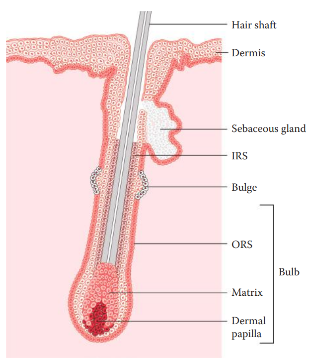
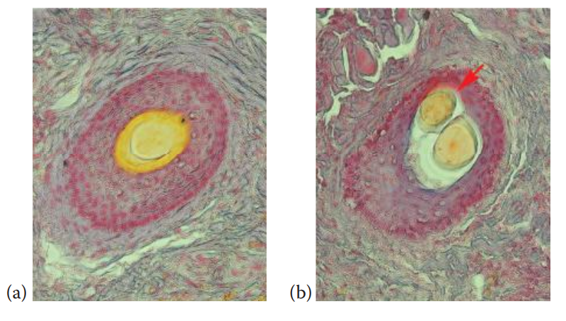
Hairs are produced in hair follicles.
Bulge: Exports the stem cells that migrate down and give rise to bulb cells.
It also produces the stem cells that migrate up to form skin cells.
Matrix Cells: These generate the hair shaft cells, which are located at the lower portion of the bulb.
Dermal Papilla: It is situated at the base of the bulb and contains cells that regulate hair growth, which is nourished by blood vessels and nerves.
The growing hair shaft is surrounded by two concentric layers of cells, which are referred to as the inner root sheath and the outer root sheath.
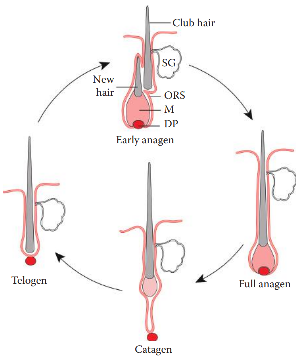
Anagen Phase: The growing phase of hair.
Catagen Phase: Occurs at the end of the anagen phase, where the matrix cells enter and undergo cell death.
Telogen Phase: Resting stage of the hair.
Hair as Source of DNA Evidence
Nuclear DNA analysis: It is usually accomplished by using freshly plucked hair roots because cells at the root region may contain nuclear DNA.
Multiple telogen hairs with roots are necessary to isolate enough nuclear DNA.
A hair follicle cell contains multiple copies of mitochondria. As a result, mtDNA can be successfully isolated from hair roots. mtDNA is embedded in the keratin matrix of hair shaft cells, which protects the mtDNA molecules from degradation.
A sequence polymorphism analysis of mtDNA from hair can be carried out. mtDNA is maternally inherited, which is useful to identify maternal relatives but cannot be used to perform paternity testing.
Heteroplasmy: The heterogeneous pool of mtDNA molecules.
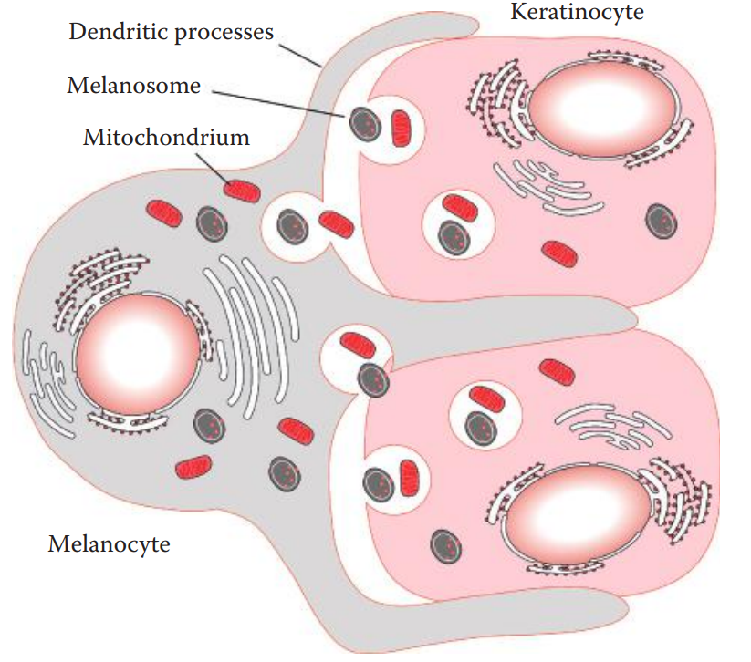
Hair follicle melanocytes are formed at the beginning of each hair cycle and die at the end of the cycle. They are located in the bulb hair follicle.
Melanosome: an organelle of melanin.
The melanosomes are transferred to neighboring keratinocytes through dendritic processes.
The melanocyte mitochondria can also be transferred to keratinocytes.
The keratinized cells in the hair shaft may carry more than one type of mitochondria, one from the keratinocytes and the other from the melanocytes.
Bones
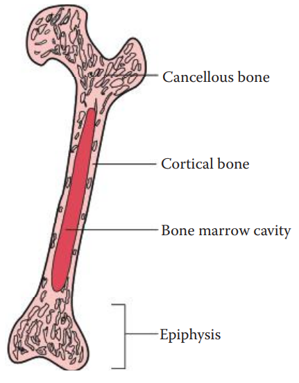
Biology of Bones
An adult human skeleton consists of 206 bones.
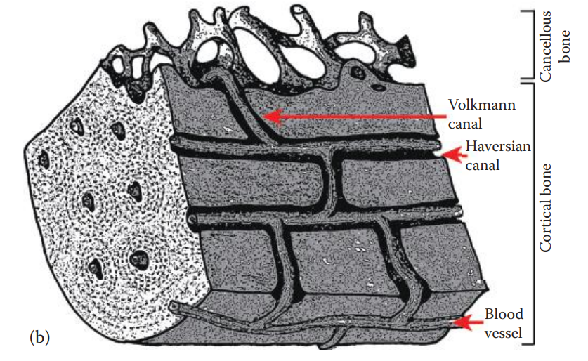
The shaft of a long bone, such as an arm or a leg bone, consists largely of an outer layer of cortical bone, which is strong and solid.
The shaft of a long bone forms a marrow cavity, which is filled with a specialized type of connective tissue called bone marrow.
Epiphysis: The portion at each end of a long bone, which is composed largely of cancellous bone and can bear the force of compression.
A flat bone can have primarily either cortical or cancellous bone.
A skull bone usually consists of largely cortical bone.
Bone, which is a connective tissue, contains a matrix and cells.
The bone matrix consists of an inorganic and an organic matrix.
Calcium and phosphorus are the major components of the inorganic bone matrix, which consists mainly of hydroxyapatite crystals.
The organic bone matrix consists of collagens, primarily type I collagen, which are insoluble fibrous proteins.
Developing bones contain small numbers of osteoprogenitor cells.
These cells can divide to produce cells that differentiate into osteoblasts.
Osteoblast cells regulate the calcification of the bone matrix.
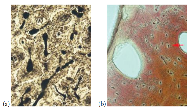
Osteoblasts that are embedded in the bone matrix are termed osteocytes and are the most abundant cells in bone.
Osteoclasts: These cells are giant cells containing 50 or more nuclei and are responsible for dissolving and recycling the bone matrix.
Bone as Source of DNA Evidence
Most DNA in cortical bone is located in the osteocytes. It has been estimated that there are approximately 20,000 osteocytes per cubic millimeter of calcified bone matrix.
A compact bone tissue should contain sufficient amounts of nuclear DNA for analysis.
The skeletal fragments recovered from burial sites are often subjected to decomposition.
During the decomposition process, both nuclear and mtDNA can be degraded.
Burial conditions with high humidity and temperature promote the degradation of DNA.
The identification of partial DNA profiles or the complete failure to obtain DNA profiles can occur after samples from decomposed remains are analyzed.
Processing bone samples for DNA extraction is a time-consuming task.
Due to the potential for commingled remains and contaminants that interfere with forensic analysis, a bone sample initially must be cleaned prior to isolating DNA.
The outer surfaces of bone fragments are usually removed by using a mechanical method such as sanding.
To obtain adequate quality and quantity of DNA from a bone sample, a high-yield DNA extraction method should be selected.
Teeth
Biology of Teeth
During embryonic development, two sets of teeth begin to form.
The first to appear are the deciduous teeth or primary teeth.
Most children have 20 deciduous teeth, which are later replaced by 32 teeth known as the secondary dentition or permanent dentition.
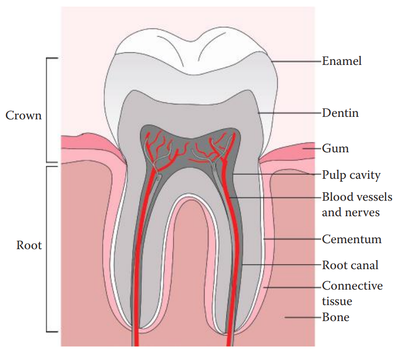
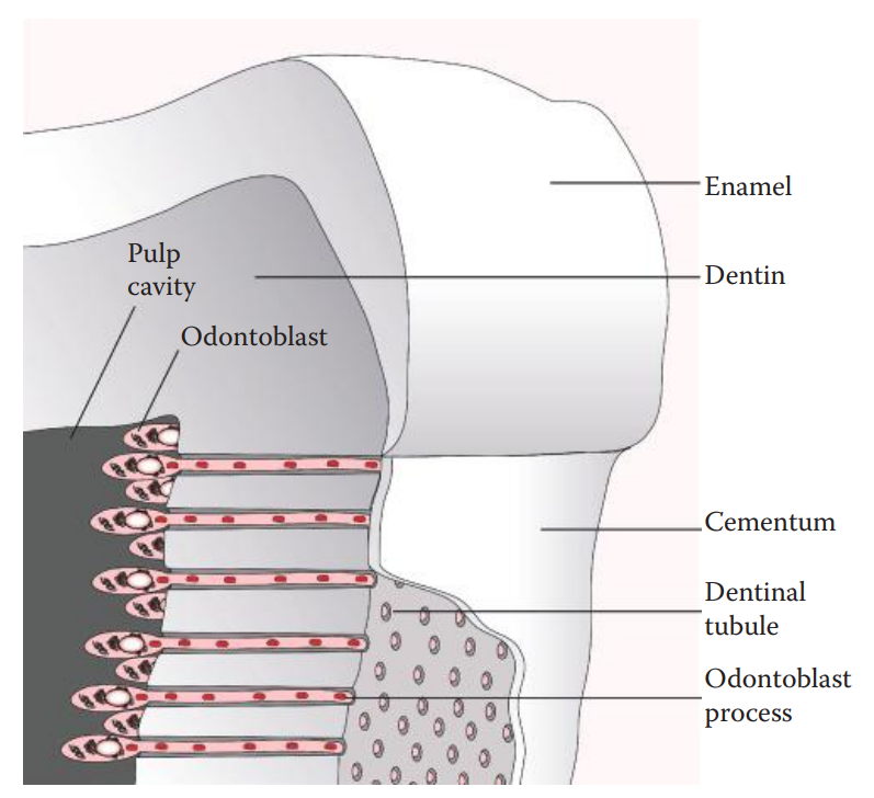
Dentin: A calcified connective tissue at the bulk of each tooth.
Enamel: A layer that covers the dentin of the crown.
Radicular Dentin: The rest of the dentin and covered by cementum.
Cementum: a layer that separates the tooth from the surrounding jawbone.
Similar to the bone, tooth tissue contains a matrix.
The inorganic matrix of the tooth contains hydroxyapatite, a calcium phosphate in a crystalline form.
The organic matrices of dentin and cementum are primarily collagens.
Amelogenin: The major protein of the organic matrix.
Pulp Cavity: The interior chamber within the tooth is surrounded by dentin.
Dental Pulp: The connective tissue made up of nerve fibers, blood vessels, and various cells.
Incisor and cuspid teeth have single roots. Bicuspids have one or two roots. Molars typically have three or more roots.
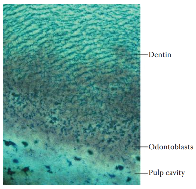
The columnar cell bodies of odontoblasts are located along the peripheral dental pulp.
A single odontoblast process, arising from each cell body of the odontoblasts, projects into the dentinal tubule.
Odontoblasts secrete collagens and ground substances that are the components of the dentinal matrix.
Cementoblasts: Cells that play roles in forming the cementum. They secrete collagens and ground substances to form the extracellular matrix of the cementum.
Cementocytes: The cementum where cementoblasts are embedded.
Ameloblasts: Cells that play a role in producing enamel and are subsequently lost during tooth eruption.
Teeth as Source of DNA Evidence
The use of dental records such as x-rays and dental casts can allow dental remains to be connected to a victim.
When no antemortem dental record is available for comparison, forensic DNA testing can be carried out for postmortem human identification.
The mineralized dental structure protects DNA from degradation in cases where it may be degraded in other tissues of the body.
Dental pulp tissue contains various cells and is a suitable source of DNA.
When tooth evidence has been exposed to high temperatures and humid environments, decomposition of the pulp tissue can occur.
Cementoblasts within the cementum, containing both nuclei and mitochondria, can then be utilized as a source of DNA.
Vertical Section: A cut along the longitudinal axis of the tooth, which allows the dissection of the pulp, dentin, and cementum tissues.
Horizontal Section: A cut through the cementum–enamel junction of the tooth.
The root portion can be pulverized to a fine powder for DNA isolation.
The crown can be preserved for forensic odontological comparisons if needed.
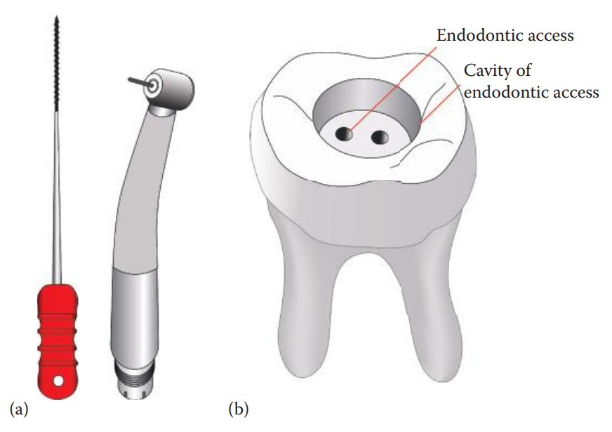
The extraction of pulp tissue can be carried out by standard endodontic access.
For calcified tissues such as dentin and cementum, a decalcification step is needed to soften the dental matrix, which facilitates DNA isolation.