


Chapter 6 - Animal Nutrition : Digestion
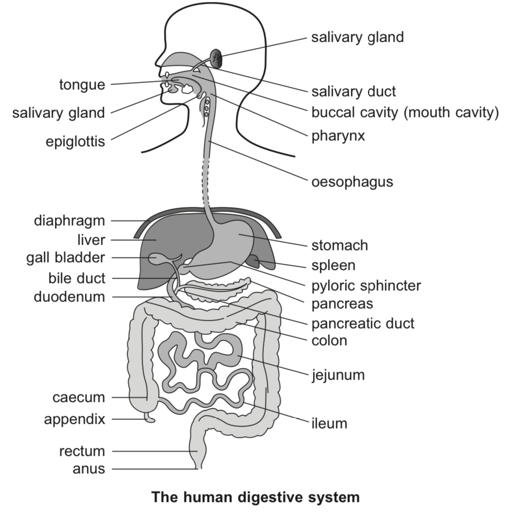
Human digestion takes place in the mouth, stomach and small intestine.
The alimentary canal consists of the mouth, the oesophagus, the stomach, the small and large intestines and the anus.
Other organs associated with digestion include the liver, pancreas, gall bladder and salivary glands.
The mouth
Food enters the body through the mouth, or buccal cavity. Physical and chemical digestion takes place in the mouth. In the mouth:
(a) Teeth start to break the food into smaller pieces. This makes food easier to swallow and also increases the surface area to volume ratio of the food for the digestive enzymes to work on more efficiently.
(b)Salivary glands secrete saliva which moistens the food and makes it easier to swallow. Saliva also contains salivary amylase, an enzyme which breaks down starch into maltose. The optimum pH of salivary amylase is 7.
(c) The tongue rolls the food into a bolus, which is then swallowed.
The oesophagus
The food passes through the pharynx and enters the oesophagus. The oesophagus is a muscular tube that leads to the stomach.
It is made up of two layers of smooth muscle. The external layer is the longitudinal muscle and the inner layer is the circular muscle. These muscles found along much of the entire length of the alimentary canal.
These muscles contract and relax alternately to cause wave-like contractions known as peristalsis.
Food moves along the oesophagus due to peristalsis.
Digestion of starch by salivary amylase continues in the oesophagus.
The stomach
The food reaches the stomach, which is a muscular bag with elastic walls.
The stomach walls form deep pits that contain gastric glands. These glands secrete mucus which protects the stomach walls. They also secrete gastric acid and pepsinogen.
Peristalsis in the stomach churns the food to break the food up and mix it thoroughly with gastric juice.
Gastric acid is hydrochloric acid with pH 2. Gastric acid (a) stops the activity of salivary amylase by denaturing it, (b) changes the inactive form of pepsin, pepsinogen, into the active form, pepsin, and (c) kills germs and bacteria.
Pepsin is a protease. The optimum pH for pepsin is about 2.
Food leaves the stomach in small quantities at regular intervals, and enters the small intestine through the pyloric sphincter as a semi-liquid mass known as chyme.
The pyloric sphincter is a ring of muscle at the base of the stomach that allows chyme to pass into the small intestine in small amounts at a time. Allowing the food to pass into the small intestine in small quantities ensures that the food can be completely digested by the enzymes in the intestines.
If the person had a heavy meal, the contents of the stomach may be emptied over a period of up to three hours.
The small intestine
The small intestine is divided into three parts: the duodenum, jejunum and ileum.
Food is moved through the small intestine by peristalsis.
In the duodenum, chyme from the stomach mixes with digestive juices from the pancreas, liver, gall bladder and intestinal glands.
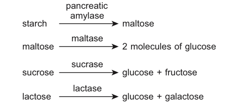
The pancreas produces pancreatic juice, which is an alkaline solution containing trypsinogen, pancreatic amylase and pancreatic lipase. Pancreatic juice enters the duodenum through the pancreatic duct.
Intestinal juice contains intestinal lipase, enterokinase, erepsin, maltase, lactase, sucrase and several other enzymes.
All enzymes in the small intestine have an optimum pH under alkaline conditions.
Bile, an alkaline greenish-yellow fluid, is produced by the liver and stored in the gall bladder. It passes into the small intestine through the bile duct. Bile breaks up large fat droplets into smaller fat droplets in a process called emulsification. This increases the surface area to volume ratio of the fats for lipases on work on and speeds up fat digestion.
Action of enzymes involved in carbohydrate digestion in the small intestine:
 Action of enzymes involved in fat digestion in the small intestine:
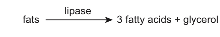
Action of enzymes involved in protein digestion in the small intestine:
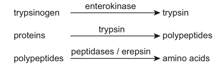
Note: Enterokinase converts the inactive form of trypsin, trypsinogen, into trypsin.
Food is completely digested in the small intestine. The jejunum and ileum function mainly to absorb nutrients and water.
Nutrients have to be absorbed into the body from the lumen of the small intestine. The small intestine is adapted for this role by having:
(a) An inner wall with large circular folds
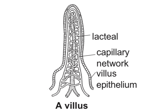
(b) Finger-like projections on the inner wall called villi
(c) Each epithelial cell on the villi has smaller projections called microvilli.
These adaptations increase the surface area of the small intestine, resulting in a larger surface for absorption.
The villi have thin walls (one-cell thick) so that food molecules diffuse over a shorter distance.
Within each villus is a network of capillaries and a small vessel called a lacteal.
Nutrients are absorbed across the wall of the small intestine and into the capillaries or lacteal. The lacteal transports fats away from the small intestine while the capillaries transport sugars and amino acids.
The transport of food away from the small intestine sets up a concentration gradient for diffusion.
Glucose and amino acids are absorbed by diffusion or active transport depending on the concentration gradient.
Fatty acids and glycerol are absorbed by the epithelial cells of the villi and recombined within those cells to form fats, which are transported into a lacteal.
Water is absorbed by passive diffusion throughout the length of the small intestine and mineral salts are absorbed in the ileum.
The food eventually leaves the small intestine and enters the large intestine.
The large intestine
The large intestine or colon is shaped like an inverted U and has the function of absorbing the remaining water and mineral salts that have not been absorbed by the small intestine. Note that most of the water that was present in the small intestine (from liquid in ingested food as well as the water content in intestinal mucus and digestive juices) had been absorbed by the small intestine.
The undigested waste matter moves along the large intestine by peristalsis, getting progressively drier.
The undigested waste matter comprises mainly cellulose, which is indigestible to humans.
The waste matter ends up at the rectum where it is stored before it can be eliminated from the body through the anus. The elimination of waste material is called egestion.
Transport of products of digestion
As absorption takes place in the small intestine, the blood in the capillaries of the villi becomes very rich in simple sugars and amino acids.
The blood capillaries of the villi converge into a large blood vessel called the hepatic portal vein, which leads to the liver.
The blood from the small intestine travels to the liver via the hepatic portal vein. The composition of blood in this vein varies greatly throughout the day depending on whether absorption of nutrients is occurring in the small intestine
Role of the liver in carbohydrate metabolism
The liver is involved in carbohydrate metabolism and regulation of blood glucose concentration.
When the glucose level in blood is high, the islets of Langerhans in the pancreas secrete insulin, which is a hormone that stimulates the liver cells to convert glucose into glycogen. The liver cells convert excess glucose in the blood from the hepatic portal vein into glycogen, which is stored in the liver.
When the glucose level in blood is low, the islets of Langerhans secrete glucagon, which is a hormone that stimulates the liver cells to convert stored glycogen in the liver back into glucose. The glucose is released into the blood leaving the liver, which supplies glucose to the body cells.
Role of the liver in fat metabolism
The liver produces bile, an alkaline liquid which helps fat digestion by emulsifying fats.
It oxidises fats to produce energy.
It converts excess carbohydrates and proteins to fatty acids and glycerol which are exported and stored as fatty tissues.
Role of the liver in breakdown of red blood cells
Aging red blood cells are removed by the spleen.
Haemoglobin from the red blood cells is brought to the liver, where it is broken down. The iron from the haemoglobin is stored in the liver while the other metabolic by-products of the breakdown form bile pigments.
Role of the liver in protein metabolism
The liver is involved in the synthesis of plasma proteins e.g. albumin, and blood clotting factors e.g. fibrinogen.
The liver is responsible for the deamination of excess amino acids, which refers to the removal of the amino group (–NH2) from an amino acid.
The amino group is converted into ammonia, which is toxic to cells, before it is further converted to urea by enzymes in the liver, and subsequently removed in urine.
The remnants of the amino acid are converted to glucose.
Role of the liver in detoxification
The liver breaks down toxic substances for excretion in urine or bile.
It also breaks down alcohol to acetaldehyde through the action of an enzyme called alcohol dehydrogenase.
Acetaldehyde is then converted to harmless acetic acid by acetaldehyde dehydrogenase.
Alcohol irritates oesophageal, stomach and intestinal linings.
Excessive alcohol consumption can lead to inflammation and ulcers.
Excessive alcohol consumption can also lead to inflammation, scarring and destruction of liver cells.
The liver cells are replaced with fibrous scar tissue in a disease called cirrhosis of the liver, leading to loss of liver function.