


Movement Analysis
4.1. Neuromuscular Function
Nervous system is made up of millions of nerve fibers, transferring electrical signals from the brain.
The central nervous system (CNS) consists of the brain and spinal cord.
The peripheral nervous system is the arrangement of nerves extending from the spinal cord to other parts of the body.
Motor neurons (motoneurons) are nerves that carry info from the CNS to the muscles and signal for contraction.
Structure of neurons
Cell body - contained in the spinal cord or in clusters just outside it called ganglia.
Dendrites - link the neuron to other neurons and information to flow.
Axon - main component to nerve signal transmission, similar to an electrical wire. Encased in myelin for insulation.
Gaps in myelin called nodes of Ranvier
Neuromuscular junction (NMJ) (motor end plate) - where the neuron meets the muscle.
Small gap between the two called the synapse.
Motor unit - a single motor neuron and the muscle it innervates.
Typically the larger the muscle the more muscle fibers are innervated by each motor neuron.
Allows a single motor neuron to generate large muscular forces
A small number of muscle fibers per motor neuron gives a small force but great precision (ex eye).
When the motor unit is innervated all the muscle fibers attached to it are contracted.
Types of motor units (fast/slow twitch)
Type I - slow twitch motor units consist of mainly slow twitch muscle fibers and have slower nerve transmission speeds and small muscle forces.
Can maintain contractions for a long period of time
Fatigue resistant
aerobic
Type IIa - fast twitch oxidative (uses oxygen) motor units consist mainly of type IIa muscle fibers and have fast nerve transmissions.
Stronger contraction forces and are more resistant to fatigue
Anaerobic and aerobic
Type IIb - fast twitch motor units with mostly fast twitch muscle fibers.
Fastest contraction times and largest forces
High fatigue rate and can’t maintain contractions for a long period of time
Anaerobic
Mechanics of Muscle Contraction
Striations - muscle fibers that appear striped due to the overlap of actin and myosin proteins within the muscle fiber.
Muscle contraction starts with electrical impulse from the brain (either voluntarily or by reflex).
Signal travels along the motor neuron to the muscle via the NMJ across the synapse.
When signal reaches here, the neurotransmitter, Acetylcholine, is released and changes the electrical state of the muscle.
The signal travels through the muscle fibers stimulating the sarcoplasmic reticulum where it releases calcium (Ca2+)
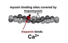
Myosin binding sites on actin are covered by tropomyosin.
Calcium binds to troponin on the tropomyosin which causes it to move and reveal the myosin binding sites on the actin.
ATP on the Myosin head is hydrolysed to form ADP + Phosphate
Cross bridge formed - myosin heads are shaped like little golf clubs and it is the ends of the heads that attach to the actin.
Myosin head remains bound until an ATP molecule releases it.
As long as there is calcium available cross bridge formation will continue until maximum contraction of the muscle fiber is reached.
The motor neuron initiates a resting potential through repolarization.
Cholinesterase, an enzyme that breaks down acetylcholine, is released and causes the muscle cell to repolarize and relax.
Calcium ions are removed from the cell and returned to the sarcoplasmic reticulum via the calcium pump
Cross bridge formation is terminated as there is no calcium which means the myosin binding sites on the actin filament are covered by tropomyosin.
Myosin heads to a resting state.
Control of Muscle force
When a muscle is signaled to contract, the force of the contraction is appropriate so the body segment moves appropriately
Quads require a large form (big muscle group), or a small force like fingers for writing.
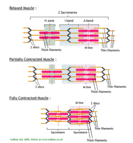
Skeletal muscle contracts by Sliding Filament Theory
Myofibril: A cylindrical organelle running the length of the muscle fibre, containing Actin and Myosin filaments.
Sarcomere: The functional unit of the Myofibril, divided into I, A and H bands.
Actin: A thin, contractile protein filament, containing 'active' or 'binding' sites. It slides past myosin casing contractions.
Myosin: A thick, contractile protein filament, with protrusions known as Myosin Heads. Pulls actin filaments towards one another by means of cross bridges.
Tropomyosin: An actin-binding protein which regulates muscle contraction.
Troponin: A complex of three proteins, attached to Tropomyosin.
Z Line: separates each sarcomere. It provides an anchor for proteins and also anchors the actin filaments to the ends of the sarcomere
M Line: is the centre of the A band and it is where adjacent myosin filaments anchor to each other
H Zone: is the centre of the sarcomere and has only myosin filaments
A Bands are also known as dark bands and has both actin and myosin microfilaments - stays the same length during contraction
I Bands are also known as light bands and have only actin microfilaments.
Sarcoplasmic reticulum stores calcium ions and releases them into the sarcoplasm for the generation of action potential during muscle contraction.
Adenosine triphosphate (ATP) is the sole fuel for muscle contraction.
Calcium triggers contraction by reaction with regulatory proteins that in the absence of calcium prevent interaction of actin and myosin.
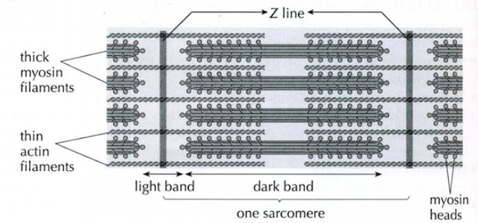
Structure of a Sarcomere
A sarcomere is a subunit of a myofibril.
At either end is a Z line to which narrow actin filaments are attached.
The actin filaments stretch inwards towards the centre of the sarcomere.
Between them, there are thicker myosin filaments, which have heads that can bind to the actin.
The part of the sarcomere containing myosin is the dark band and the part containing only actin filament is the light band.
Slow and Fast twitch fibre types differ in structure and function
Slow-twitch, or type I, fibres have more mitochondria, store oxygen in myoglobin, rely on aerobic metabolism, have a greater capillary to volume ratio and are associated with endurance; these produce ATP more slowly.
Fast-twitch, or type II, fibers have fewer mitochondria, are capable of more powerful (but shorter) contractions, metabolize ATP more quickly, have a lower capillary to volume ratio, and are more likely to accumulate lactic acid.
Fast Twitch (Type 2)
Contract quickly
Give sharp, powerful muscle contractions
Don't use oxygen
Suited to activities with bursts of strength and power
Tire quickly
Slow Twitch (Type 1)
Take longer to contract
Give long sustained muscle contractions
Not as powerful
Have a good oxygen supply
Suited to activities which require long term energy
Fast Twitch Type 2
Fast Twitch 2a: Fast Twitch High
Oxidative Glycolytic (FOG)
Have a greater resistance to fatigue due to endurance training
Fast Twitch 2b: Fast Twitch Glycolytic (FTG)
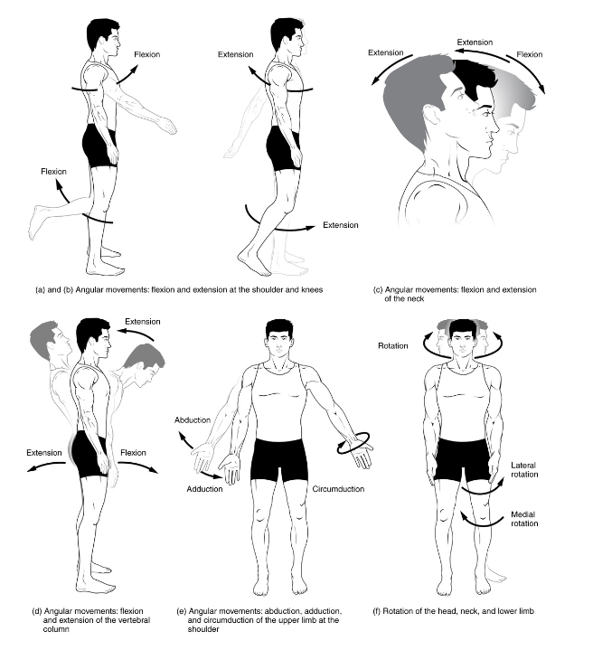
4.2 Joint and Movement Type
Plane | Motion | Axis | Example |
Sagittal | Plantar flexion and dorsi flexion / extension | Frontal | Walkin, squatting, overhead press |
Frontal | Abduction Side flexion Inversion / eversion | Sagittal | Star jump Lateral arm raise Side bending |
Transverse | Internal rotation / external rotation Horizontal flexion / extension Supination / pronation / circumduction | Vertical | Throwing Baseball swing Golf swing |
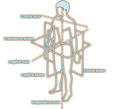
Axes
An axis is a straight line around which an object rotates.
Movements at joints take place in a plane about an axis.
The three axis of rotation are:
Sagittal axis - passes horizontally from posterior to anterior and its formed by the intersection of the sagittal and transverse planes.
Frontal axis - passes horizontally from left to right and is formed by the intersection of the frontal and transverse planes.
Vertical axis - passes vertically from inferior to superior and is formed by the intersection of the frontal and sagittal planes.
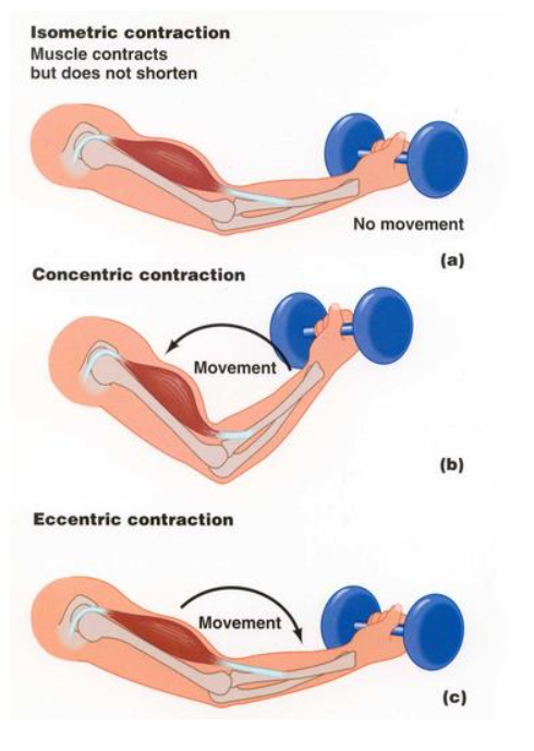
Types of Muscle Contraction
Isometric Contraction
In general in this form of contraction the muscle length remains constant. It occurs when muscle force balances resistance and no joint movement occurs
there is generally no movement resulting from this type of contraction
pushing against a fixed object
planking
Isotonic contraction
an increase in tension (load) results in changes in skeletal muscle length.
i.e. lengthening and shortening of the muscle.
Concentric contraction
Concerns muscle actions that produce a force to overcome the load being acted upon.
The work done is referred to as positive work.
Eccentric contraction
Refers to muscle action in which the muscle force yields to the imposed load.
The work done during a concentric contraction is referred to as negative
Isokinetic contraction
The term is used in two contexts.
First, as a specific muscle contraction and second as a testing and rehabilitation machine.
When a muscle contracts so that the body segment to which it is attached moves at a constant speed around the joint, rarely found in sport.
Reciprocal Inhibition
When an agonist contracts to move a body segment, it is usual for the antagonist (the muscle with the opposite concentric contraction action) to relax.
This means that the agonist is not being opposed by any muscle torque acting in the opposite direction of the motion.
This is an automatic action controlled by neurons.
When the agonist motor neuron is stimulated the motoneuron to the antagonist is inhibited preventing it from contacting strongly.
Analyze movements in relation to Joint action and Muscle contraction
Joints Involved | Action | Agonist Muscle |
Hip | Extension and Hyperextension | Gluteal muscles (gluteus maximus and gluteus minimus) and Hamstrings (biceps femoris, semimembranosus, semitendinosus) |
Knee | Extension | Quadriceps group of muscles (rectus femoris, vastus medialis, vastus lateralis and vastus intermedialis) |
Ankle | Plantar flexion | Gastrocnemius |
Delayed onset muscle soreness (DOMS) in relation to eccentric and concentric muscle contractions
The pain and stiffness felt in muscles several hours to days after unaccustomed or strenuous exercise.
Brought on by eccentric contractions of the muscle causing pressure at the nerve endings.
DOMS results primarily from eccentric muscle action and is associated with structural muscle damage, inflammatory reactions in the muscle, overstretching and overtraining.
DOMS is prevented/minimized by reducing the eccentric component of muscle actions during early training, starting training at a low intensity and gradually increasing the intensity, and warming up before exercise, cooling down after exercise.
4.3. Fundamentals of biomechanics
Force: a push or pull on an object
Speed: maximum rate at which a person is able to move their body
Velocity: rate at which an object changes position
Displacement: distance measured in a stated direction
Acceleration: rate of change of velocity (speed/direction) per second
Momentum: the amount of motion possessed by a moving object
Impulse: force x time. The motion (momentum) of a body depends not only on the force, but also the duration (time) the force is applied.
Scalar
length
mass
area
volume
speed
density
pressure
Vector
displacement
direction
velocity
acceleration
momentum
force
impulse
weight
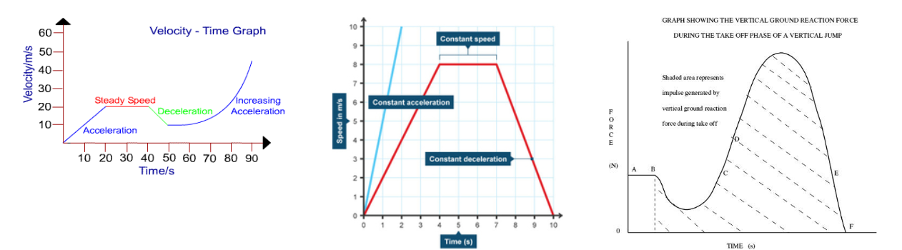
Velocity-time, distance-time, and Force-time graphs of sporting actions
Center of Mass
The point at which the body is balanced in all directions.
A change in body position during sporting activities can change the position of the center of mass
The center of mass can change when the body is moving dynamically.
The center of mass is not always inside the body, it can be outside of the body depending on position.
Sporting Example: High Jump
The Scissor Kick
The center of mass is within the pelvic girdle. The center of mass is within the body
The action involves clearing the bar one leg at a time
As the center of mass is within the body, it is more likely that the bar will be hit and the jump will be invalid.
Frosbery Flop
The center of mass in this jump is externally placed.
The arch in the back allows the mass to be shifted to the outside of the body, and there is greater opportunity for clearance.
The greater the arch of the back the lower the center of mass is
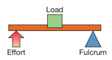
Components of a Lever System
The parts that make up a lever system, for example, in the body this would be a bone, a joint, a muscle and the body weight
4 components:
the load: The object that needs to be moved.
the fulcrum: Muscular force applied to move the load.
the effort: Joint around which the movement takes place.
the lever: Bones in the body serving as the structures for movement.
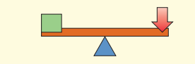
First Class Levers
Fulcrum is between the effort and the load.
Examples in the Body:
Limited instances in the body.
Triceps' attachment to the elbow joint makes elbow extension a first-class lever.
Elbow serves as the fulcrum.
Triceps provide the effort.
Load is the object being thrown (e.g., javelin).
Nodding of the head is another example.
Load: Weight of the head.
Fulcrum: Joint allowing nodding.
Effort: Muscular force for nodding.
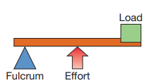
Second Class Levers
Significant Application in Sport:
Notable example with great relevance for sport.
Formed between:
Ball of the foot.
Gastrocnemius muscle.
Load of the body weight.
Example: Pointing toes or going onto toes.
Foot acts as the lever bar.
Third Class Lever
Memory Aid for Lever Systems: 1-2-3 = F-L-E
1 (First Class): Fulcrum is between components.
2 (Second Class): Load is between components.
3 (Third Class): Effort is between components.
Common Lever System in the Body: Third Class
Most prevalent in body movements.
Examples:
Biceps curl.
Hitting a ball with a racket or bat.
Knee during kicking.
Hip during running.
Newton’s three Laws of Motion
First Law (Law of Inertia)
An object will remain at rest or constant velocity unless acted upon by an external force.
Example: An athlete at a starting block will not move unless a force acts upon them. The external force comes from the block and this propels the sprinter out of the blocks when they exert a downward and backward force against the blocks.
Second Law (Law of Acceleration)
The rate of change of acceleration of an object is proportional to the force applied and acts in the direction of the force.
The acceleration of an object is directly proportional to the force causing it and is inversely proportional to the mass of the object.
Example: Two athletes at a starting block both push off, one is lighter (and has a lesser mass) and therefore accelerates quicker. Two athletes at a starting block of the same mass both push off, the one who applied greater force accelerates faster.
Third Law (Law of Reaction)
For every action there is an equal and opposite reaction.
Example: The sprinter applied downward and backward force on the immovable starting blocks, they exert back with a forwards and upward reaction force on the sprinter, pushing the sprinter forwards. The harder the sprinter pushes, the greater the reaction force will be.
The relationship between angular momentum, moment of inertia and angular velocity
Angular momentum: the product of the body's moment of inertia, and its angular velocity.
M=I*V
Moment of inertia: It determines the torque (force that causes rotation) needed for a desired angular acceleration about a rotational axis.
It depends on the mass of the object, its shape and its relative point of rotation.
Angular velocity: is a ratio of the change of angular displacement and the time during which the change occurred. The rate of which a body spins/rotates/turns through an angle.
Angular velocity = angular displacement ÷ time
Concept of angular momentum in relation to sporting activities
Angular Momentum
Definition: Angular momentum refers to the rotational equivalent of linear momentum. It represents the quantity of rotation of a body and is dependent on the mass distribution and the speed of rotation.
Expression: It is mathematically expressed as the product of an object's moment of inertia and its angular velocity.
Moment of Inertia
Definition: This term describes an object's resistance to change in its rotational motion. It is analogous to mass in linear motion.
Factors Affecting Moment of Inertia: The moment of inertia depends on the mass of the object and how this mass is distributed relative to the axis of rotation. For instance, a spread-out mass (like extended arms) increases the moment of inertia.
Projectiles
objects or athletes that are propelled in the air
Influences
Height of release
the higher the release = the greater distance covered
the higher the release = the longer spent in the air
the higher the release = the longer the horizontal component will be acting
Angle of release
ideal angle of release is 45 degrees
the angle changes the relationship between the horizontal and vertical components of projectile
Speed of release (most influential)
speed is directly related to the distance
greater the speed = greater the distance
initial vertical velocity increases the height of the trajectory, creating a longer flight path
initial horizontal velocity will increase the length of flight time and distance
Bernoulli Principle
Fundamental Concept: The Bernoulli Principle, formulated by Daniel Bernoulli, is a principle in fluid dynamics that states that for an inviscid flow, an increase in the speed of the fluid occurs simultaneously with a decrease in pressure or a decrease in the fluid's potential energy.
Fluid Dynamics Application: It is used to explain the behavior of non-viscous fluids in motion and is pivotal in aerodynamics and hydrodynamics.
Relevance to Projectile Motion: The principle is key to understanding how variations in air pressure can affect the trajectory and velocity of objects, such as balls in sports.
The Bernoulli Principle in Action: Spinning Golf Ball
Impact of Spin on Airflow:
Top Surface of the Ball: The spin causes the air to move faster over the top surface, reducing pressure.
Bottom Surface of the Ball: Conversely, the air moves slower under the ball, creating higher pressure.
Generation of Lift Force: This difference in pressure on either side of the ball creates a lift force, causing the ball to deviate from its initial path.
Magnus Effect: This phenomenon is also known as the Magnus effect, where the spin of an object in a fluid medium alters its trajectory.
Trajectory Alteration in Golf:
Distance and Direction: The spin can increase the range and alter the direction of the ball.
Skill Application: Golfers leverage this knowledge to control the ball's flight for different shots.