


Movement Analysis
Lever Systems in the Body
Lever systems
Created in the body by the musculo-skeletal system.
Purpose of Levers:
Facilitate physical work by making it easier to move heavy objects or perform quick movements.
Application in Exercise:
Running, lifting weights, kicking, throwing a ball involve the use of levers.
Examples of Lever Systems in the Body:
Lever (bone) is used to move objects:
Running: Body is the object being moved.
Kicking a ball: Ball is the object being moved.
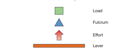
Components of a Lever System
The parts that make up a lever system, for example, in the body this would be a bone, a joint, a muscle and the body weight
4 components:
the load: The object that needs to be moved.
the fulcrum: Muscular force applied to move the load.
the effort: Joint around which the movement takes place.
the lever: Bones in the body serving as the structures for movement.
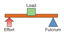
First Class Levers
Fulcrum is between the effort and the load.
Examples in the Body:
Limited instances in the body.
Triceps' attachment to the elbow joint makes elbow extension a first-class lever.
Elbow serves as the fulcrum.
Triceps provide the effort.
Load is the object being thrown (e.g., javelin).
Nodding of the head is another example.
Load: Weight of the head.
Fulcrum: Joint allowing nodding.
Effort: Muscular force for nodding.
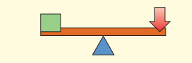
Second Class Levers
Significant Application in Sport:
Notable example with great relevance for sport.
Formed between:
Ball of the foot.
Gastrocnemius muscle.
Load of the body weight.
Example: Pointing toes or going onto toes.
Foot acts as the lever bar.
Third Class Lever
Memory Aid for Lever Systems: 1-2-3 = F-L-E
1 (First Class): Fulcrum is between components.
2 (Second Class): Load is between components.
3 (Third Class): Effort is between components.
Common Lever System in the Body: Third Class
Most prevalent in body movements.
Examples:
Biceps curl.
Hitting a ball with a racket or bat.
Knee during kicking.
Hip during running.
Mechanical Advantage and Disadvantage
Lever Systems Functions:
Move a heavier load.
Move a load further and faster.
Mechanical Advantage:
Lift heavier loads with less effort.
Example: Car jack requires little effort to lift a car a short distance.
Mechanical Disadvantage:
Greater force needed than the load.
Used in sports for moving loads over a large distance with speed.
Examples:
Tennis forehand.
Drive in badminton.
Position Determines Function:
Relative position of lever components determines mechanical advantage or disadvantage.
Influences the ease of lifting heavy loads or achieving speed and distance in movement.
Analysis of Movement
Detailed analysis of movement is a complex activity requiring sophisticated equipment.
However, the fundamental analysis of motion can be done visually and should involve the following:
A description of the actual actions which occur at the joints involved
The plane(s) in which the movement occurs
The muscles producing the movement
The function of the muscles involved (agonists, antagonists, synergists & fixators)
The type of contraction (isotonic - concentric or eccentric, isometric)
The range of the muscle action (inner, middle, outer)
Analysis of Sprinting
The running leg action occurs in a sagittal plane about a frontal axis and involves the hip, knee and ankle joints.
The hip's bones are the femur and pelvic girdle, which form a ball and socket joint.
The knee bones involved are the femur and tibia, which form a hinge joint.
The ankle bones are the tibia and calcaneus, which form a modified joint.
Each of these joints produces two actions, one when the leg is in contact with the ground (driving phase) and one when the leg is not in contact with the ground (recovery phase).
Driving Phase
Joints Involved | Action | Agonist Muscle |
|---|---|---|
Hip | Extension and Hyperextension | Gluteal muscles (gluteus maximus and gluteus minimus) and Hamstrings (biceps femoris, semimembranosus, semitendinosus) |
Knee | Extension | Quadriceps group of muscles (rectus femoris, vastus medialis, vastus lateralis and vastus intermedialis) |
Ankle | Plantar Flexion | Gastrocnemius |
Recovery Phase
Joints Involved | Action | Agonist Muscle |
|---|---|---|
Hip | Flexion | Iliopsoas |
Knee | Flexion | Hamstrings (biceps femoris, semimembranosus, semitendinosus) |
Ankle | Dorsiflexion | Tibialis anterior |
Analysis of Throwing
Throwing comprises two phases, the preparatory phase and the throwing phase.
Most actions are rotational in the transverse plane and longitudinal axis and the two joints primarily involved are the elbow and shoulder.
The elbow is a hinge joint formed by the humerus and ulna.
The shoulder is a ball and socket joint formed between the humerus and the scapula.
Preparatory Phase
Joints Involved | Articulating Bones | Action | Agonist Muscle |
|---|---|---|---|
Shoulder | Humerus & scapula | Horizontal Hyperextension | Posterior deltoids and latissimus dorsi |
Elbow | Humerus & ulna | Extension | Triceps brachii |
Throwing Phase
Joints Involved | Articulating Bones | Action | Agonist Muscle |
|---|---|---|---|
Shoulder | Humerus & scapula | Horizontal Flexion | Anterior deltoids and Pectoralis major |
Elbow | Humerus & ulna | Flexion | Biceps brachii |
Analysis of Racket Strokes
There are two phases to striking a ball with a racket, the preparatory phase and the striking phase.
Most actions are rotational in the transverse plane, and longitudinal axis and the three joints concerned are the wrist, elbow and shoulder.
The elbow is a hinge joint formed by the humerus and ulna.
The shoulder is a ball and socket joint formed between the humerus and the scapula.
The wrist forms a condyloid joint between the ulna and carpal bones.
Preparatory Phase
Joints involved | Articulating Bones | Action | Agonist Muscle |
|---|---|---|---|
Wrist | Ulna & carpal Radius & ulna | Supination | Supinator |
Elbow | Humerus & ulna | Extension | Triceps brachii |
Shoulder | Humerus & scapula | Horizontal Hyperextension | Posterior deltoid and latissimus dorsi |
Striking Phase
Joints involved | Articulating Bones | Action | Agonist Muscle |
|---|---|---|---|
Wrist | Ulna & carpal Radius & ulna | Pronation | Pronator teres |
Elbow | Humerus & ulna | Flexion | Biceps brachii |
Shoulder | Humerus & scapula | Horizontal Flexion | Pectoralis major and Anterior deltoid |
Trunk | Rotation | External obliques |
Analysis of Jumping
The action in jumping takes place in a sagittal plane about a transverse axis and involves the hip, knee and ankle joints.
The hip's bones are the femur and pelvic girdle, which form a ball and socket joint.
The bones of the knee involved are the femur and tibia which form a hinge joint.
The bones of the ankle involved are the tibia and calcaneus which form a modified joint.
Joints Involved | Action | Agonist Muscle |
|---|---|---|
Hip | Extension and hyperextension | Gluteal muscles (gluteus maximus and gluteus minimus) and Hamstrings (biceps femoris, semimembranosus, semitendinosus) |
Knee | Extension | Quadriceps group of muscles (rectus femoris, vastus medialis, vastus lateralis and vastus intermedialis) |
Ankle | Plantar Flexion | Gastrocnemius |
Analysis of Kicking
The kicking action takes place in a sagittal plane about a frontal axis and involves the hip, knee and ankle joints.
The hip's bones are the femur and pelvic girdle, which form a ball and socket joint.
The bones of the knee involved are the femur and tibia which form a hinge joint.
The bones of the ankle involved are the tibia and calcaneus which form a modified joint.
Kicking comprises two phases, the preparatory phase and the kicking phase.
Preparatory Phase
Joints Involved | Action | Agonist Muscle |
|---|---|---|
Hip | Extension and hyperextension | Gluteal muscles (gluteus maximus and gluteus minimus) |
Knee | Extension | Hamstrings (biceps femoris, semimembranosus, semitendinosus) |
Ankle | Plantar Flexion | Gastrocnemius |
Kicking Phase
Joints Involved | Action | Agonist Muscle |
|---|---|---|
Hip | Flexion | Iliopsoas |
Knee | Extension | Quadriceps group of muscles (rectus femoris, vastus medialis, vastus lateralis and vastus intermedialis) |
Ankle | Plantar Flexion | Gastrocnemius |
Agonist, Antagonist, Fixator & Synergist Muscles
When kicking the ball then:
Agonist - Quadricep muscles
Antagonist - Hamstring muscles
Fixator - Gluteus Maximus
Synergist - Abdominal muscles
Planes and Axes of movement
Movement is defined by reference to a plane or axis.
The Four Planes
Median or Sagittal Plane - a vertical plane which passes from front to rear, dividing the body into right and left sections.
Coronal or Frontal or Lateral Plane - which passes from side to side at right angles to the sagittal plane which divides the body into a front and back section.
Transverse or Horizontal Plane - a horizontal plane which divides the body into an upper and lower section.
Oblique Plane - any plane through the body that is not parallel to one of the former three.
The Three Axis
Frontal Axis - passes from side to side at right angles to the sagittal plane.
Sagittal or Transverse Axis - passes horizontally from front to rear, lying at right angles to the frontal plane.
Longitudinal or Vertical Axis - passes from head to foot at right angles to the transverse plane.
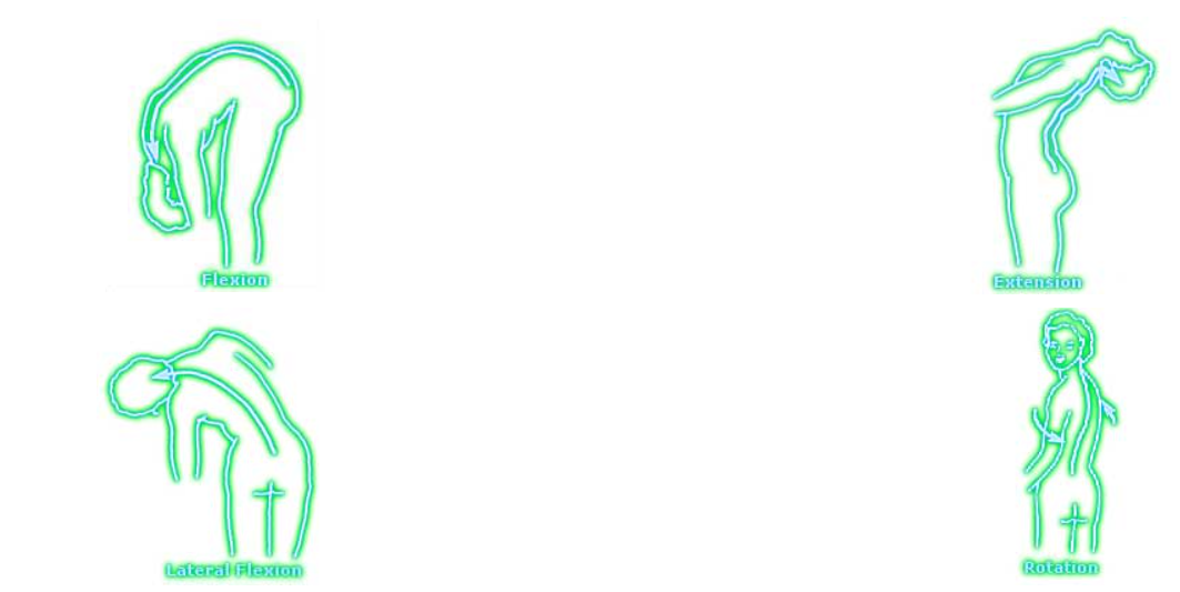
Spinal Column
The vertebral column has the following normal ranges of movement: Flexion, Extension, Lateral Flexion and Rotation.
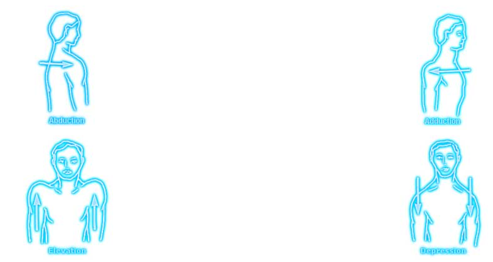
Shoulder Girdle
The shoulder girdle has the following normal ranges of movement: Elevation, Depression, Adduction and Abduction.
Shoulder Joint
The shoulder joint has the following normal ranges of movement: Flexion, Extension, Adduction, Abduction and Medial Rotation.
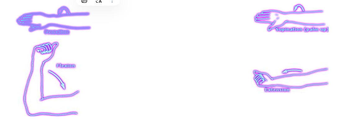
Elbow Joint
The elbow joint has the following normal ranges of movement: Flexion, Extension, Pronation and Supination.
Wrist Joint
The wrist joint has the following normal ranges of movement: Flexion, Extension, Adduction, Abduction and Circumduction.
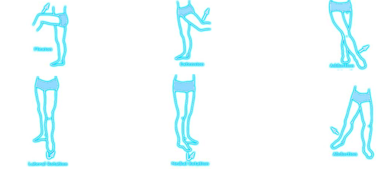
Hip Joint
The hip joint has the following normal ranges of movement: Flexion, Extension, Adduction, Abduction, Medial Rotation and Lateral Rotation.
Knee Joint
The knee joint has the following normal ranges of movement: Flexion and Extension.
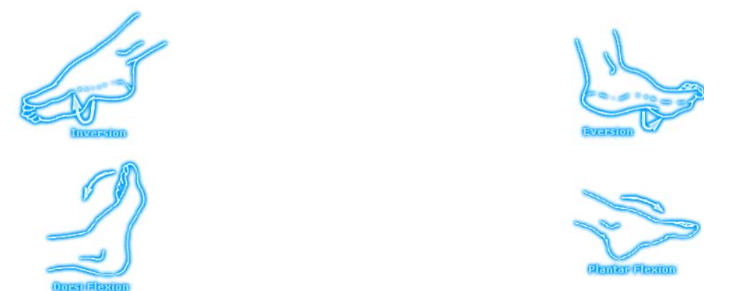
Ankle Joint
The ankle joint has the following normal ranges of movement: Plantar Flexion, Dorsi Flexion, Inversion and Eversion.