Cell Structure and Organisation
Cell Theory:
-The cell theory states that all organisms are composed of cells; the cell is the basic unit of life.
-Organisms can be unicellular, such as amoeba and bacteria, or multicellular such as plants and animals.
-New cells arise from pre-existing cells; specialised cells arise from undifferentiated stem cells.
-Advances in microscopy have allowed us to understand the ultrastructure of cells.
Eukaryotic Cells:
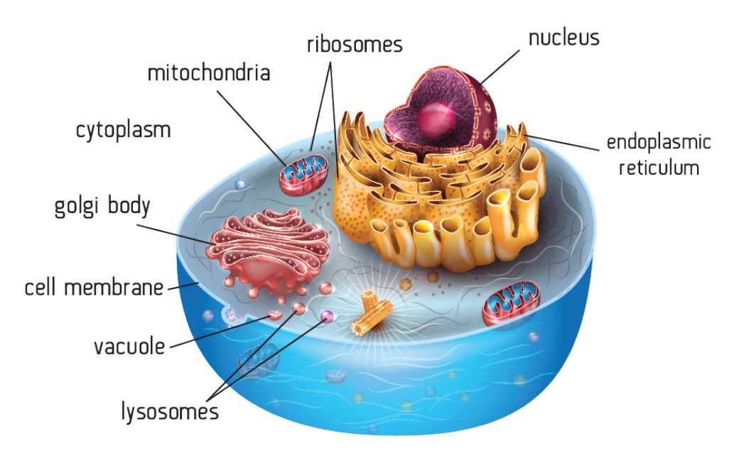
Eukaryotic cells have a nucleus and membrane bound organelles. Eukaryotic cells include plant and animal cells.
Plant cells have additional organelles and structures e.g. chloroplasts for photosynthesis and cellulose cell walls for support and to maintain turgor pressure.
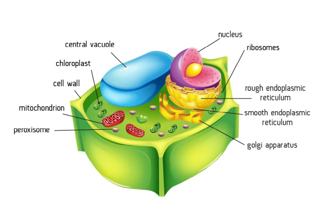
Structure | Function |
|---|---|
Nucleus | Contains DNA which codes for or controls protein synthesis. DNA replication occurs here. Transcription produces mRNA templates. |
Nuclear pores | Allow the transport of mRNA and ribosomes out of the nucleus. |
Nuclear envelope | Separates the contents of the nucleus form the cytoplasm. |
Nucleolus | Produces rRNA, tRNA and ribosomes. |
Chromatin | Condenses before cell division to form chromosomes. |
RER | Packaging and storing proteins. Producing transport vesicles which merge to form the Golgi body. |
SER | Produce, package and transport steroids and lipids. |
Golgi body | Packaging proteins for secretion from the cell. Modification of proteins e.g. by adding carbohydrate chains to form glycoproteins. Producing lysosomes and digestive enzymes (tertiary structure). |
Lysosomes | Contain powerful digestive enzymes to break down worn out organelles or cells. Phagocytes use lysosomes to digest engulfed bacteria. |
Centrioles | Form the spindle during cell division. They are not present in higher plant cells. |
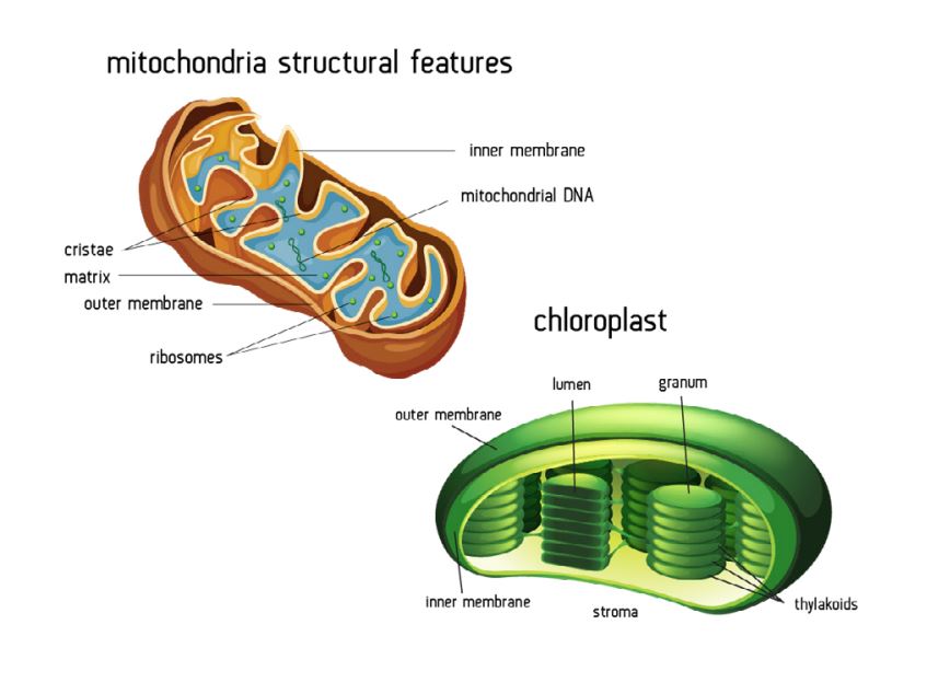
Mitochondria | ATP synthesis by aerobic respiration. |
Chloroplasts | Contain photosynthetic pigments which trap light energy for photosynthesis. |
Vacuole | Contains cell sap and stores solutes such as glucose. Swells due to osmosis for turgidity. |
Ribosomes | Protein synthesis. Primary protein structure is formed at the ribosome. |
Plasmodesmata | Connects cells via cytoplasm filled canals, which pass through cell walls. Allows transport via the symplastic pathway. |
Cell wall | Mechanical strength due to the high tensile strength of cellulose microfibrils. Transport of solutes via the apoplastic pathway. Cell to cell communication via the plasmodesmata. |
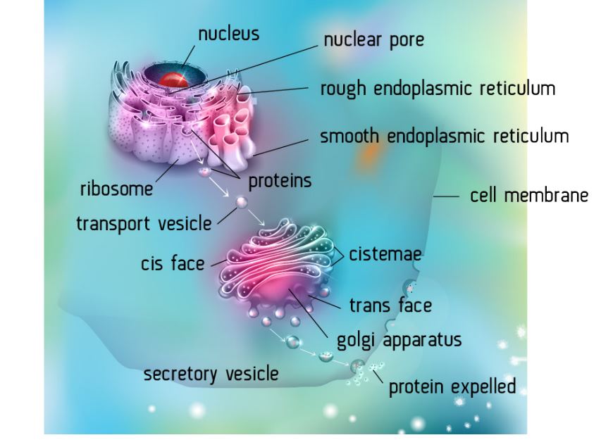
How organelles work together in eukaryotes:
Although organelles are described separately, their functions are interrelated. Protein synthesis and secretion is a great example of how organelles work together. - Ribosomes are produced in the nucleolus; they leave the nucleus via the nuclear pores and take up their positions on the rough endoplasmic reticulum (ER). --The nuclear pores also allow mRNA molecules (formed from DNA templates by transcription) to leave the nucleus. The mRNA molecules attach to the ribosomes on the rough ER.
-Protein synthesis takes place at the ribosome. The mRNA molecule contains the code for the primary structure of a protein; the order of amino acids in a polypeptide chain.
-The rough ER transports the polypeptides via transport vesicles, which merge with the Golgi body.
-The polypeptides are modified in the Golgi body and converted to their tertiary structure e.g. enzymes.
-The enzymes are packaged into secretory vesicles and transported to the cell membrane.
-The secretory vesicles merge with the cell membrane and release the enzymes by exocytosis.
Mitochondria v Chloroplast:
Similarities include:
-Both have double membrane
-Both have highly folded inner membranes -Both have a circle of DNA for self-replication
-Both have ribosomes
-Both produce ATP
Differences include:
-Mitochondria have cristae, but chloroplasts have thylakoid membranes.
-Chloroplasts contain photosynthetic pigments to absorb light energy, mitochondria do not.
-Mitochondria have an inner matrix, but chloroplasts have a stroma.
Prokaryotic Cells:
Bacteria do not have membrane bound organelles in their cells – no nucleus, rough endoplasmic reticulum, Golgi apparatus, mitochondria or chloroplasts; these cells are prokaryotic cells.
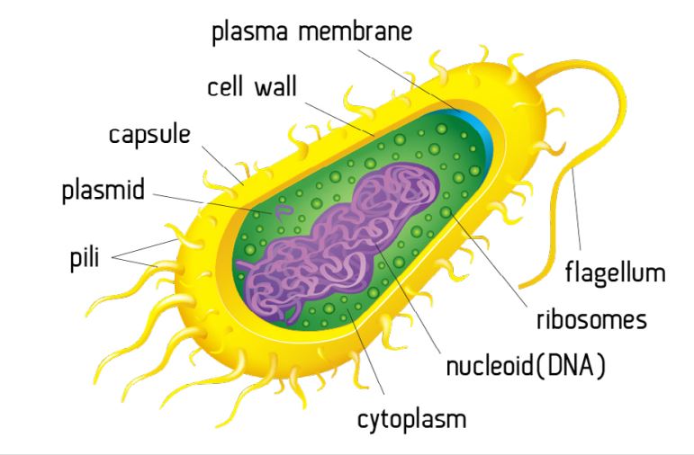
Prokaryote v Eurkaryote:
Prokaryotic | Eukaryotic |
|---|---|
Small cells 1-10µm | Larger cells at 10-100µm |
Ribosomes are smaller and free in cytoplasm (70S) | Ribosomes are larger and bound to the rough endoplasmic reticulum (80S) |
No membrane bound organelles | Membrane bound organelles present |
DNA free in cytoplasm | DNA contained within the nucleus |
No nuclear envelope (double membrane | Nucleus has a double membrane |
Plasmids present | No plasmids |
Cell wall is composed of peptidoglycan (murein) | Cell wall (when present) is composed of cellulose |
No mitochondria, uses a mesosome ( folded region of the cell membrane) for aerobic respiration | Mitochondria are used for aerobic respiration. There is no mesosome |
Viruses:
Viruses do not fit the cell theory; they have no cell membrane, no cytoplasm, no organelles and no chromosomes. Viruses can only reproduce with the help of a host cell. Viruses are composed of a protein coat or capsid which surrounds DNA, RNA or simply a few genes; the HIV virus has only 9 genes.
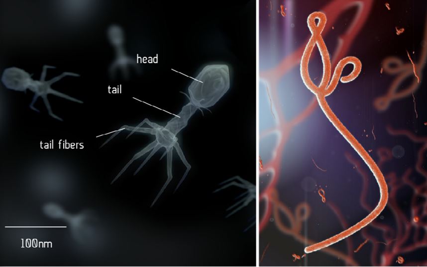
A bacteriophage and an Ebola virus- they can only be viewed using an electron microscope. Outside a living cell a virus exists as an inert viron. When they invade the cell they are able to take over the cell’s metabolism and reproduce within the cell. Viruses cause a variety of infectious disease in humans, animals and plants.
Levels of organisation:
Atoms to systems:
-Atoms are arranged into molecules.
-Molecules form cells.
-Cells work together to form tissues.
-Tissues form organs and organs form systems.
Cells adjacent to each other in the embryo often differentiate in the same way to form a tissue. A tissue is defined as a group of similar cells working together to perform a particular function. Mammals have several different tissue types.
Type of Tissue | Example |
|---|---|
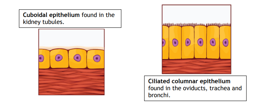
Epithelial tissue – This type of tissue forms a continuous layer, covering or lining the internal or external surfaces of the body. Epithelia have no blood vessels, but may have nerve endings. The cells sit on a basement membrane, made of collagen and protein and they vary in shape and complexity. The often have a protective or secretory function. | Cuboidal epithelium lines the kidney tubules and the small intestine. Cuboidal epithelium cells are cube shaped. |
Ciliated epithelium is composed of cells which transport substances like mucus in the bronchi and ova in the fallopian tubes/oviducts. The cilia move and sweep substances along. These cells are columnar (they look like columns). | |
Squamous epithelium consists of flattened cells on a basement membrane. They form the walls of the alveoli and line Bowman’s capsule in the kidney nephron. |
Type of tissue | Example |
|---|---|
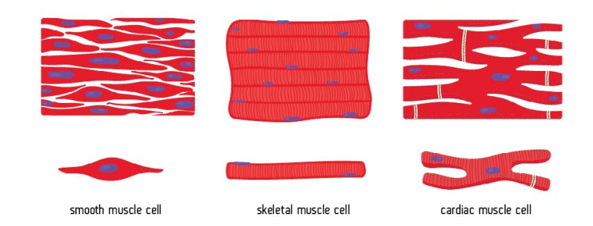
Muscle tissue – Muscle tissue comes in three main types, each with a different structure and function. | Skeletal muscle is attached to bones and moves the skeleton. It has bands of long cells called fibres, which can contract powerfully, but tire easily. You can choose whether or not to contract these muscles, so they are called voluntary muscles. The fibres form a striped pattern which can be viewed under the microscope; this is why skeletal muscle is often referred to as striped or striated muscle. |
Smooth muscle has individual spindle shaped cells that can contract rhythmically, but less powerfully than skeletal muscle. They occur in the skin, in the walls of the blood vessels and in the digestive and respiratory tracts. You cannot control these muscles, so they are called involuntary muscles. They do not have stripes and so are also called unstriped or unstriated muscle. | |
Cardiac muscle is only found in the heart. Its structure and properties are somewhat in between skeletal and smooth muscle. The cells have stripes, but lack the long fibres of skeletal muscle. They contract rhythmically, without any stimulation from nerves or hormones, although these can modify their contraction. Cardiac muscle does not tire. |
Not all cells differentiate to become specialised tissue cells, some remain unspecialised and are called stem cells. Stem cells can divide by mitosis to become new stem cells or they differentiate into new specialised cells. For example, stem cells in the bone marrow can differentiate into any type of blood cell. Specialised cells can no longer divide; when a specialised cell needs to be replaced a stem cell must differentiate into a new specialised cell.
Type of tissue | Example |
|---|---|
Connective tissue – Connective tissue connect, support or separate tissues and organs. It contains elastic and collagen fibres in an extracellular fluid or matrix. Between the fibres are fat storing cells (adipocytes) and cells of the immune system. | Areolar tissue is found under the skin and connects organs and tissues together. |
Collagen forms tendons which connect muscles to bones. | |
Ligaments which connect bones are elastic tissues. | |
Adipose tissue is composed of fatty cells and is found just under the skin and around organs. It functions as an energy store, thermal insulator and protects delicate organs. |
Organs are comprised of several tissues working together, performing a specific function, in humans, for example, the eye contains nervous, connective, muscle and epithelial tissue. In plants, the leaf contains epidermal tissue, vascular tissue, mesophyll (photosynthetic) tissue and parenchyma (packing) tissue.
Organ systems are groups of organs working together with a particular role.
Calculating the size of a structure from a magnified image:
Step by step guide:
-Measure the A to B with a ruler in mm.
-Divide this by the magnification.
-Then multiply by 1000 to convert mm to μm.