


Reproduction & Development
Why do Cells Divide?
Growth/Differentiation: Mitosis enables organisms to grow from a single-celled zygote into a mature organism that might contain hundreds of trillions of specialized cells
Maintenance: new cells produced to replace worn out/dead cells
Repair: They can regenerate damaged tissues (finger cut → new skin). Some organisms can regenerate entire body parts.
Single Cell Reproduction (asexual)
No new combination of cellular material occurs (all new cells contain same DNA as original cell)
Occurs in all somatic (body) cells, unicellular organisms, and simple multicellular organisms (budding, runners)
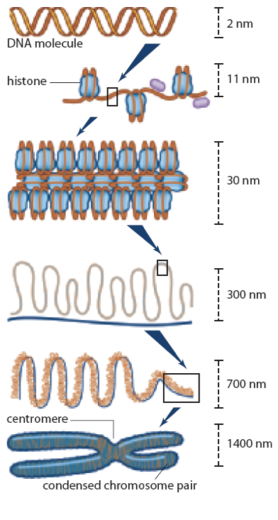
DNA Organization
DNA Molecule: two sugar-phosphate backbones with nitrogen base “rungs”
Histones: DNA molecule wraps around histones, forming a bead-like structure
Chromatin Strands: The bead-like structure is packed tightly, producing chromatin strands
Chromatin Fibres: Stands from loops which are attached to a supporting protein scaffold
Chromosomes: Protein scaffold folds further to condense the genetic material into chromosomes (duplicate during replication)
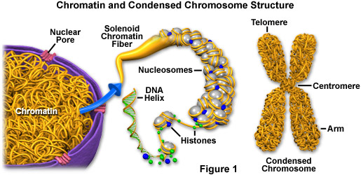
Chromosomes
Small, sausage-like structure of nucleic acids
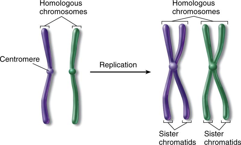
May be found as individual chromatids (during late stages of cell division) or as paired/sister chromatids (connected at the centromere)
Sister chromatids are identical to each other (exact copies)
All somatic cells contain homologous pairs of chromosomes
one from the mother’s egg (maternal)
one from the father’s sperm (paternal)
Humans have 23 pairs of chromosomes (46 chromosomes total)
Each homologous pair is similar in shape and length and is responsible for the same types of characteristics
The last “pair” of chromosomes (#23), determines gender (sex chromosomes)
Homologous pair (XX chromosomes) = female
Heterologous pair (XY chromosomes) = male
Homologs
Homologous chromosomes carry the same genes, in the same order
Despite this, homologous chromosomes often have slightly different DNA sequences resulting in different alleles (different form of the same gene)
Share several other characteristics, including:
Length
Centromere location
Banding pattern
Spontaneous Generation
Spontaneous generation was the theory that living organisms could arise from non-living matter
Most scientists accepted this theory until the mid-1800’s when advances in mircoscope maganification allowed for obervations of cell division
These oberservations led scientists to propose an alternative; new cells arise only from the division of other cells
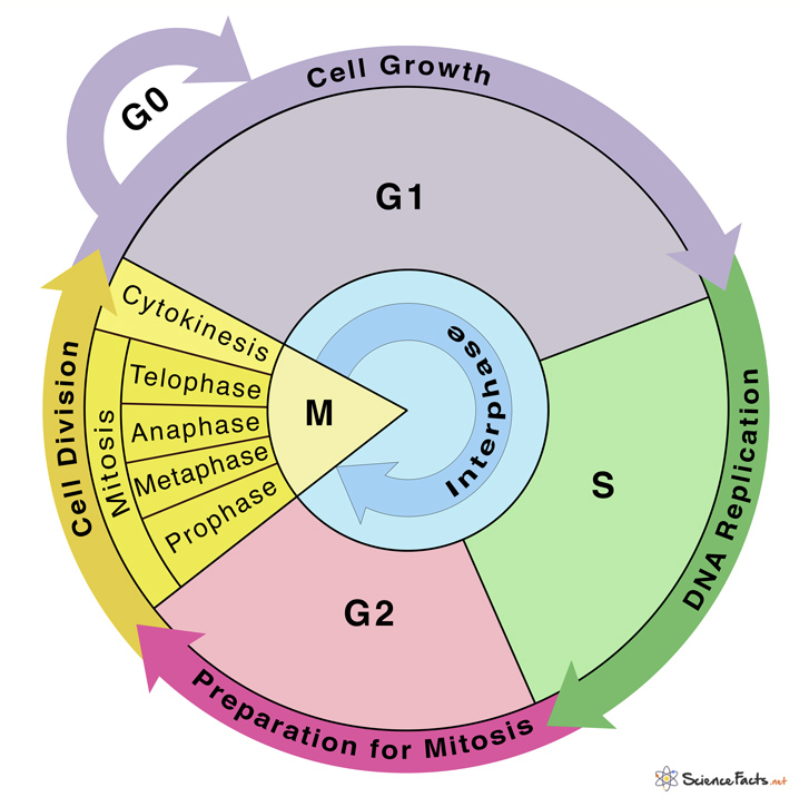
The Cell Cycle
Does not start and stop, but continues → different cells may cycle at different pace
Stages of the Cycle
Interphase: growth stage consists of G1, S & G2
G1 (first cell growth stage): organelle replication
S (synthesis phase): DNA is replicated
G2 (second cell growth stage): rebuilds energy reserves and prepares for mitosis
Cell Division (Mitosis): division of genetic material & nucleus
Prophase
Metaphase
Anaphase
Telophase
Cytokinesis
Mitosis
Cell division in somatic cells
All the cells produced by mitosis are IDENTICAL in genetic makeup to the original cells (particularly important that the chromosome # doesn’t change)
The unique appearance and functionality found in different cells of the body (except the sex cells) is NOT due to difference in cellular content, but a difference in the way that content is expressed (differentiation)
Prophase (Step 1)
Nuclear envelope breaks down; contents of nucleus become visible
DNA strands shorten and thicken, causing chromatin to condense into visible chromosomes
Centrioles separate and move to opposite poles of cell
Centrioles start growing spindle fibres
Nucleolus becomes invisible
Metaphase (Step 2)
Chromosomes move to centre of cell
Centromeres align across equator
Spindle fibres attach to the centromeres
Anaphase (Step 3)
Spindle fibres shorten and start pulling the sister chromatids apart
Chromatids separate at centromeres
Chromatids move to opposite poles of cell (same number of single-copy chromosomes should be at each pole)
Telophase (Step 4)
Chromosomes at opposite ends of cell
Chromosomes un-condense to form chromatin
Nuclear envelope and nucleolus reappears
Cytokinesis (Step 5)
Cytoplasm division
In plant cells, a cell-plate forms first, separating two cells by forming cell wall
In animal cells, cell membrane pinches in at the cleavage furrow to form two distinct daughter cells

Asexual vs. Sexual Reproduction
Asexual
Single individual is the sole parent
Single parent passes on all genes to its offspring
Offspring are genetically identical to the parent, resulting in a clone
Genetic differences rarely occur, but are the result of a mutation
Sexual Reproduction
Two parents (mother and father)
Each parent passes on half of their genes, resulting in the offspring having a unique combination of genes
Increases genetic variation
Haploid vs. Diploid Cells
The life cycle of all sexually reproducing organisms alternates between haploid and diploid cells
Somatic Cells
Somatic cells are diploid cells
They have DNA from maternal and paternal sides combined
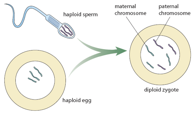
Gamete Cells
Gametes (sperm or eggs) are haploid cells
They only hold half the DNA from somatic cells from which they came
When an ovum is fertilized by a sperm, the original number of chromosomes (46 = 2n) is restored, forming a zygote
Zygote
A diploid cell that results from the fusion of two haploids gametes
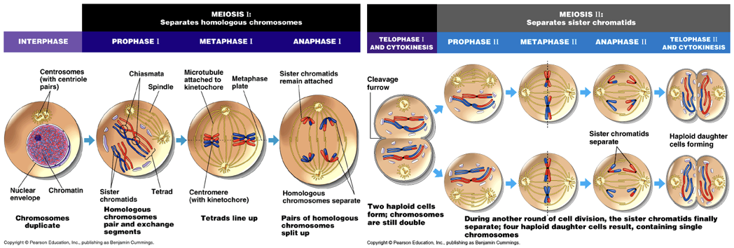
Meiosis
Creates gamete cells by reducing the number of chromosomes from 46 to 23 by copying chromosomes once, but dividing twice
Meiosis I: separates homologous chromosomes (first division)
Meiosis II: separates sister chromatids (second division)
Step 1: Prophase I
Same as prophase of mitosis:
Chromatin condenses into chromosomes
Centrioles move to opposite poles
Spindle fibres appear
Nuclear envelope begins to disappear
Nucleolus becomes invisible
Homologous chromosomes pair up side by side (synapsis) by corresponding genes forms tetrad (4 chromatids)
Homologous chromosomes overlap and occasionally break, exchanging identical sized segments (crossing over)
Crossing over leads to more genetic variation!
Step 2: Metaphase I
Homologous pairs move to centre → centromeres on either side of equator
Spindle fibres attach to centromeres only on exposed sides
Step 3: Anaphase I
Homologous pairs separate (not sister chromatids) at the centromere
Chromosomes move to opposite poles = segregation
There should be 23 double chromosomes at each pole (sister chromatids remain intact)
Step 4: Telophase I
Chromosomes at opposite poles
Chromosomes do not uncoil to form chromatin
Nuclear envelope occasionally reappears (in some cells)
Step 5: Cytokinesis
Division of the cytoplasm & organelles
Step 6: Prophase II
Centrioles move to opposite poles
New spindle fibres form
Note: Meiosis II is very similar to Mitosis
Step 7: Metaphase II
Cell moves directly to metaphase → no DNA replication and no formal organization of nucleus
Chromosomes move to centre, centromeres align on equator (metaphase plate)
Spindle fibres attach to outside of centromeres
Step 8: Anaphase II
Spindle fibres shorten → chromatids separate at centromeres
Chromatids move to opposite poles
There should be 23 single stranded chromosomes at each pole
Step 9: Telophase II
Chromosomes at opposite ends un-condense to form chromatin
Nuclear envelope reappears
Step 10: Cytokinesis
Division of the cytoplasm & organelles
Meiosis in Oocytes
In oocytes (female germ cell involved in reproduction; an immature ovum/egg cell) meiosis I is put on hold at the end of prophase I
Once the female reaches puberty, meiosis I is completed
Meiosis II is completed if/when the oocyte is fertilized
Gametogenesis
The formation of ova and sperm follow the process of meiosis, specializations dependent on their function
Sperm are designed for movement (little cytoplasm), lots of cell division, produce 4 small sperm
Eggs are designed to nourish the zygote – only one ovum is produced per oocyte → the other 3 polar bodies sacrifice their cytoplasm to produce one large egg
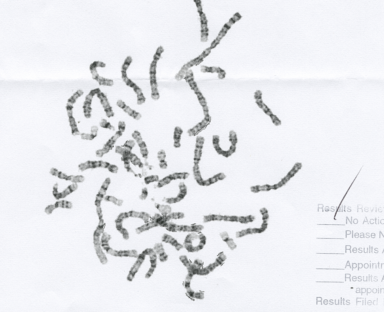
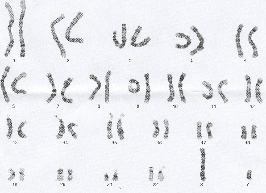
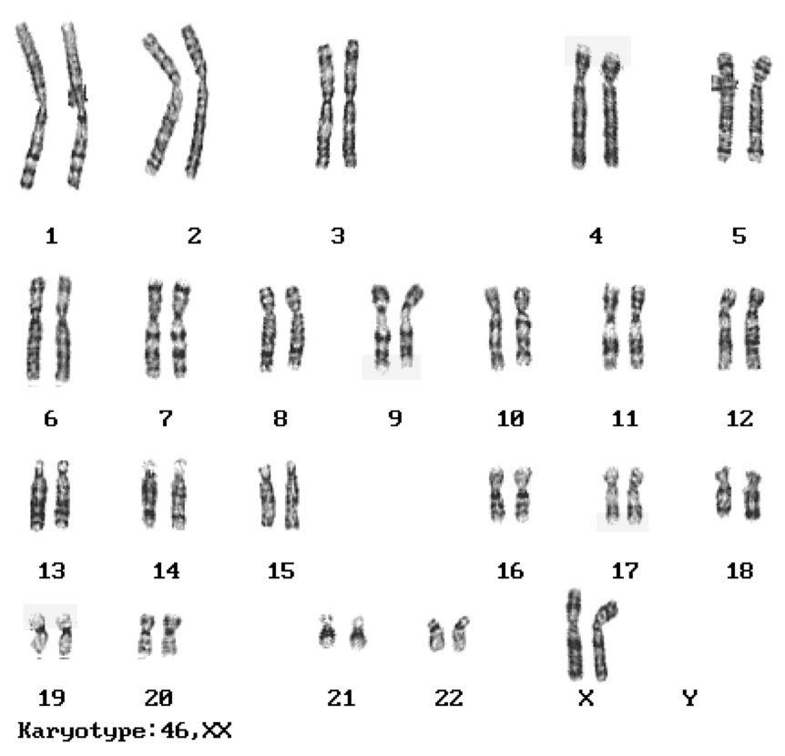
Karyotyping
A method of identification of chromosomes
Pictures of chromosomes are taken as cell undergoes mitosis → picture enlarged
Individual chromosomes are cut out
Chromosomes are matched up based on:
Size (largest to smallest)
Centromere position
G-banding
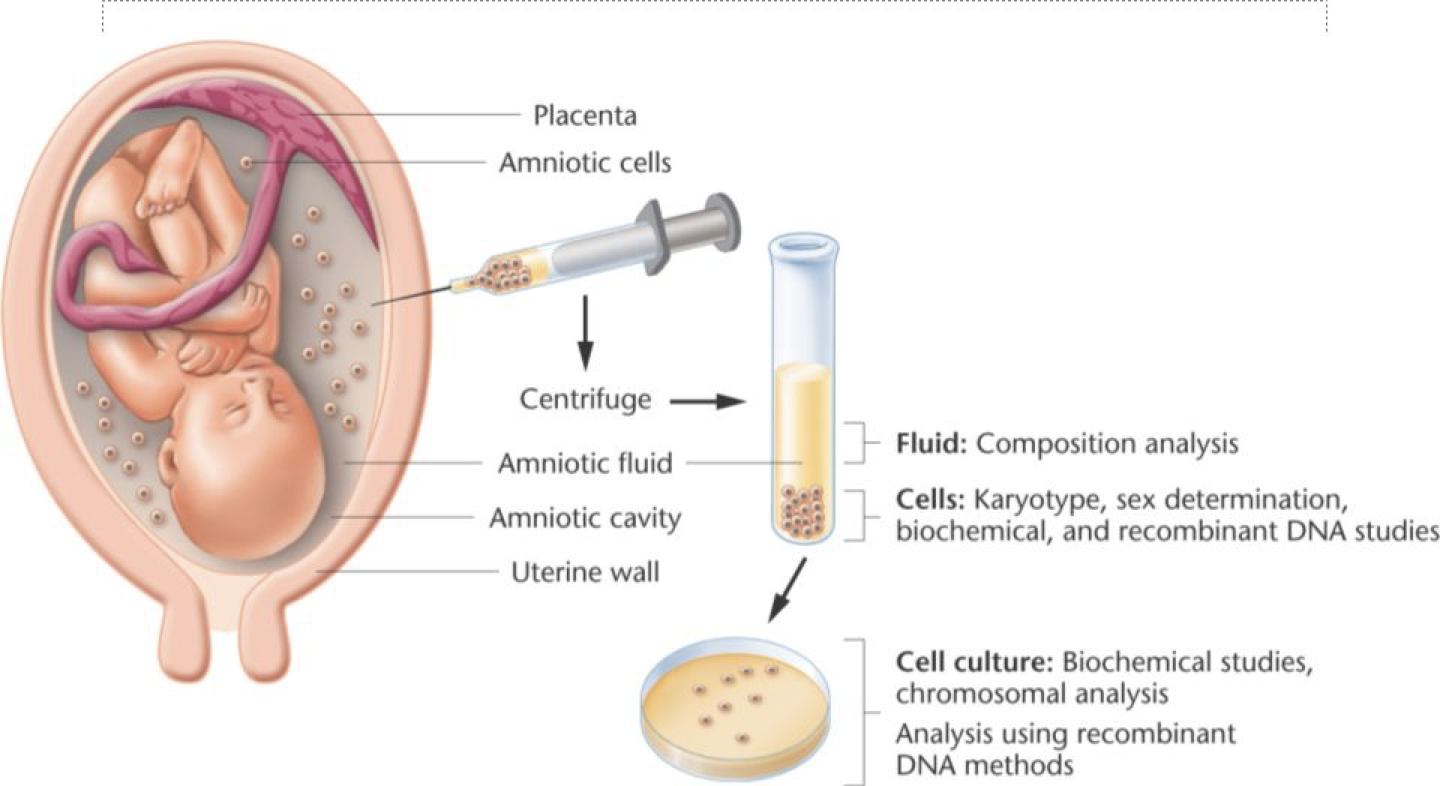
Fetal Tests
Amniocentesis
Procedure where a small amount of amniotic fluid is removed from the sac surrounding the fetus
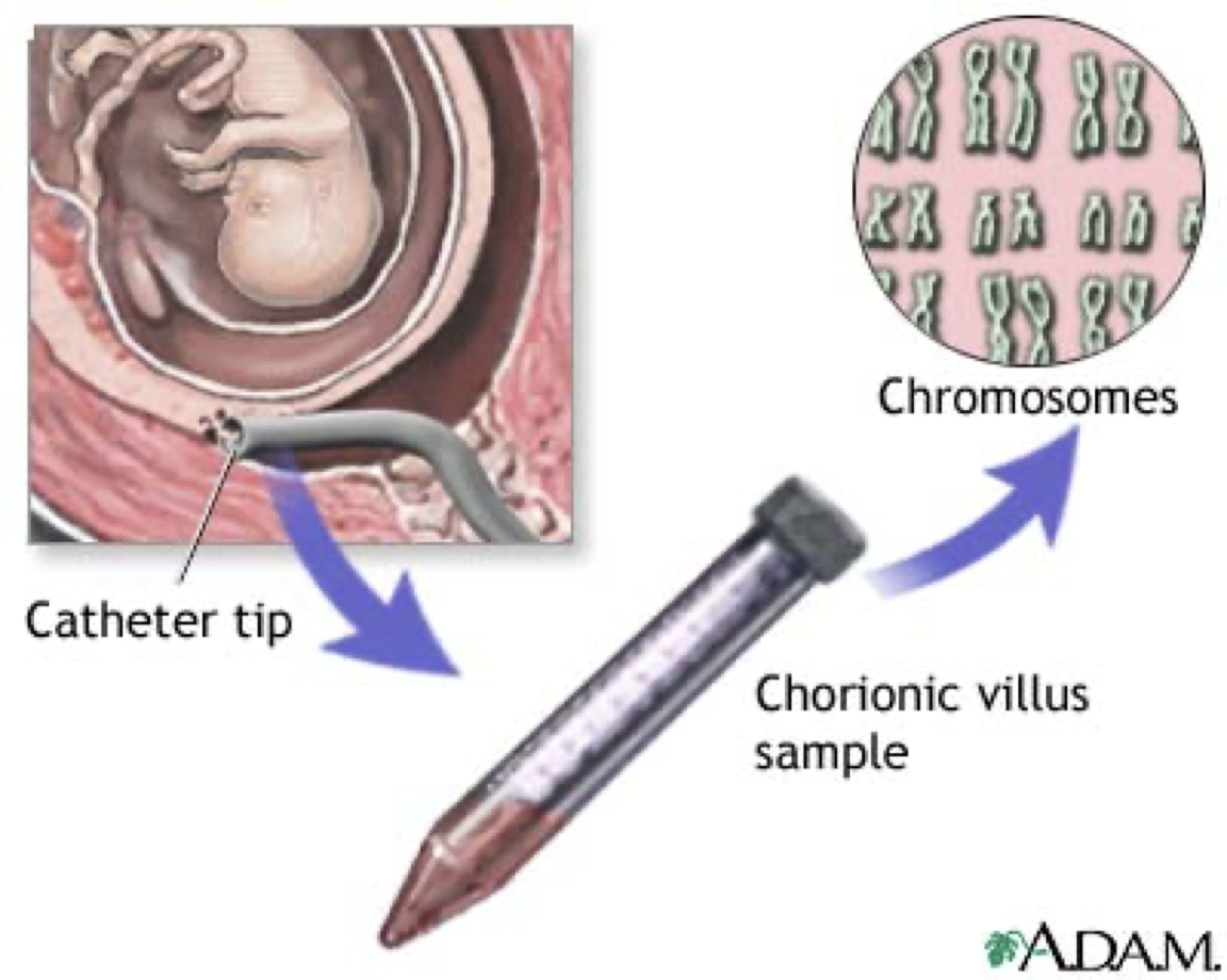
Chorionic Villi Sampling
A sample of chorionic villi is removed from the placenta for testing
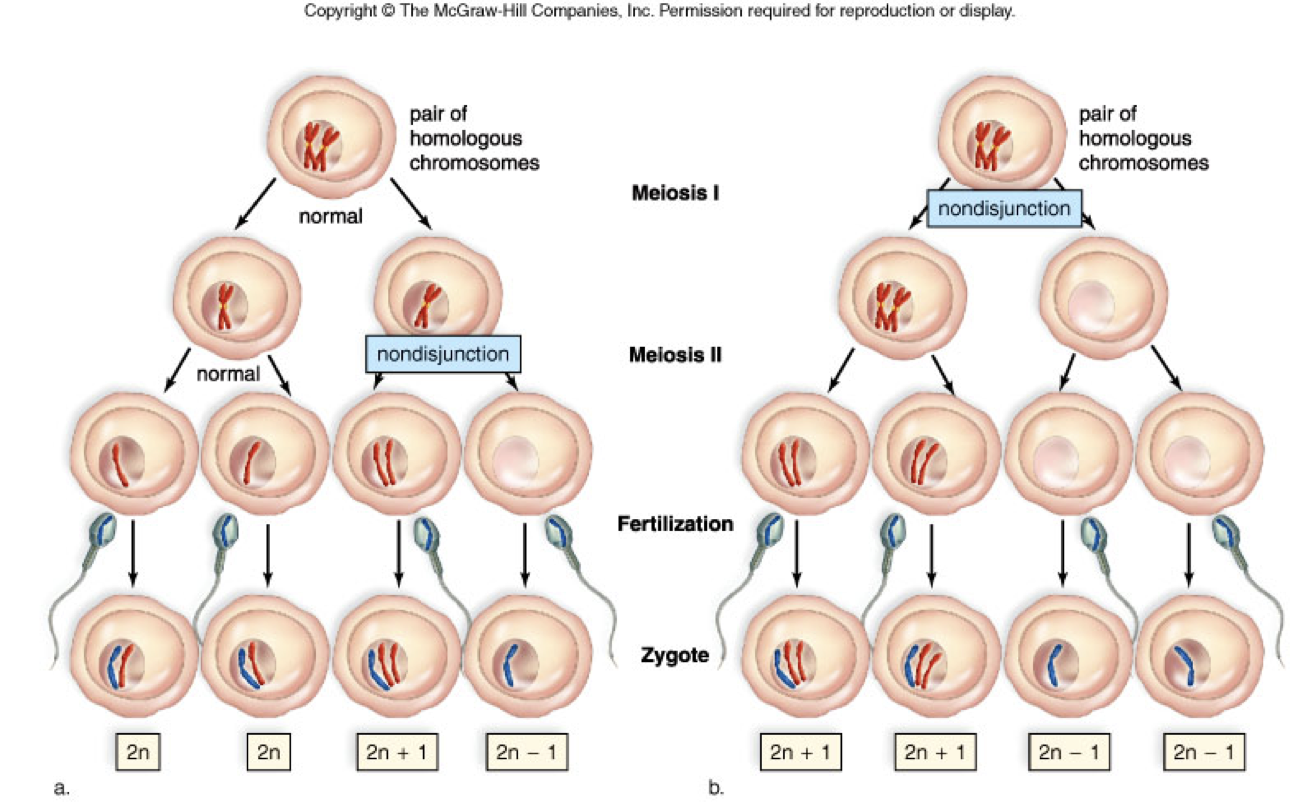
Nondisjunction of Autosomes
Chromosomes don’t separate properly during anaphase I or II
One daughter cell produced during separation will be lacking information, the other will have too much
If one too many chromosomes, one pair will be a triplet (trisomy)
if one too few chromosomes, one pair will be a singlet (monosomy)
Non-disjunction occurs quite often among humans
The impact is usually so severe to zygote that miscarriage occurs very early in pregnancy
If the baby survives, the set of traits is called a syndrome
Trisomy 21, 13 and 18 are the only known trisonomic autosomal genetic disorders that result in offspring surviving for a short period of time
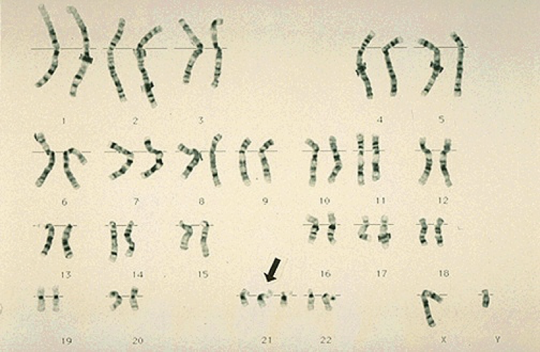
Down’s Syndrome (Trisomy 21)
Most commonly known trisomy
1 out of 700 births
Life expectancy has raised in recent decades (from 25 in 1983 to 60 today)
Short stature, fingers, and toes
Large tongue – makes speech difficult
Mental Disability Prone to heart defects, respiratory problems & leukemia
Risk of Down Syndrome
Odds of having a Down’s child increases with the age of the mother
At age 25, the risk is 1 in 1250
At age 30, the risk is 1 in 1000
At age 35, the risk is 1 in 400
At age 40, the risk is 1 in 100
At age 45, the risk is 1 in 30
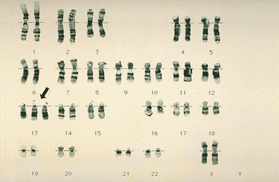
Patau’s Syndrome (Trisomy 13)
1:15,000 births as most fetuses die before term
Of those that survive, 5% live to age 3; 45% die within the first month
Serious eye, brain, and circulatory defects, malformations, kidney/heart defects
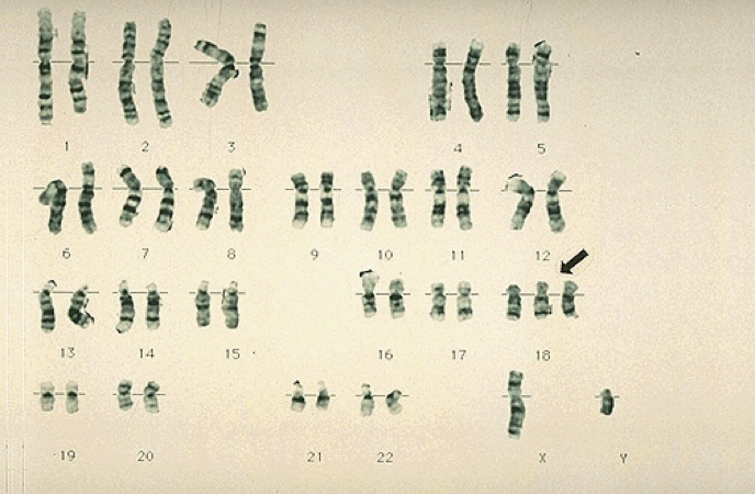
Edward’s Syndrome (Trisomy 18)
Only 10% survive past one year
All die early in infancy
Many complications
(Babies are small, small heads, intellectual disabilities)
Nondisjunction of the Sex Chromosomes
These can be fatal, but most survive just fine
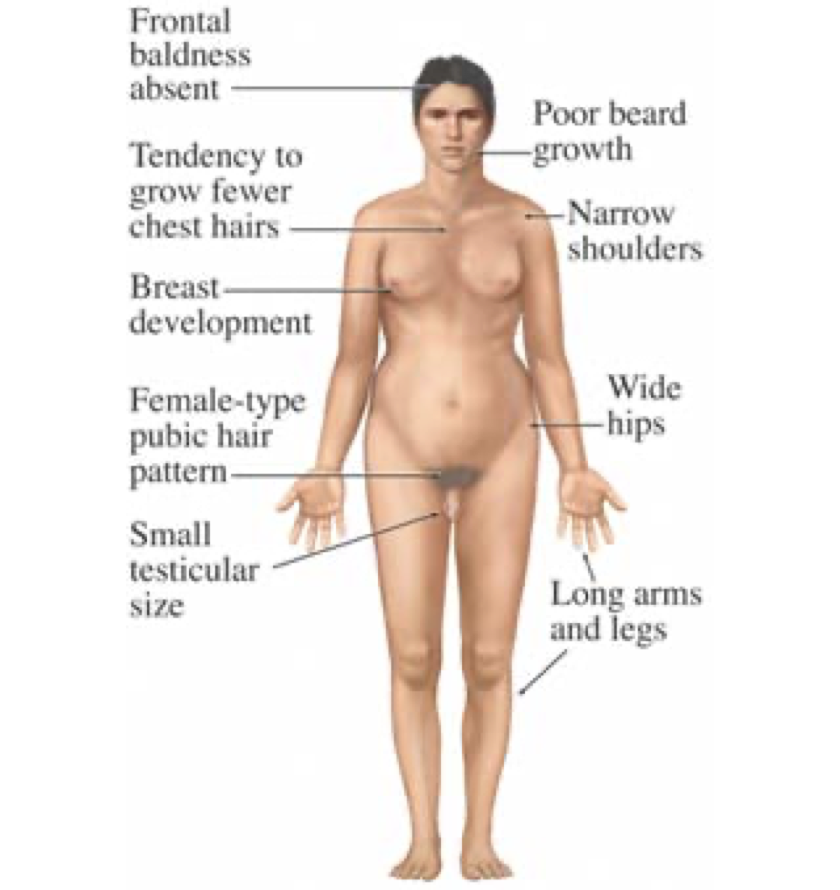
Klinefelter’s Syndrome (XXY Chromosomes)
Affects 1:500 males
Tall, sterile males
Normal intelligence
Has female characteristics
Jacob’s Syndrome (XYY super male)
Genetic conditions where a male has an extra male chromosome (Y)
1 out of 1000 males
Physical features:
Somewhat taller than average
Slightly below normal intelligence
Delayed emotional development
Learning problems in school
Extra testosterone
Other common symptoms include immaturity, acne, swollen joints, arthritis, and many more

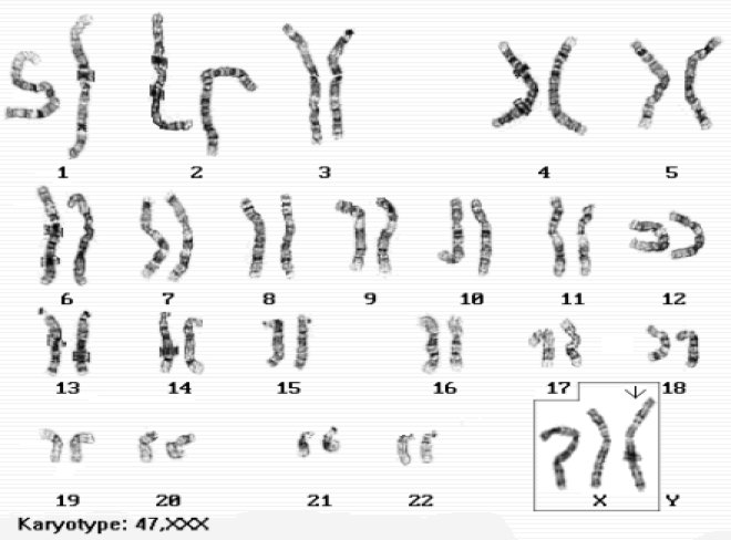
Super Female (XXX)
1:1000 live births
Normal intelligence
Fertile
No physical problems
There are some women who are XXXX and XXXXX – each increasing X results in lesser intelligence and fertility
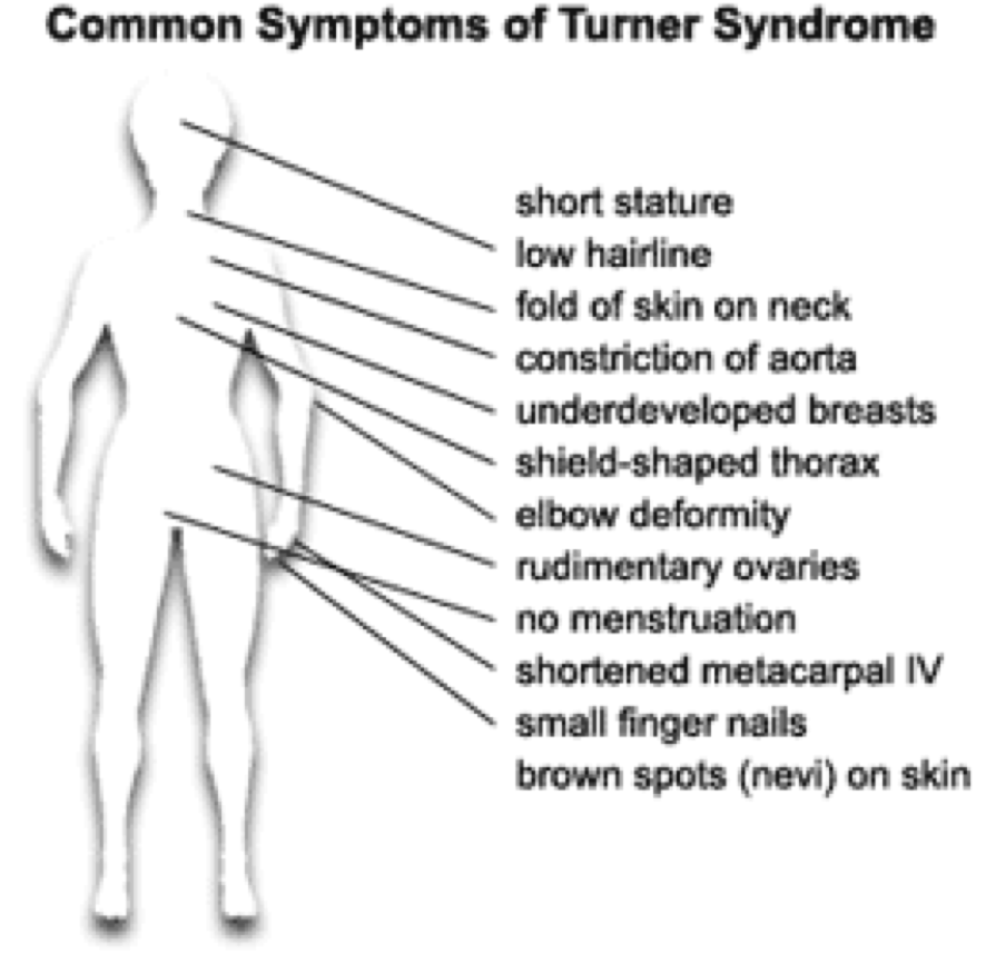
Turner’s Syndrome (X)
The ONLY surviving monosomy
1:2700 births
Live normal lives but do not mature sexually at puberty
Sterile
Physical features:
Short stature
Short broad neck
Broad chest
Other Chromosomeal Issues
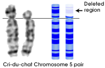
Deletion
A segment of the chromosome is missing
Example: Cri-du-chat (1:1,000,000)
Improperly developed larynx
Severely mentally handicapped
Duplication
Ex. Fragile X 1:1500 males, 2500 females
Most common form of mentally handicapped offspring
Prader-Willi Syndrome
Part of chromosome 15 is missing
Obese
Reduced muscle tone
Reduced mental ability produce little or no sex hormones
Polyploidy
nondisjunction is actually a desired characteristic in the development of large luscious fruit – big strawberries might be 4n or even 6n (polyploidy)
An estimated 30-80% of living plant species are polyploid