Chapter 17: Nucleic Acids and Protein Synthesis
17.1: Components of Nucleic Acids
Nucleic Acids
Deoxyribonucleic acid (DNA)
Ribonucleic acid (RNA)
Nucleotides: These are the repeating monomer units.
Each nucleotide has three components:
A base
A five-carbon sugar
A phosphate group
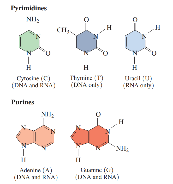
The nitrogen-containing bases in nucleic acids are derivatives of pyrimidine or purine.
In DNA, the purine bases with double rings are adenine (A) and guanine (G); and the pyrimidine bases with single rings are cytosine (C) and thymine (T).
RNA contains the same bases, except thymine (T) is replaced by uracil (U).
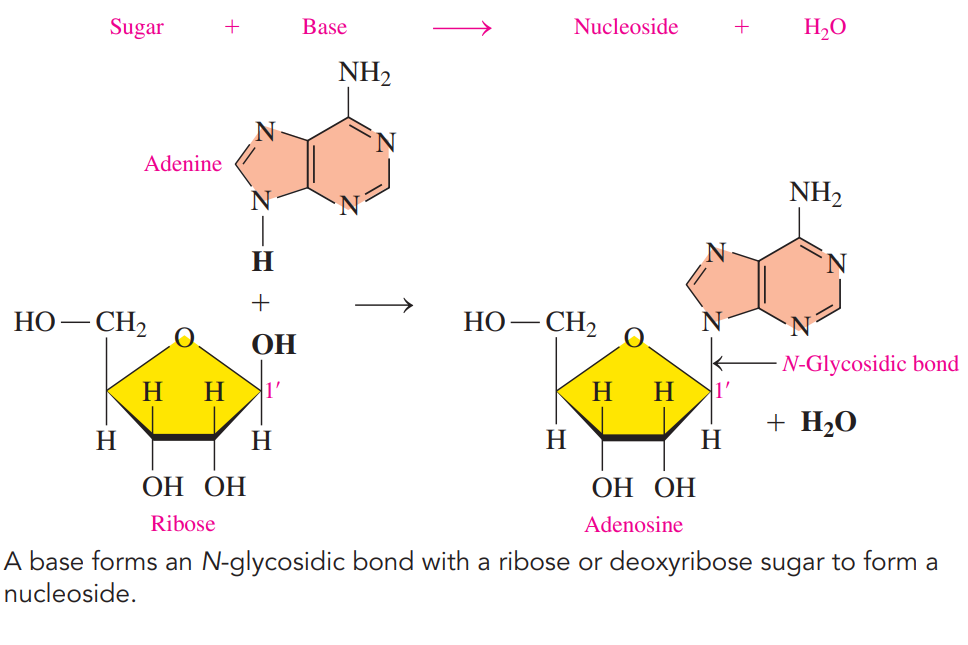
Ribose: The five-carbon sugar in RNA which gives the letter R in the abbreviation of RNA.
Deoxyribose: The five-carbon sugar in DNA, is similar to ribose except that there is no hydroxyl group.
Nucleosides: A combination of sugar and a base, is produced when the nitrogen atom in a pyrimidine or a purine base forms an N-glycosidic bond to carbon 1 of sugar, either ribose or deoxyribose.
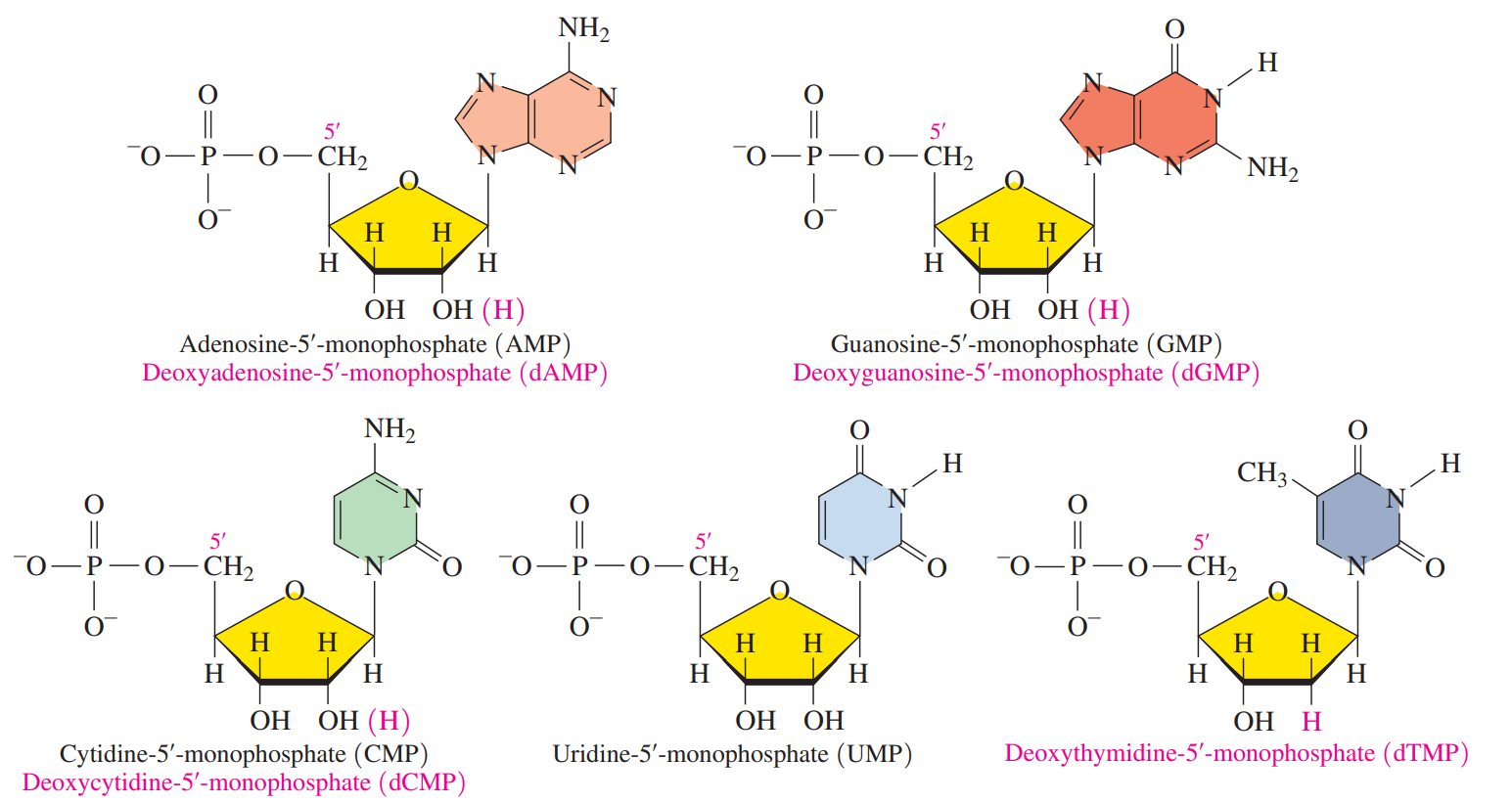
Nucleotides: These are nucleosides in which a phosphate group bonds to the —OH group on carbon 5 of ribose or deoxyribose.
Naming Nucleosides and Nucleotides
Base | Nucleoside | Nucleotide |
|---|---|---|
DNA | ||
Adenine (A) | Deoxyadenosine | Deoxyadenosine-5’ - monophosphate (dAMP) |
Guanine (G) | Deoxyguanosine | Deoxyguanosine-5’ - monophosphate (dGMP) |
Cytosine (C) | Deoxycytidine | Deoxycytidine-5’ - monophosphate (dCMP) |
Thymine (T) | Deoxythymidine | Deoxythymidine-5’ - monophosphate (dTMP) |
RNA | ||
Adenine (A) | Adenosine | Adenosine-5’ - monophosphate (AMP) |
Guanine (G) | Guanosine | Guanosine-5’ - monophosphate (GMP) |
Cytosine (C) | Cytidine | Cytidine-5’ - monophosphate (CMP) |
Uracil (U) | Uridine | Uridine-5’ - monophosphate (UMP) |
17.2: Primary Structure of Nucleic Acids
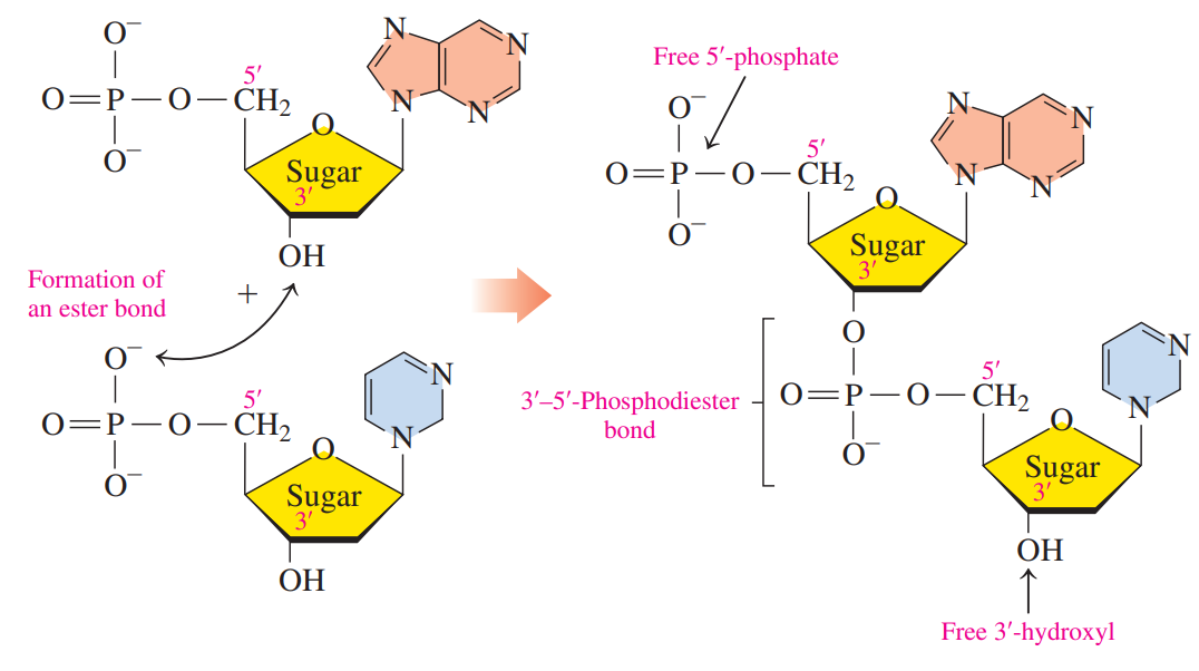
Nucleic Acids: These are polymers of many nucleotides in which the 3′-hydroxyl group of the sugar in one nucleotide bonds to the phosphate group on the 5′-carbon atom in the sugar of the next nucleotide.
Phosphodiester bond: The link between the sugars in adjacent nucleotides.
Primary Structure of Nucleic Acid: It is this sequence of bases that carries the genetic information from one cell to the next.
In any nucleic acid, the sugar at the one end has an unreacted or free 5′-phosphate terminal end, and the sugar at the other end has a free 3′-hydroxyl group.
17.3: DNA Double Helix
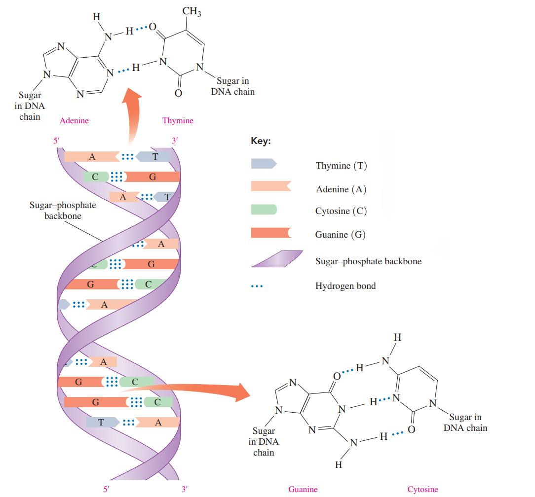
During the 1940s, biologists determined that the bases in DNA from a variety of organisms had a specific relationship: the amount of adenine (A) was equal to the amount of thymine (T), and the amount of guanine (G) was equal to the amount of cytosine (C).
Eventually, scientists determined that adenine is always paired (1:1) with thymine, and guanine is always paired (1:1) with cytosine.
In 1953, James Watson and Francis Crick proposed that DNA was a double helix that consisted of two polynucleotide strands winding about each other like a spiral staircase.
Complementary Base Pairs: The pairs AT and GC;
These are the specific pairing of the bases occur because adenine and thymine form only two hydrogen bonds, while cytosine and guanine form three hydrogen bonds.
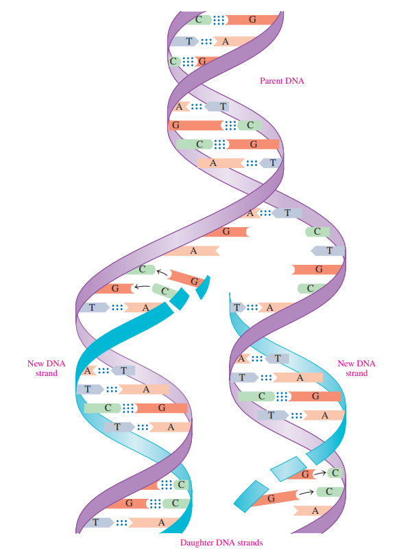
DNA Replication
The strands in the original or parent DNA molecule separate to allow the synthesis of complementary DNA strands.
The process begins when helicase catalyzes the unwinding of a portion of the double helix by breaking the hydrogen bonds between the complementary bases.
The resulting single strands act as templates for the synthesis of new complementary strands of DNA.
17.4: RNA and the Genetic Code
RNA: It makes up most of the nucleic acid found in the cell, is involved with transmitting the genetic information needed to operate the cell.
RNA molecules are polymers of nucleotides as well.
RNA differs from DNA in several important ways:
The sugar in RNA is ribose rather than the deoxyribose found in DNA.
In RNA, the base uracil replaces thymine
RNA molecules are single-stranded, not double-stranded.
RNA molecules are much smaller than DNA molecules.
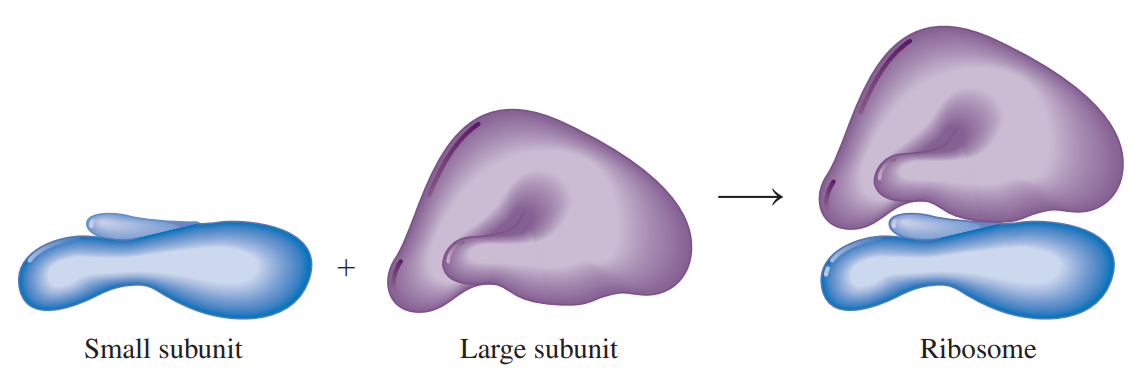
Ribosomal RNA (rRNA): The most abundant type of RNA is combined with proteins to form ribosomes.
Ribosomes, which are the sites for protein synthesis, consist of two subunits: a large subunit and a small subunit.
Messenger RNA (mRNA): It carries genetic information from the DNA, located in the nucleus of the cell, to the ribosomes located in the cytoplasm.
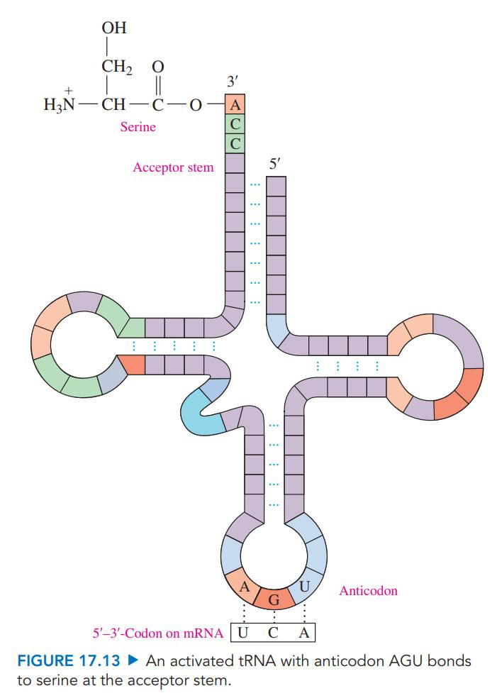
Transfer RNA (tRNA): The smallest of the RNA molecules interprets the genetic information in mRNA and brings specific amino acids to the ribosome for protein synthesis.
Anticodon: A series of three bases that complements three bases on mRNA.
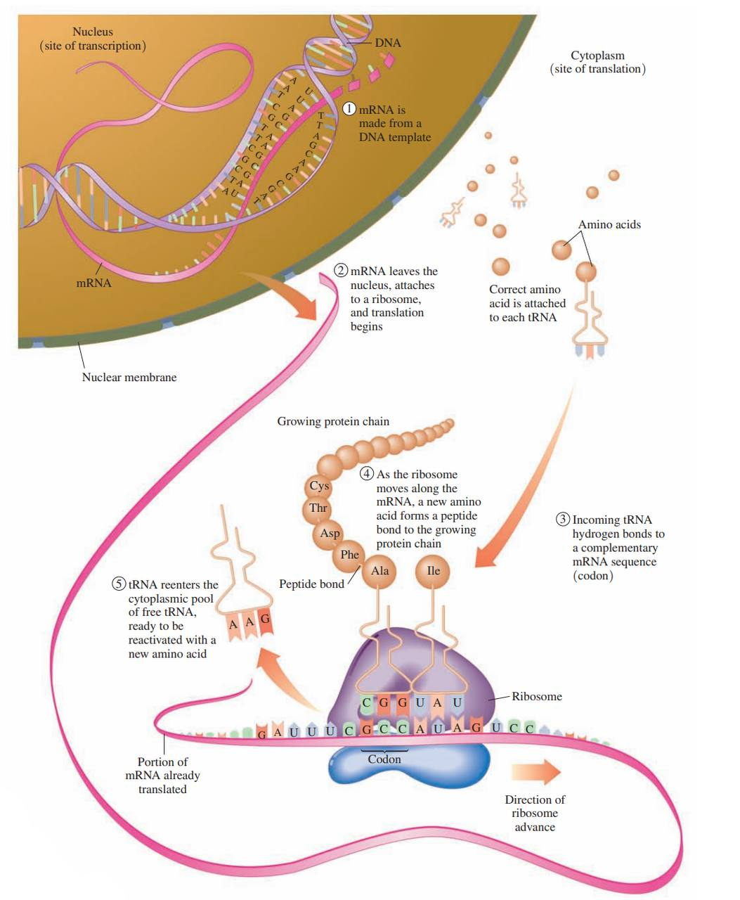
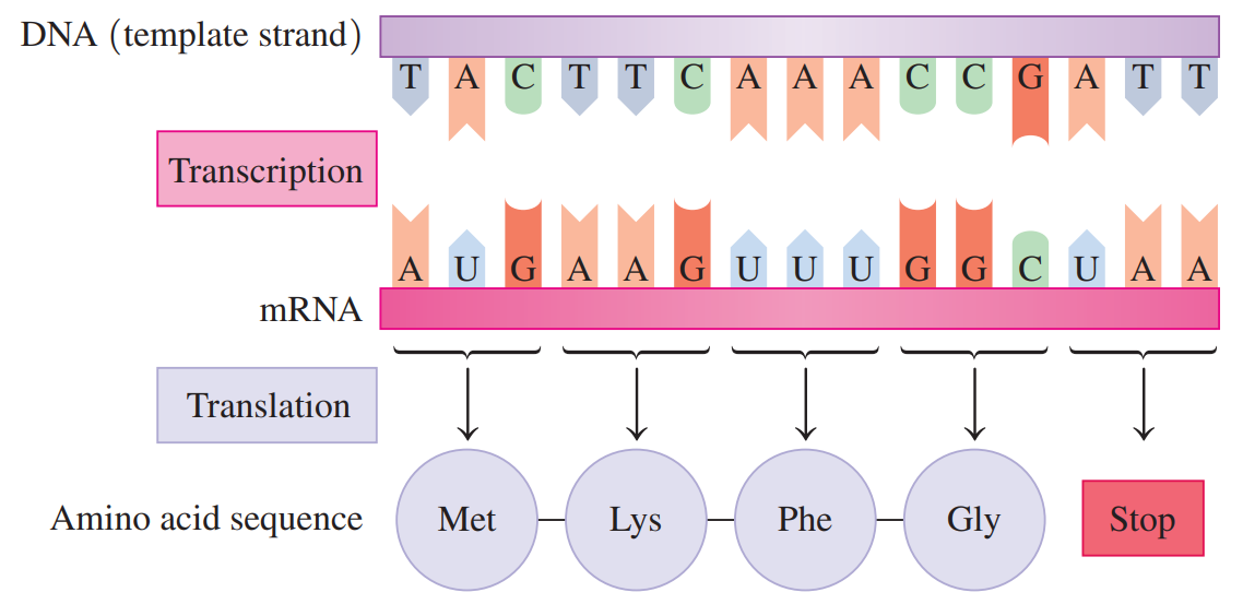
Transcription: Genetic information for the synthesis of a protein is copied from a gene in DNA to make mRNA.
Translation: tRNA molecules convert the information in the mRNA into amino acids, which are placed in the proper sequence to synthesize a protein.
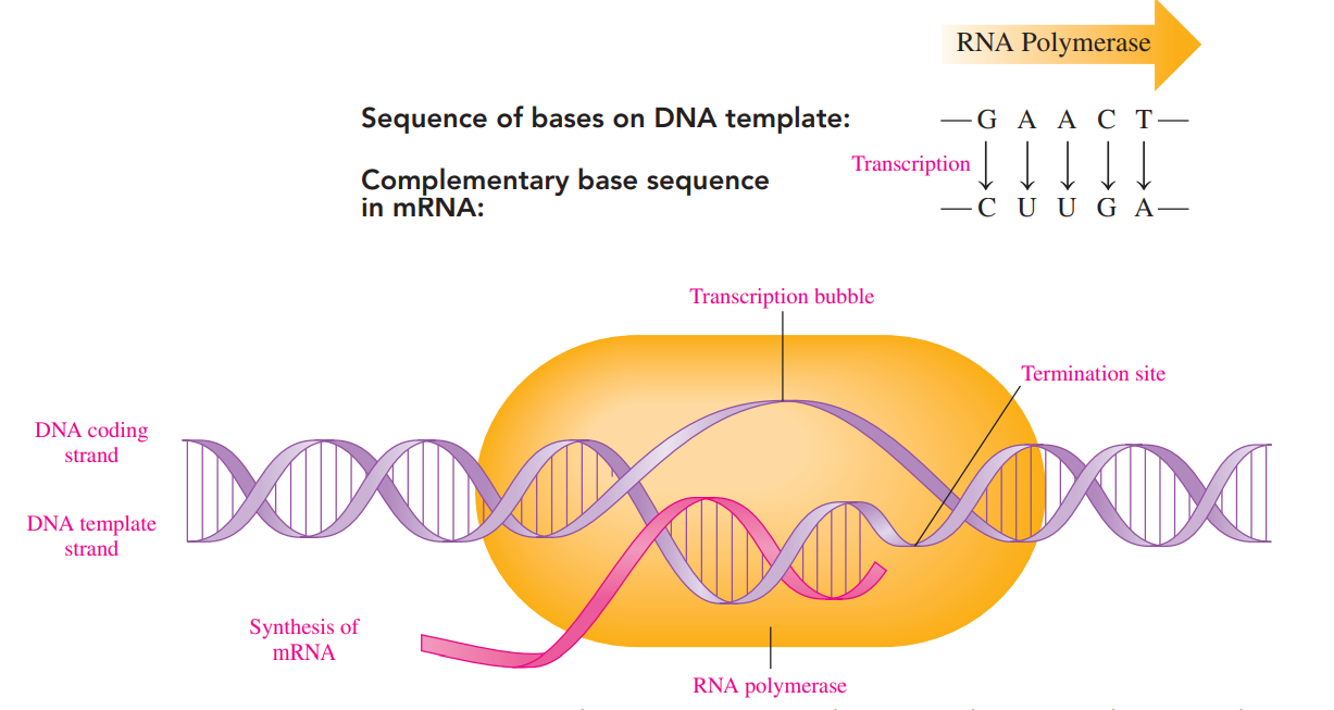
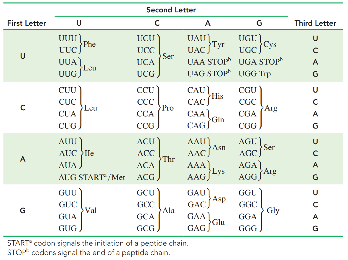
Genetic Code: It consists of a series of three nucleotides in mRNA called codons that specify the amino acids and their sequence in the protein.
Genetic Codes of Amino Acid
17.5: Protein Synthesis
Protein Synthesis
Once the mRNA is synthesized, it migrates out of the nucleus into the cytoplasm to the ribosomes.
In the translation process, tRNA molecules, amino acids, and enzymes convert the codons on mRNA to build a protein.
Activation of tRNA: It occurs when aminoacyl–tRNA synthetase forms an ester bond between the carboxylate group of its amino acid and the hydroxyl group on the acceptor stem.
Initiation and Chain Elongation
The first codon in mRNA is a start codon, AUG, which forms hydrogen bonds with methionine–tRNA.
Another tRNA hydrogen bonds to the next codon, placing a second amino acid adjacent to methionine.
A peptide bond forms between the C-terminal of methionine and the N-terminal of the second amino acid
Translocation: The initial tRNA detaches from the ribosome, which shifts to the next available codon.
During chain elongation, the ribosome moves along the mRNA from codon to codon, so that the tRNAs can attach new amino acids to the growing protein chain.
Sometimes, polysome translates the same strand of mRNA to produce several copies of the protein at the same time.
Chain Termination
Stop codons: These are encountered which the termination of protein synthesis and the release of the protein chain from the ribosome.
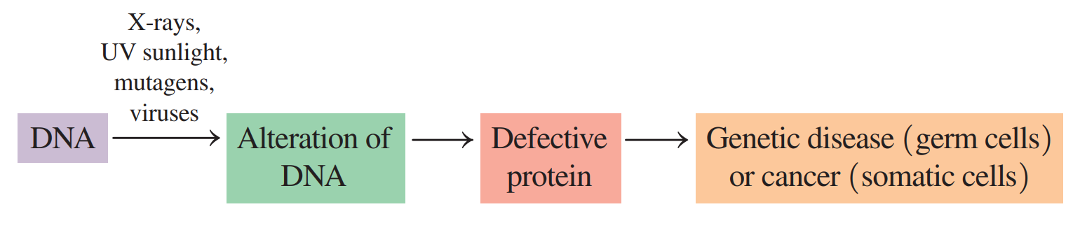
17.6: Genetic Mutations
Mutation: A change in the nucleotide sequence of DNA.
Such a change may alter the sequence of amino acids, affecting the structure and function of a protein in a cell.
If a mutation occurs in a somatic cell, the altered DNA will be limited to that cell and its daughter cells.
If the mutation causes uncontrolled growth, cancer could result.
If a mutation occurs in a germ cell, then all the DNA produced in a new individual will contain the same genetic change.
When a mutation severely alters the function of structural proteins or enzymes, the new cells may not survive or the person may exhibit a genetic disease.
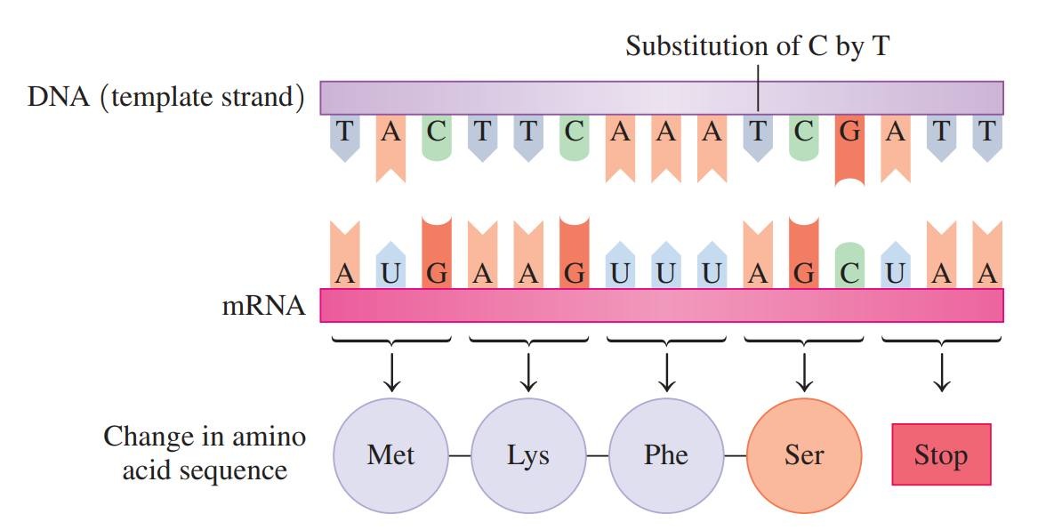
Point Mutation: The replacement of one base in the template strand of DNA with another.
Silent Mutation: This occurs if a substitution gives a codon for the same amino acid, and there is no change in the amino acid sequence in the protein.
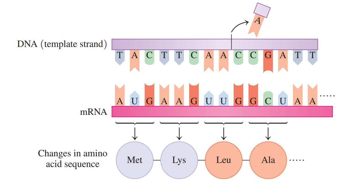
Frameshift Mutation: A base is inserted into or deleted from the normal order of bases in the template strand of DNA.
Genetic Disease: It is the result of a defective enzyme caused by a mutation in its genetic code.
Normal DNA and protein synthesis
Substitution of one base
Frameshift mutation caused by the deletion of a base
List of some Common Genetic Diseases
Galactosemia: The transferase enzyme required for the metabolism of galactose-1-phosphate is absent, resulting in the accumulation of galactose-1-phosphate.
It leads to cataracts and mental retardation.
Cystic fibrosis: It is caused by a mutation in the gene for the protein that regulates the production of stomach fluids and mucus
It is one of the most common inherited diseases in children, in which thick mucus secretions make breathing difficult and block pancreatic function.
Down syndrome: It is the leading cause of mental retardation, occurring in about 1 of every 800 live births; the mother’s age strongly influences its occurrence.
Mental and physical problems, including heart and eye defects, are the result of the formation of three chromosomes, usually under number 21, instead of a pair.
Familial hypercholesterolemia: It occurs when there is a mutation of a gene on chromosome 19, which produces high cholesterol levels.
This usually lead to early coronary heart disease in people 30 to 40 years old
Muscular dystrophy: It is caused by a mutation in the X chromosome.
This muscle-destroying disease appears at about age 5, with death by age 20, and occurs in about 1 of 10 000 males.
Huntington’s disease: It affects the nervous system, leading to total physical impairment.
It is the result of a mutation in a gene on chromosome 4, which can now be mapped to test people in families with a history of HD.
Sickle-cell anemia: It is caused by a defective form of hemoglobin resulting from a mutation in a gene on chromosome 11.
It decreases the oxygen-carrying ability of red blood cells, which take on a sickled shape, causing anemia and plugged capillaries from red blood cell aggregation.
Hemophilia: It is the result of one or more defective blood-clotting factors that lead to poor coagulation, excessive bleeding, and internal hemorrhages.
Tay–Sachs disease: It is the result of defective hexosaminidase A, which causes an accumulation of gangliosides and leads to mental retardation, loss of motor control, and early death.
17.7: Viruses
Viruses
These are small particles of 3 to 200 genes that cannot replicate without a host cell
It does not have the necessary material such as nucleotides and enzymes to make proteins and grow.
The only way a virus can replicate is to invade a host cell and take over the machinery and materials necessary for protein synthesis and growth.
Viral Infection
It begins when an enzyme in the protein coat of the virus makes a hole in the host cell, allowing the viral nucleic acids to enter and mix with the materials in the host cell.
If the virus contains DNA, the host cell begins to replicate the viral DNA in the same way it would replicate normal DNA.

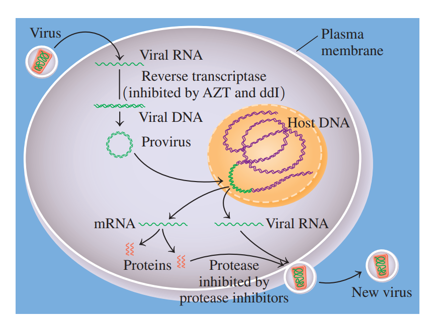
Reverse Transcription
It is a process that occurs once inside the host cell, it must first make viral DNA.
Retrovirus: A virus that contains RNA as its genetic material.
Reverse transcriptase: An polymerase enzyme in a retrovirus that uses the viral RNA template to synthesize complementary strands of DNA.
Provirus: A newly formed DNA that integrates with the DNA of the host cell.
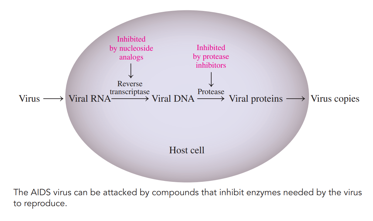
Acquired Immune Deficiency Syndrome
HIV-1 Virus: Known to be the AIDS-causing agent.
HIV: A retrovirus that infects and destroys T4 lymphocyte cells, which are involved in the immune response.
AIDS is characterized by opportunistic infections such as:
Pneumocystis carinii
Kaposi’s sarcoma
Treatment of AIDS often combines reverse transcriptase inhibitors with protease inhibitors such as saquinavir, indinavir, fosamprenavir, nelfinavir, and ritonavir.