


Chapter 4: Inside the Cell
4.1 Cells Under the Microscope
Cell
The fundamental unit of life.
The body is composed of several hundred cell types, and each type is present in billions of copies.
While cells are complex, they are tiny and require a microscope to be seen.
A microscope enables us to see the surface features and fine details of cells and even some of the larger molecules within them.
Cells need to be able to rapidly exchange materials with the external environment.
It is because of this that a cell needs a surface area large enough to allow efficient movement of nutrients into the cell and waste materials out of the cell.
Small cells are most likely to have an adequate surface area for exchanging wastes and nutrients.
Many cells also possess adaptations that increase the surface-area-to-volume ratio.
4.2 The Plasma Membrane
All cells have an outer membrane called the plasma membrane, which acts as the boundary between the outside and inside of a cell.
The membrane acts as a gatekeeper, making it vital to the cell.
The plasma membrane is in charge of regulating the passage of molecules and ions into and out of the cell.
The structure of the plasma membrane plays a vital role in its function and is broken down here:
A phospholipid bilayer with numerous proteins embedded in it.
A bilayer where the polar (hydrophilic) heads of the phospholipids are oriented in two directions.
An outer layer of the membrane, where the phospholipid heads face the external environment.
An interior layer of the membrane, where the heads of phospholipid molecules are directed toward the interior cytoplasm of the cell.
The nonpolar (hydrophobic) tails of the phospholipids point toward each other in the space between the layers, where there is no water.
Cholesterol molecules present in the plasma membrane of some cells lend support to the membrane, giving it the general consistency of olive oil.
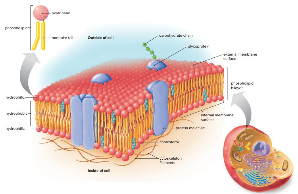
The structure of the plasma membrane is often referred to as the fluid-mosaic model since the protein molecules embedded in the membrane have a pattern (a mosaic) within the fluid phospholipid bilayer.
The actual pattern of proteins varies according to the type of cell, but it may also vary within the membrane of an individual cell over time.
Some glycoproteins are involved in establishing the identity of the cell; they often play an important role in the immune response against disease-causing agents entering the body.
Functions of Membrane Proteins
Channel Proteins
Channel proteins form a tunnel across the entire membrane, allowing only one or a few types of specific molecules to move readily through the membrane
Transport Proteins
Transport proteins are also involved in the passage of molecules and ions through the membrane. They often combine with a substance and help it move across the membrane with an input of energy.
Cell Recognition Proteins
Cell recognition proteins are glycoproteins; Among other functions, these proteins enable our bodies to distinguish between our own cells and the cells of other organisms. Without this distinction, pathogens would be able to invade the body freely.
Receptor Proteins
A receptor protein has a shape that allows a specific molecule, called a signal molecule, to bind to it; The binding of a signal molecule causes the receptor protein to change its shape and thereby bring about a cellular response.
Enzymatic Proteins
Some plasma membrane proteins are enzymatic proteins that directly participate in metabolic reactions; Without enzymes, some of which are attached to the various membranes of a cell, the cell would never be able to perform the degradative and synthetic reactions that are important to its function.
Junction Proteins
Proteins are also involved in forming various types of junctions between cells; The junctions assist cell-to-cell adhesion and communication. The adhesion junctions in your bladder keep the cells bound together as the bladder swells with urine.
4.3 The Two Main Types of Cells
The idea that all organisms are composed of cells and that cells come only from preexisting cells are the two central tenets of the cell theory.
Characteristics common in all cells include the following:
A plasma membrane made of phospholipids that regulates the movement of materials into and out of the cell
A semifluid interior called the cytoplasm, where chemical reactions occur
Genetic material (DNA) that provides the information needed for cellular activities, including growth and reproduction.
Cells are divided into two main types according to the way their genetic material is organized, those being:
Prokaryotic cells
Lack a membrane-bound nucleus.
DNA is located in a region of the cytoplasm called the nucleoid.
Eukaryotic cells
Have a nucleus that houses their DNA.
Prokaryotic Cells
The first cells to appear on Earth were prokaryotes.
Prokaryotes are classified as being part of either domain Archaea or domain Bacteria.
Prokaryotic cells are generally much smaller in size and simpler in structure than eukaryotic cells.
The prokaryotic cell’s small size and simple structure allow them to reproduce very quickly and effectively; they exist in great numbers in the following areas:
Air
Bodies of water
Soil
on us
Even with the slightly destructive nature of bacteria, the biosphere would not long continue without bacteria.
Significant contributions bacteria has are the following:
Contribute to ecological cycles.
Use in industrial chemicals, drugs, and foods.
We are dependent on bacteria for many things, including the synthesis of some vitamins we can’t make ourselves.
Knowledge about how DNA specifies the sequence of amino acids in proteins was significantly advanced by experiments utilizing Escherichia coli (E.coli).
Bacterial Structure
Capsule
gel-like coating outside the cell wall.
A protective layer of polysaccharides that lies outside the cell wall.
Nucleoid
The location of the bacterial chromosome
DNA is located in a single circular, coiled chromosome that resides in a region of the cell called the nucleoid.
Ribosome
The site of protein synthesis.
The many proteins specified by bacterial DNA are synthesized on tiny structures called ribosomes.
Plasma Membrane
The sheet that surrounds the cytoplasm and regulates the entrance and exit of molecules.
Cell wall
The structure that provides support and shapes the cell.
Cytoplasm
Semifluid solution surrounded by the plasma membrane; contains nucleoid and ribosomes.
Flagellum
Rotating filament that propels the cell.
4.4 Eukaryotic Cells
Eukaryotic cells are highly compartmentalized.
These compartments are formed by membranes that create internal spaces that divide the labor necessary to conduct life functions.
Organelles
The compartments of a eukaryotic cell.
Organelles carry out specialized functions that together allow the cell to be more efficient and successful.
A membrane surrounds nearly all organelles with embedded proteins, many of which are enzymes.
Enzymes
Molecules that speed up chemical reactions
Nucleus
A compartment that houses the genetic material within eukaryotic
chromosomes, which contain hereditary information.
The nucleus communicates with ribosomes in the cytoplasm and the organelles of the endomembrane system.
Vesicles
Membranous sacs that enclose the molecules and keep them separate from the cytoplasm.
Vesicles move around by means of an extensive network of protein fibers called the cytoskeleton, which also maintains cell shape and assists with cell movement.
Nucleus and Ribosomes
The nucleus stores genetic information, and the ribosomes in the cytoplasm use this information to carry out the manufacture of proteins.
The Nucleus
The nucleus is one of the most noticeable structures in the eukaryotic cell.
The nucleus contains chromatin within a semifluid matrix called the nucleoplasm.
Chromatin
A network of DNA, protein, and a small amount of RNA.
Just before the cell divides, the chromatin condenses and coils into rodlike structures called chromosomes.
All the cells of an organism contain the same number of chromosomes except for the egg and sperm, which usually have half this number.
The DNA within a chromosome is organized into genes, each of which has a specific sequence of nucleotides.
These nucleotides may code for a polypeptide or sometimes regulatory RNA molecules.
mRNA acts as a messenger between the DNA and the ribosome, where polypeptide chains are formed.
Because proteins are important in determining the structure and function of a cell, the nucleus may be thought of as the command center of the cell.
Nucleolus
A dark structure within the nucleus that is a type of RNA called ribosomal RNA (rRNA) is produced.
Proteins join with rRNA to form the subunits of ribosomes.
The assembled ribosomal subunits are then sent out of the nucleus into the cytoplasm, where they join and assume their role in protein synthesis.
The nucleus is separated from the cytoplasm by a double membrane of phospholipids known as the nuclear envelope.
Ribosomes
Ribosomes are found in both prokaryotes and eukaryotes.
In both types of cells, ribosomes are composed of two subunits, one large and one small.
Each subunit has its own mix of proteins and rRNA.
The ribosome acts as a workbench, and it is here that the information contained within the mRNA from the nucleus is used to synthesize a polypeptide chain.
Proteins may contain one or more polypeptide chains.
In eukaryotic cells, some ribosomes occur freely within the cytoplasm; Other ribosomes are attached to the endoplasmic reticulum (ER), an organelle of the endomembrane system.
After the ribosome binds to a receptor at the ER, the polypeptide being synthesized enters the lumen (interior) of the ER, where it may be further modified and then assume its final shape.
Endomembrane System
Endomembrane system
Consists of the nuclear envelope, the membranes of the endoplasmic reticulum (ER), the Golgi apparatus, and numerous vesicles.
This system helps compartmentalize the cell so that particular enzymatic reactions are restricted to specific regions.
Transport vesicles carry molecules from one part of the system to another.
Endoplasmic Reticulum
Endoplasmic reticulum (ER)
Consists of an interconnected system of membranous channels and saccules (flattened vesicles).
It is physically continuous with the outer membrane of the nuclear envelope.
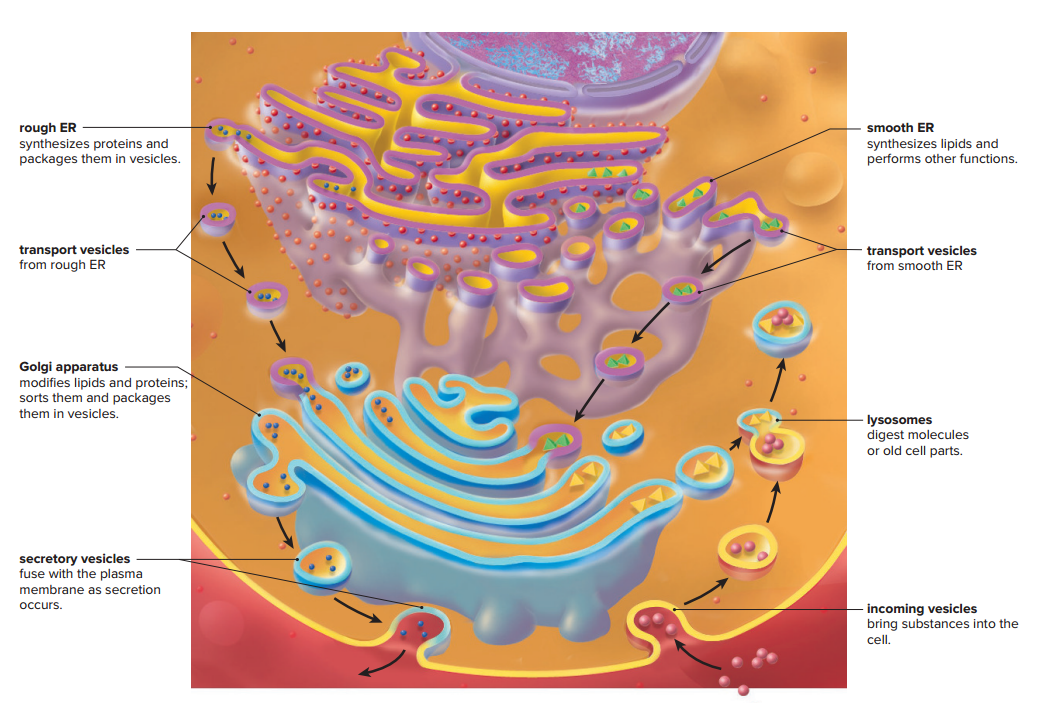
Rough ER is studded with ribosomes on the side of the membrane that faces the cytoplasm; therefore, rough ER is able to synthesize polypeptides.
It also modifies the polypeptides after they have entered the central enclosed region of the ER, called the lumen, where proteins take shape.
The rough ER forms transport vesicles, which take proteins to other parts of the cell.
Smooth ER**,** which is continuous with rough ER, does not have attached ribosomes.
Smooth ER synthesizes lipids, such as phospholipids and steroids; The functions of smooth ER are dependent on the particular cell.
Smooth ER produces testosterone, and in the liver, it helps detoxify drugs.
Smooth ER also forms transport vesicles that carry molecules to other parts of the cell.
Golgi Apparatus
Golgi apparatus
Consists of a stack of slightly curved, flattened saccules resembling pancakes.
The golgi apparatus First receives transport vesicles sent to it by rough and smooth ER; The molecules within the vesicles are modified as they move between saccules.
In animal cells, some of the vesicles that leave the Golgi are lysosomes.
Lysosomes
Lysosomes
Vesicles, produced by the Golgi apparatus, that digest molecules and even portions of the cell itself.
In Tay-Sachs disease, a genetic disorder, lysosomes in nerve cells are missing an enzyme for a particular lipid molecule.
The cells become so full of storage lipids that they lose their ability to function.
In all cases, the individual dies, usually in childhood.
Vesicles and Vacuoles
Vacuoles
Membranous sacs larger than vesicles and more specialized.
Vacuoles contain not only water, sugars, and salts but also pigments and toxic molecules.
Energy-Related Organelles
Chloroplasts and mitochondria are the two eukaryotic organelles that specialize in energy conversion.
Chloroplasts
Use solar energy to synthesize carbohydrates.
Mitochondria
Break down carbohydrates to produce adenosine triphosphate (ATP) molecules.
The production of ATP is significant because ATP serves as a carrier of energy in cells.
Without a constant supply of ATP, no cell could exist for long.
Chloroplasts
Chloroplast
An organelle found in plants and algae and is the location where carbon dioxide gas, water, and energy from the sun are used to produce carbohydrates by the process of photosynthesis.
Chloroplasts are very large, having twice the width and as much as five times the length of a mitochondrion.
Chloroplasts are bound by a double membrane, which includes an outer membrane and an inner membrane.
The large inner space, called the stroma, contains a concentrated mixture of enzymes and disclike sacs called thylakoids.
Granum
A stack of thylakoids.
The pigments that capture solar energy are located in the membrane of the thylakoids, and the enzymes that synthesize carbohydrates are in the stroma.
The carbohydrates produced by chloroplasts serve as organic nutrient molecules for plants and, ultimately, for all living organisms on the planet.
The discovery that chloroplasts have their own DNA and ribosomes supports an accepted theory that chloroplasts are derived from photosynthetic bacteria that entered a eukaryotic cell in the distant past.
This process is called endosymbiosis.
Mitochondria
Mitochondria
Smaller than chloroplasts.
Usually visible only under an electron microscope.
Often change shape.
A double membrane binds mitochondria.
The inner membrane is highly convoluted into folds, called cristae, that project into the interior space called the matrix.
Cristae increase the surface area of the inner membrane so much that, in a liver cell, they account for about one-third of the total membrane in the cell.
Mitochondria are often called the powerhouses of the cell because they produce most of the ATP the cell utilizes.
The matrix contains a highly concentrated mixture of enzymes that assists in the breakdown of carbohydrates and other nutrient molecules.
Cellular respiration
The complete breakdown of carbohydrates which also involves cytoplasm
The matrix contains mitochondrial DNA and ribosomes.
The presence of mitochondrial DNA and ribosomes is evidence that mitochondria and chloroplasts have similar origins and are derived from bacteria that took up residence in an early eukaryotic cell.
The origin of mitochondria is an example of endosymbiosis.
All eukaryotic cells (with a few rare exceptions) have mitochondria, but only photosynthetic organisms (plants and algae) have chloroplasts.
The Cytoskeleton and Motor Proteins
Cytoskeleton
A network of interconnected protein filaments and tubules that extends from the nucleus to the plasma membrane in eukaryotic cells.
Much as bones and muscles give an animal structure and produce movement, the elements of the cytoskeleton maintain cell shape and, along with motor proteins, allow the cell and its organelles to move.
Unlike an animal’s skeleton, the cytoskeleton is highly dynamic—its elements can be quickly assembled and disassembled as appropriate.
The cytoskeleton includes microtubules, intermediate filaments, and actin filaments.
Motor Proteins
Motor proteins associated with the cytoskeleton are instrumental in allowing cellular movements.
The major motor proteins are the following:
Myosin
Interacts with actin filaments when movement occurs.
Kinesin & Dynein
Move along microtubules much as a car travels along a highway.
Microtubules
Microtubules
Small, hollow cylinders composed of 13 long chains of tubulin dimers.
Microtubules are dynamic; they can easily change their length by removing tubulin dimers.
This process is controlled by the centrosome, a microtubule organizing center that lies near the nucleus.
Microtubules radiating from the centrosome help maintain the shape of the cell and act as tracks along which organelles and other materials can move.
Intermediate Filament
Intermediate filaments are intermediate in size between actin filaments and microtubules.
Intermediate filaments are ropelike assemblies of proteins that typically run between the nuclear envelope and the plasma membrane.
The network filaments form supports both the nucleus and the plasma membrane.
The protein making up intermediate filaments differs according to the cell type.
Intermediate filaments made of the protein keratin give great mechanical strength to skin cells.
Actin Filaments
Each actin filament consists of two chains of globular actin monomers twisted about one another in a helical manner to form a long filament.
Actin filaments support the cell, forming a dense, complex web just under the plasma membrane.
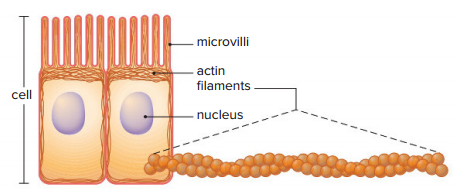
Actin filaments support projections of the plasma membrane, such as microvilli.
Centrioles
Centrioles
Short, barrel-shaped organelles composed of microtubules located in the centrosome
It is possible that centrioles give rise to basal bodies, which are located at the base of cilia and flagella and are believed to organize the microtubules in these structures.
The centrioles are also involved in organizing microtubules during cell division.
Some eukaryotes, such as plants and fungi, lack centrioles (although they have centrosomes), suggesting that centrioles are not necessary for the assembly of cytoplasmic microtubules.
Cilia and Flagella
Cilia and flagella
whiplike projections of cells.
Cilia move stiffly, like an oar, and flagella move in an undulating, snakelike fashion.
Cilia are short (2–10 μm), while flagella are longer (usually no more than 200 μm).
Some single-celled protists utilize cilia or flagella to move about.
In our bodies, ciliated cells are critical to respiratory health and our ability to reproduce.
The ciliated cells that line our respiratory tract sweep debris trapped within mucus back up into the throat, which helps keep the lungs clean.
Ciliated cells also move an egg along the uterine tube, where it can be fertilized by a flagellated sperm cell.
4.5 Outside the Eukaryotic Cell
A cell does not consist only of its plasma membrane and internal contents; in fact, Most cells also have extracellular structures formed from materials the cell produces and transports across its plasma membrane.
These structures may either provide support or allow for interaction with other cells.
Cell Walls
Cell wall
Provides support to the cell.
Cell walls are found in many eukaryotic cells, including those of plants, fungi, and most protists but not those of animals.
The composition of the cell wall differs between plants and fungi.
A primary cell wall contains cellulose fibrils and noncellulose substances, and these allow the wall to stretch when the cell is growing.
Adhesive substances are abundant outside the cell wall in the middle lamella, a layer that holds two plant cells together.
For added strength, some plant cells have a secondary cell wall that forms inside the primary cell wall.
The secondary wall has a greater quantity of cellulose fibrils, which are laid down at right angles to one another. Lignin, a substance that adds strength, is a common ingredient of secondary cell walls.
Extracellular Metrix
Animal cells do not have a cell wall, but they do have an extracellular matrix outside the cell.
Extracellular matrix (ECM)
A meshwork of fibrous proteins and polysaccharides in close association with the cell that produced them.
Collagen and elastin are two well-known proteins in the extracellular matrix.
Collagen resists stretching, and elastin provides resilience.
Polysaccharides play a dynamic role by directing the migration of cells along collagen fibers during development.
Other ECM proteins bind to receptors in a cell’s plasma membrane, permitting communication between the extracellular matrix and the cytoskeleton within the cytoplasm of the cell.
The extracellular matrices of tissues vary greatly; they may be quite flexible, as in cartilage, or rock solid, as in bone. The rigidity of the extracellular matrix is influenced mainly by the number and types of protein fibers present and how they are arranged.
The extracellular matrix of bone is very hard because, in addition to the components already mentioned, mineral salts, notably calcium salts, are deposited outside the cell.
Junctions Between Cells
Three types of junctions are found between certain cells, those being the following:
Junctions
Internal cytoplasmic plaques, firmly attached to the cytoskeleton within each cell, are joined by intercellular filaments.
The result is a sturdy but flexible sheet of cells.
In some organs, junctions hold the cells together.
Tight junctions
The plasma membrane proteins attach to each other, producing a zipperlike fastening.
The cells of tissues that serve as barriers are held together by tight junctions.
Gap junctions
Allows cells to communicate
A gap junction is formed when two identical plasma membrane channels join; The channel of each cell is lined by six plasma membrane proteins that allow the junction to open and close.
A gap junction lends strength to the cells, but it also allows small molecules and ions to pass between them.
Gap junctions are important in heart muscle and smooth muscle because they permit the flow of ions that is required for the cells in these tissues to contract as a unit.
The type of junction between two cells depends on whether or not the cells need to be able to exchange materials and whether or not they need to be joined together very tightly.
In a plant, living cells are connected by plasmodesmata.
Plasmodesmata
Numerous narrow, membrane-lined channels that pass through the cell wall.
Cytoplasmic strands within these channels allow the direct exchange of some materials between adjacent plant cells and eventually among all the cells of a plant.
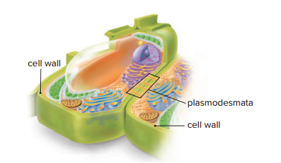
The plasmodesmata allow only water and other small molecules to pass freely from cell to cell.