


Chapter 13: Anatomy of the Nervous System
The Neural Tube
Molecular signals induce cells in this region to differentiate into the neuroepithelium, forming a neural plate.
A neural groove forms, visible as a line along the dorsal surface of the embryo.
The ridge-like edge on either side of the neural groove is referred as the neural fold.
As the neural folds come together and converge, the underlying structure forms into a tube just beneath the ectoderm called the neural tube.
Cells from the neural folds then separate from the ectoderm to form a cluster of cells referred to as the neural crest, which runs lateral to the neural tube.
Primary Vesicles
The prosencephalon (pros- = “in front”) is the forward-most vesicle, and the term can be loosely translated to mean forebrain.
The mesencephalon (mes- = “middle”) is the next vesicle, which can be called the midbrain.
The third vesicle at this stage is the rhombencephalon.
Secondary Vesicles
The three primary vesicles become five secondary vesicles.
The prosencephalon enlarges into two new vesicles called the telencephalon and the diencephalon.
Relating Embryonic Development to the Adult Brain
The neural tube establishes the anterior–posterior dimension of the nervous system, which is called the neuraxis.
There is a major curve between the brain stem and forebrain, which is called the cephalic flexure.
The Cerebrum
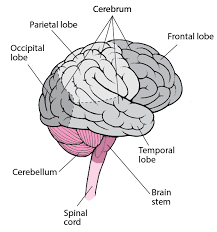
The iconic gray mantle of the human brain, which appears to make up most of the mass of the brain, is the cerebrum.
The wrinkled portion is the cerebral cortex, and the rest of the structure is beneath that outer covering.
There is a large separation between the two sides of the cerebrum called the longitudinal fissure.
It separates the cerebrum into two distinct halves, a right and left cerebral hemisphere.
Deep within the cerebrum, the white matter of the corpus callosum provides the major pathway for communication between the two hemispheres of the cerebral cortex.
The basal nuclei are responsible for cognitive processing, the most important function being that associated with planning movements.
The basal forebrain contains nuclei that are important in learning and memory.
The limbic cortex is the region of the cerebral cortex that is part of the limbic system, a collection of structures involved in emotion, memory, and behavior.
Cerebral Cortex
A gyrus (plural = gyri) is the ridge of one of those wrinkles, and a sulcus (plural = sulci) is the groove between two gyri.
The lateral sulcus that separates the temporal lobe from the other regions is one such landmark.
Superior to the lateral sulcus are the parietal lobe and frontal lobe, which are separated from each other by the central sulcus.
The posterior region of the cortex is the occipital lobe, which has no obvious anatomical border between it and the parietal or temporal lobes on the lateral surface of the brain.
From the medial surface, an obvious landmark separating the parietal and occipital lobes is called the parietooccipital sulcus.
The main sensation associated with the parietal lobe is somatosensation, meaning the general sensations associated with the body.
Posterior to the central sulcus is the postcentral gyrus, the primary somatosensory cortex, which is identified as Brodmann’s areas 1, 2, and 3.
All of the tactile senses are processed in this area, including touch, pressure, tickle, pain, itch, and vibration, as well as more general senses of the body such as proprioception and kinesthesia, which are the senses of body position and movement.
The precentral gyrus is the primary motor cortex.
The premotor area is responsible for thinking of a movement to be made.
Broca’s area is responsible for the production of language, or controlling movements responsible for speech; in the vast majority of people, it is located only on the left side.
Anterior to these regions is the prefrontal lobe, which serves cognitive functions that can be the basis of personality, short-term memory, and consciousness.
Subcortical structures
Beneath the cerebral cortex are sets of nuclei known as subcortical nuclei that augment cortical processes.
The hippocampus and amygdala are medial-lobe structures that, along with the adjacent cortex, are involved in long-term memory formation and emotional responses.
The major structures of the basal nuclei that control movement are the caudate, putamen, and globus pallidus, which are located deep in the cerebrum.
The caudate and putamen are called the striatum.
The direct pathway is the projection of axons from the striatum to the globus pallidus internal segment (GPi) and the substantia nigra pars reticulata (SNr).
The indirect pathway is the projection of axons from the striatum to the globus pallidus external segment (GPe), then to the subthalamic nucleus (STN), and finally to GPi/SNr.
The direct pathway causes the disinhibition of the thalamus (inhibition of one cell on a target cell that then inhibits the first cell), whereas the indirect pathway causes, or reinforces, the normal inhibition of the thalamus.
The switch between the two pathways is the substantia nigra pars compacta, which projects to the striatum and releases the neurotransmitter dopamine.
The Diencephalon
The single exception is the system associated with olfaction, or the sense of smell, which connects directly with the cerebrum.
The diencephalon can be described as any region of the brain with “thalamus” in its name.
There are other structures, such as the epithalamus, which contains the pineal gland, or the subthalamus, which includes the subthalamic nucleus that is part of the basal nuclei.
Thalamus: The thalamus is a collection of nuclei that relay information between the cerebral cortex and the periphery, spinal cord, or brain stem.

Hypothalamus
Inferior and slightly anterior to the thalamus is the hypothalamus, the other major region of the diencephalon.
The hypothalamus is a collection of nuclei that are largely involved in regulating homeostasis.

Brain Stem: The midbrain and hindbrain (composed of the pons and the medulla) are collectively referred to as the brain stem.
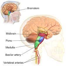
Midbrain
It is separated into the tectum and tegmentum, from the Latin words for roof and floor, respectively.
The inferior colliculus is the inferior pair of these enlargements and is part of the auditory brain stem pathway.
The superior colliculus is the superior pair and combines sensory information about visual space, auditory space, and somatosensory space.
Pons
The word pons comes from the Latin word for bridge.
It is visible on the anterior surface of the brain stem as the thick bundle of white matter attached to the cerebellum.
Medulla
The medulla is the region known as the myelencephalon in the embryonic brain.
The initial portion of the name, “myel,” refers to the significant white matter found in this region—especially on its exterior, which is continuous with the white matter of the spinal cord.
A diffuse region of gray matter throughout the brain stem, known as the reticular formation, is related to sleep and wakefulness, such as general brain activity and attention.
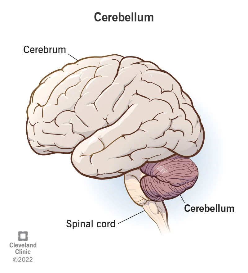
The Cerebellum
The cerebellum, as the name suggests, is the “little brain.”
It is covered in gyri and sulci like the cerebrum, and looks like a miniature version of that part of the brain.
Sensory information from the periphery, which enters through spinal or cranial nerves, is copied to a nucleus in the medulla known as the inferior olive.
The Spinal Cord
The description of the CNS is concentrated on the structures of the brain, but the spinal cord is another major organ of the system.
The anterior midline is marked by the anterior median fissure, and the posterior midline is marked by the posterior median sulcus.
Axons enter the posterior side through the dorsal (posterior) nerve root, which marks the posterolateral sulcus on either side.
This comes from the initial development of the spinal cord, which is divided into the basal plate and the alar plate.

White Columns
Ascending tracts of nervous system fibers in these columns carry sensory information up to the brain, whereas descending tracts carry motor commands from the brain.
Between the two posterior horns of gray matter are the posterior columns.
Between the two anterior horns, and bounded by the axons of motor neurons emerging from that gray matter area, are the anterior columns.
The white matter on either side of the spinal cord, between the posterior horn and the axons of the anterior horn neurons, are the lateral columns.
Blood Supply to the Brain
The next branches give rise to the common carotid arteries, which further branch into the internal carotid arteries.
The orthostatic reflex is a reaction to this change in body position, so that blood pressure is maintained against the increasing effect of gravity (orthostatic means “standing up”).
The internal carotid artery enters the cranium through the carotid canal in the temporal bone.
A second set of vessels that supply the CNS are the vertebral arteries, which are protected as they pass through the neck region by the transverse foramina of the cervical vertebrae.
The vertebral arteries enter the cranium through the foramen magnum of the occipital bone.
Branches off the left and right vertebral arteries merge into the anterior spinal artery supplying the anterior aspect of the spinal cord, found along the anterior median fissure.
The two vertebral arteries then merge into the basilar artery,which gives rise to branches to the brain stem and cerebellum.
Venous Return
After passing through the CNS, blood returns to the circulation through a series of dural sinuses and veins.
The superior sagittal sinus runs in the groove of the longitudinal fissure, where it absorbs CSF from the meninges.
The superior sagittal sinus drains to the confluence of sinuses, along with the occipital sinuses and straight sinus, to then drain into the transverse sinuses.
The transverse sinuses connect to the sigmoid sinuses, which then connect to the jugular veins.
Protective Coverings of the Brain and Spinal Cord
The outer surface of the CNS is covered by a series of membranes composed of connective tissue called the meninges, which protect the brain.
The dura mater is a thick fibrous layer and a strong protective sheath over the entire brain and spinal cord. It is anchored to the inner surface of the cranium and vertebral cavity.
The arachnoid mater is a membrane of thin fibrous tissue that forms a loose sac around the CNS.
Beneath the arachnoid is a thin, filamentous mesh called the arachnoid trabeculae, which looks like a spider web, giving this layer its name.
Directly adjacent to the surface of the CNS is the pia mater, a thin fibrous membrane that follows the convolutions of gyri and sulci in the cerebral cortex and fits into other grooves and indentations.
The Ventricular System

The ventricles are the open spaces within the brain where CSF circulates.
There are four ventricles within the brain, all of which developed from the original hollow space within the neural tube, the central canal.
The first two are named the lateral ventricles and are deep within the cerebrum.
These ventricles are connected to the third ventricle by two openings called the interventricular foramina.
The third ventricle is the space between the left and right sides of the diencephalon, which opens into the cerebral aqueduct that passes through the midbrain.
The aqueduct opens into the fourth ventricle, which is the space between the cerebellum and the pons and upper medulla.
The single median aperture and the pair of lateral apertures connect to the subarachnoid space so that CSF can flow through the ventricles and around the outside of the CNS.
Cerebrospinal fluid is produced within the ventricles by a type of specialized membrane called a choroid plexus.
The Peripheral Nervous System
Many of the neural structures that are incorporated into other organs are features of the digestive system; these structures are known as the enteric nervous system and are a special subset of the PNS.
The most common type of sensory ganglion is a dorsal (posterior) root ganglion.
Another type of sensory ganglion is a cranial nerve ganglion.
This is analogous to the dorsal root ganglion, except that it is associated with a cranial nerve instead of a spinal nerve.
The sympathetic chain ganglia constitute a row of ganglia along the vertebral column that receive central input from the lateral horn of the thoracic and upper lumbar spinal cord.
Superior to the chain ganglia are three paravertebral ganglia in the cervical region.
Three other autonomic ganglia that are related to the sympathetic chain are the prevertebral ganglia, which are located outside of the chain but have similar functions.
Another group of autonomic ganglia are the terminal ganglia that receive input from cranial nerves or sacral spinal nerves and are responsible for regulating the parasympathetic aspect of homeostatic mechanisms.
Terminal ganglia below the head and neck are often incorporated into the wall of the target organ as a plexus.
Nerves
The outer surface of a nerve is a surrounding layer of fibrous connective tissue called the epineurium.
Within the nerve, axons are further bundled into fascicles, which are each surrounded by their own layer of fibrous connective tissue called perineurium.
Individual axons are surrounded by loose connective tissue called the endoneurium.
Cranial Nerves
The olfactory nerve and optic nerve are responsible for the sense of smell and vision, respectively.
The oculomotor nerve is responsible for eye movements by controlling four of the extraocular muscles.
The trochlear nerve and the abducens nerve are both responsible for eye movement, but do so by controlling different extraocular muscles.
The trigeminal nerve is responsible for cutaneous sensations of the face and controlling the muscles of mastication.
The facial nerve is responsible for the muscles involved in facial expressions, as well as part of the sense of taste and the production of saliva.
The vestibulocochlear nerve is responsible for the senses of hearing and balance.
The glossopharyngeal nerve is responsible for controlling muscles in the oral cavity and upper throat, as well as part of the sense of taste and the production of saliva.
The vagus nerve is responsible for contributing to homeostatic control of the organs of the thoracic and upper abdominal cavities.
The spinal accessory nerve is responsible for controlling the muscles of the neck, along with cervical spinal nerves.
The hypoglossal nerve is responsible for controlling the muscles of the lower throat and tongue.
Spinal Nerves
Axons from different spinal nerves will come together into a systemic nerve.
This occurs at four places along the length of the vertebral column, each identified as a nerve plexus, whereas the other spinal nerves directly correspond to nerves at their respective levels.
The cervical plexus is composed of axons from spinal nerves C1 through C5 and branches into nerves in the posterior neck and head, as well as the phrenic nerve, which connects to the diaphragm at the base of the thoracic cavity.
The other plexus from the cervical level is the brachial plexus.
A large nerve from this plexus is the radial nerve from which the axillary nerve branches to go to the armpit region.
The radial nerve continues through the arm and is paralleled by the ulnar nerve and the median nerve.
The lumbar plexus arises from all the lumbar spinal nerves and gives rise to nerves enervating the pelvic region and the anterior leg.
The femoral nerve is one of the major nerves from this plexus, which gives rise to the saphenous nerve as a branch that extends through the anterior lower leg.
The sacral plexus comes from the lower lumbar nerves L4 and L5 and the sacral nerves S1 to S4.
The most significant systemic nerve to come from this plexus is the sciatic nerve, which is a combination of the tibial nerve and the fibular nerve.
Spinal nerves of the thoracic region, T2 through T11, are not part of the plexuses but rather emerge and give rise to the intercostal nerves found between the ribs, which articulate with the vertebrae surrounding the spinal nerve.