Tags & Description
Anatomical Position
Standard reference point in medicine that is used to increase accuracy
Anatomical Position
To stand erect, facing forward, arms at the side, palms and toes directed forward
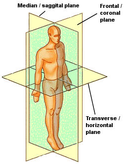
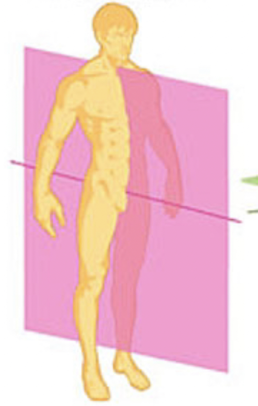
Sagittal Plane
Cardinal Plane that divides the body into left and right halves
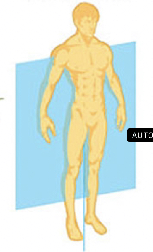
Frontal or Coronal Plane
Cardinal Plane that divides the body intro front and back halves
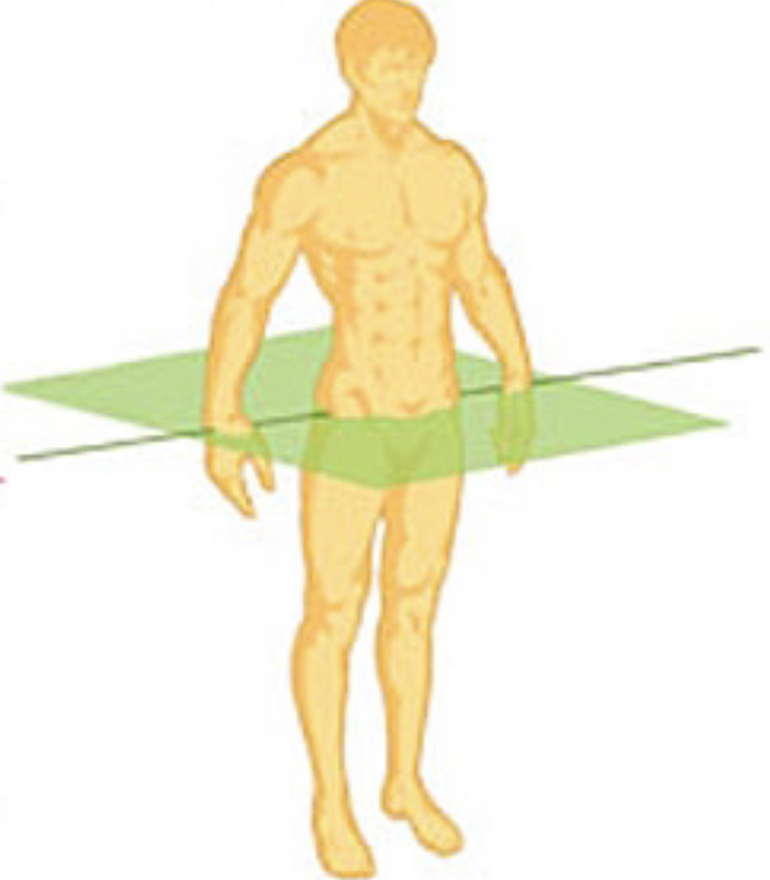
Transverse plane
Cardinal plane that divides the body into upper and lower halves
Claudial or inferior
Located in the tail of the body
Cranial or cephalic or superior
Located at the head
Pelvic
Located at the hips
Oblique Section
Cuts made diagonally
Pectoral
Located at the chest
Anterior
Frontal or near the front region of the body
Posterior
hind or near the hind region of the body
Dorsal
Toward the back or near the back region of the body
Ventral
toward the belly or near the belly region of the body
Lateral
Located at the side or near the side relative to the center of the body
Medial or central
located or towards the middle part of the body relative to its center
Proximal
near the origin or point of attachment in the body
Distal
Far from the origin or point of attachment in the body
Superficial
Surface of the body
Deep
Within the body
Ipsilateral
On the same side
Contralateral
On opposite sides
Flexion
Movement of the body that refers to decreasing joint angle
Extension
Movement of the body that refers to Increasing joint angle
Abduction
Movement of the body that refers to Moving away from the midline
Adduction
Movement of the body that refers to moving toward the midline
Hyperflexion
Movement of the body that refers to flexion beyond normal range
Hyperextension
Movement of the body that refers to extension beyond normal range
Hyperabduction
Movement of the body that refers to Abduction past 180 degrees point
Hyperadduction
Movement of the body that refers to Adduction past 0 degree point
Circumduction
Movement in a conic fashion
Plantarflexion
Movement of the body that refers to increasing angle between foot and shank
Dorsiflexion
Movement of the body that refers to decreasing angle between foot and shank
Inversion
Movement of the body that refers to lifting the medial edge of foot
Eversion
Movement of the body that refers to lifting the lateral edge of foot
Median Rotation
Internal or inward rotation
Lateral Rotation
External or outward rotation
Midsagittal Plane
Sagittal plane that lies on the midline
Integumentary system
Set of organs that forms the external covering of the body and protects it from external environment
Integumentum
Latin word that means "to cover"
Skin
Main organ of the integumentary system
36 degrees celsius
Normal temperature of human body
presence of vocal sacs size of the body shape of snout or head enlarge innermost digit presence of dark spots in the neck or throat
Differences of male and female frogs
Rana vittigera
Scientific name of frog
Parts are similar to man they are small, can be easily handles they are readily available and cheaper they have a well documented life cycle
Reasons why frogs are used as representative sample in zoology
Rainy season
Breeding season of frogs
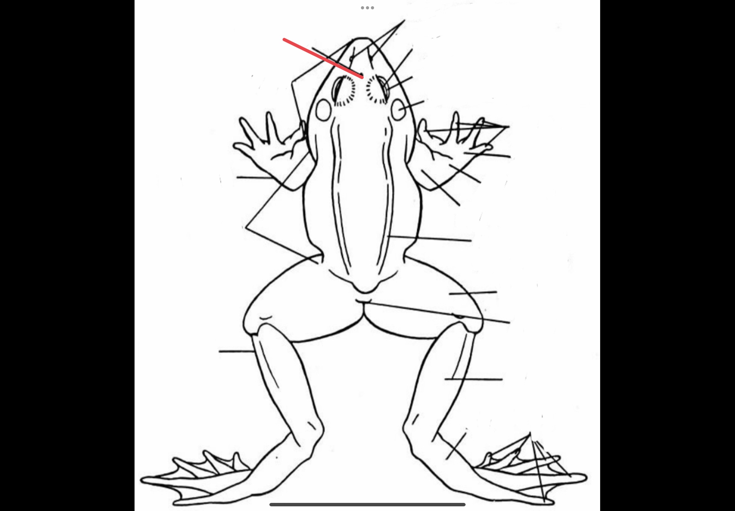
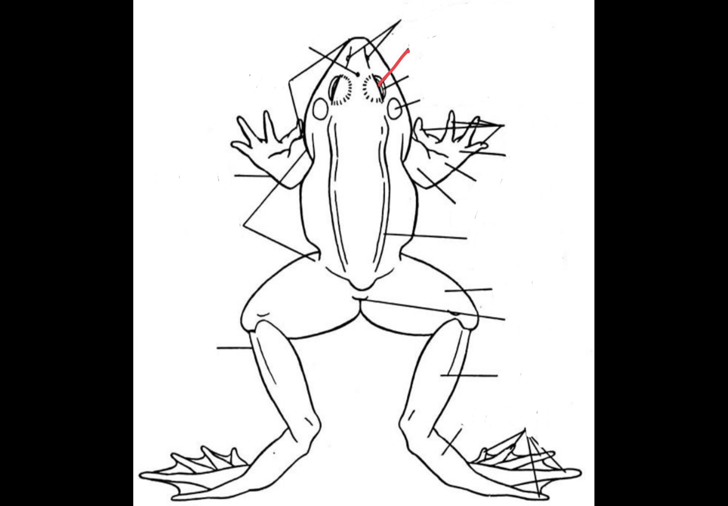
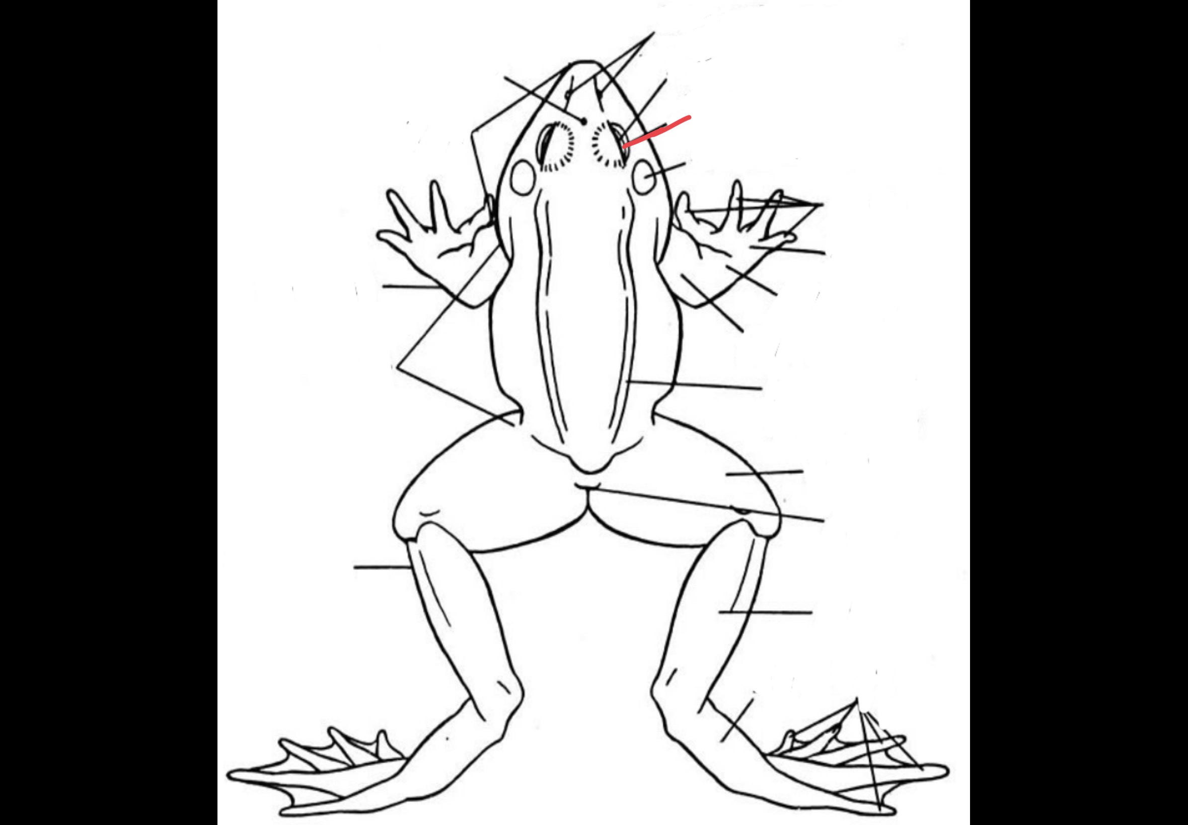
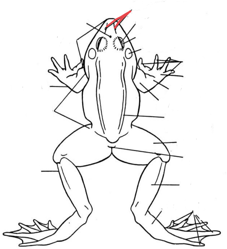
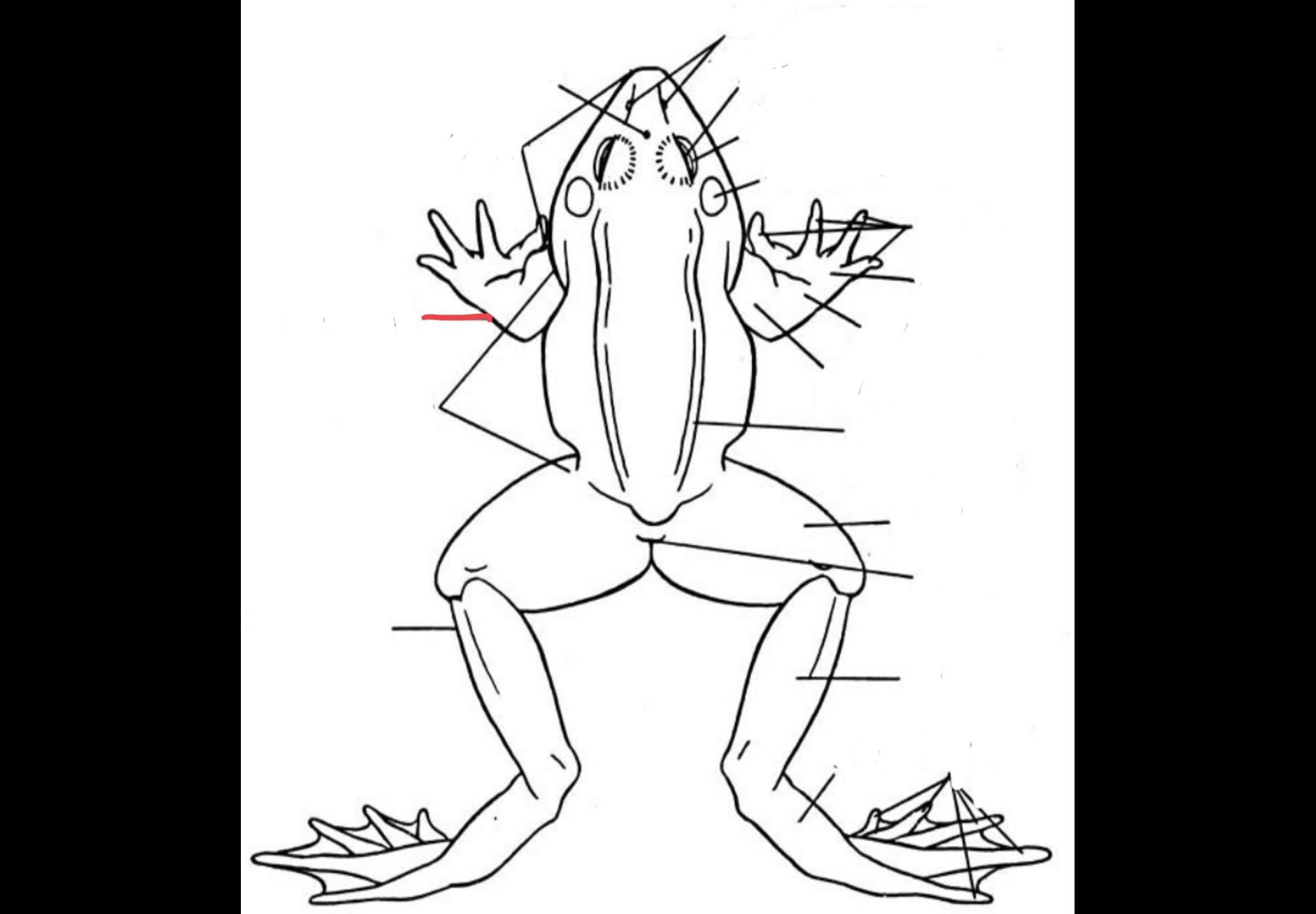
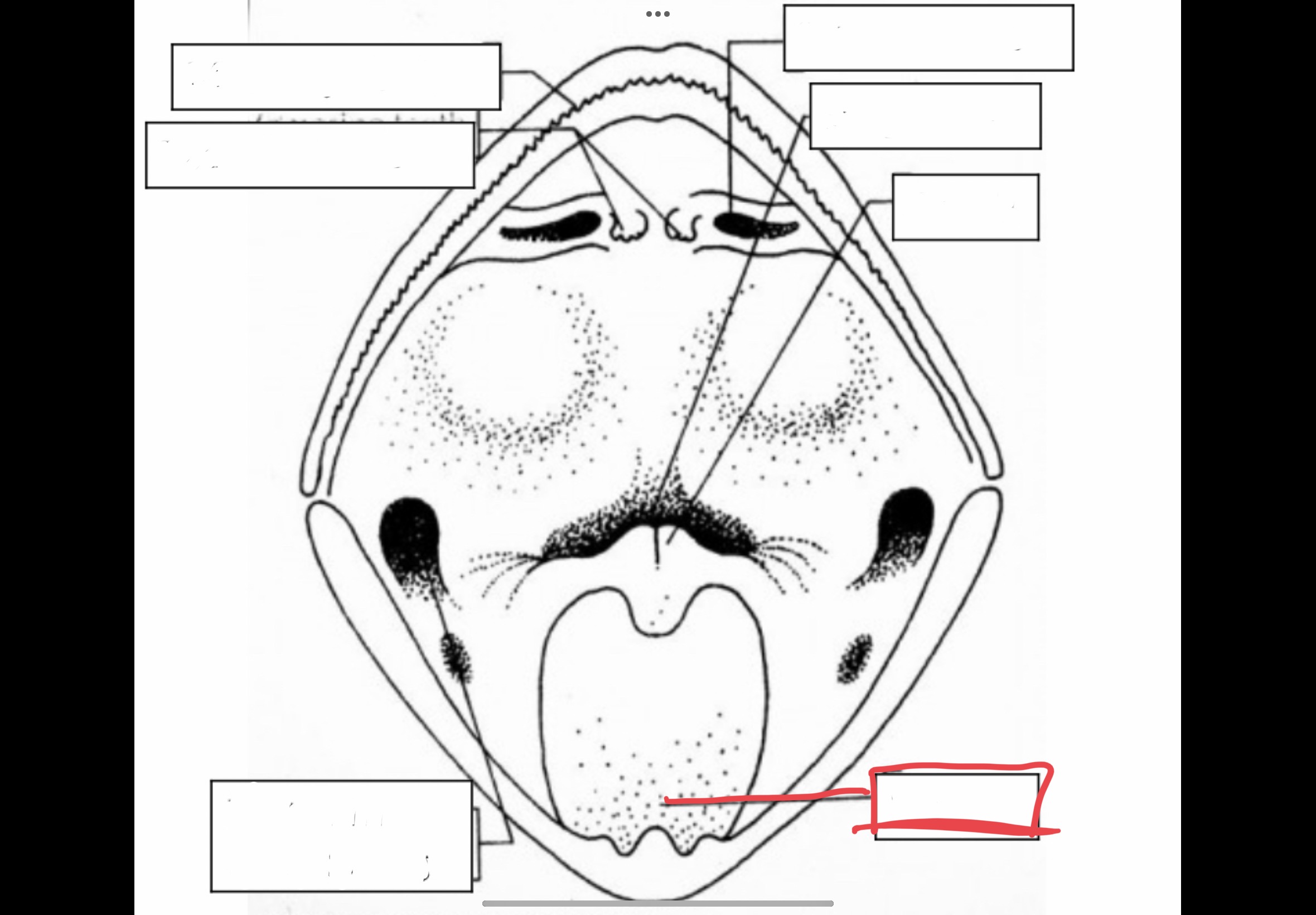
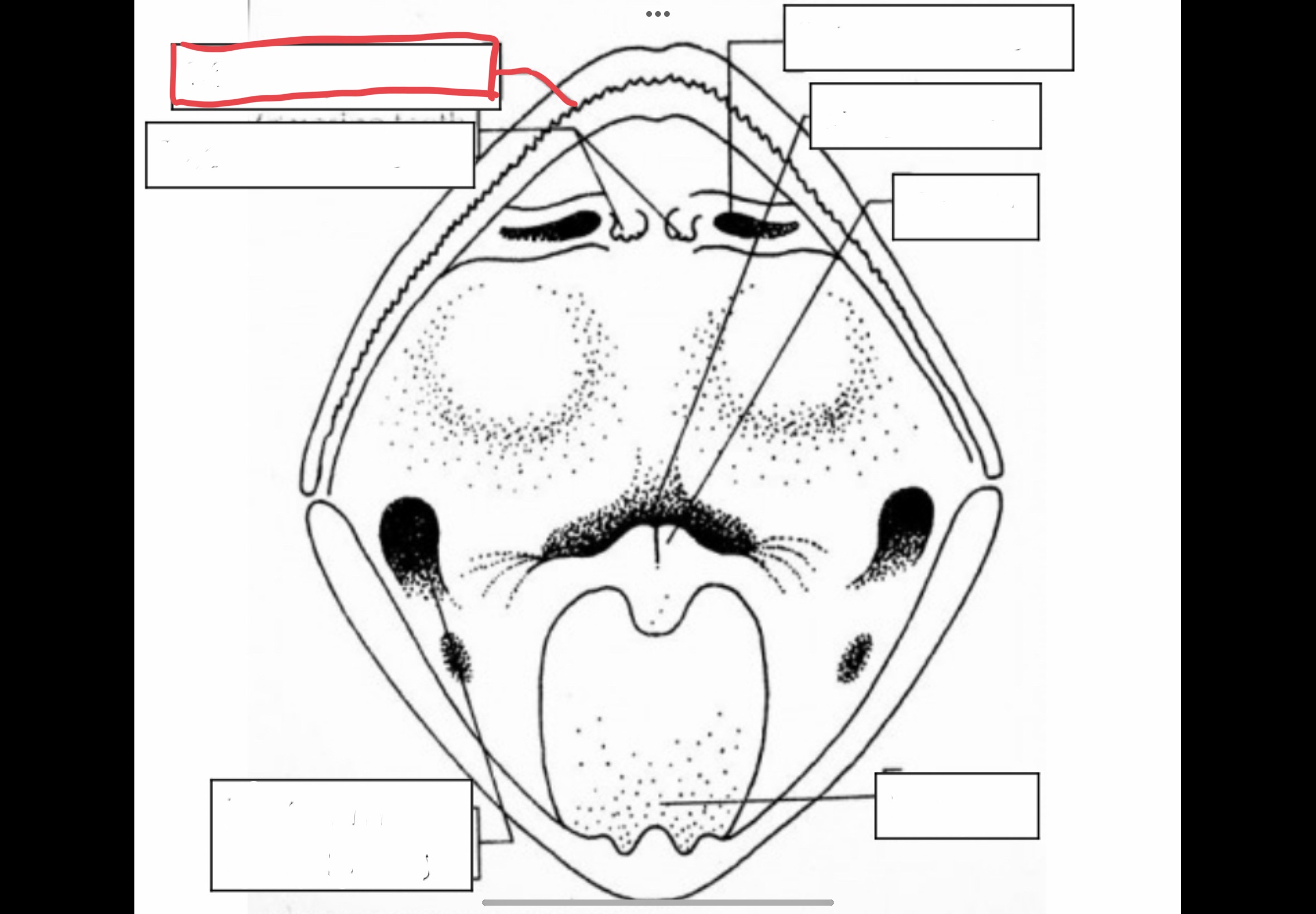
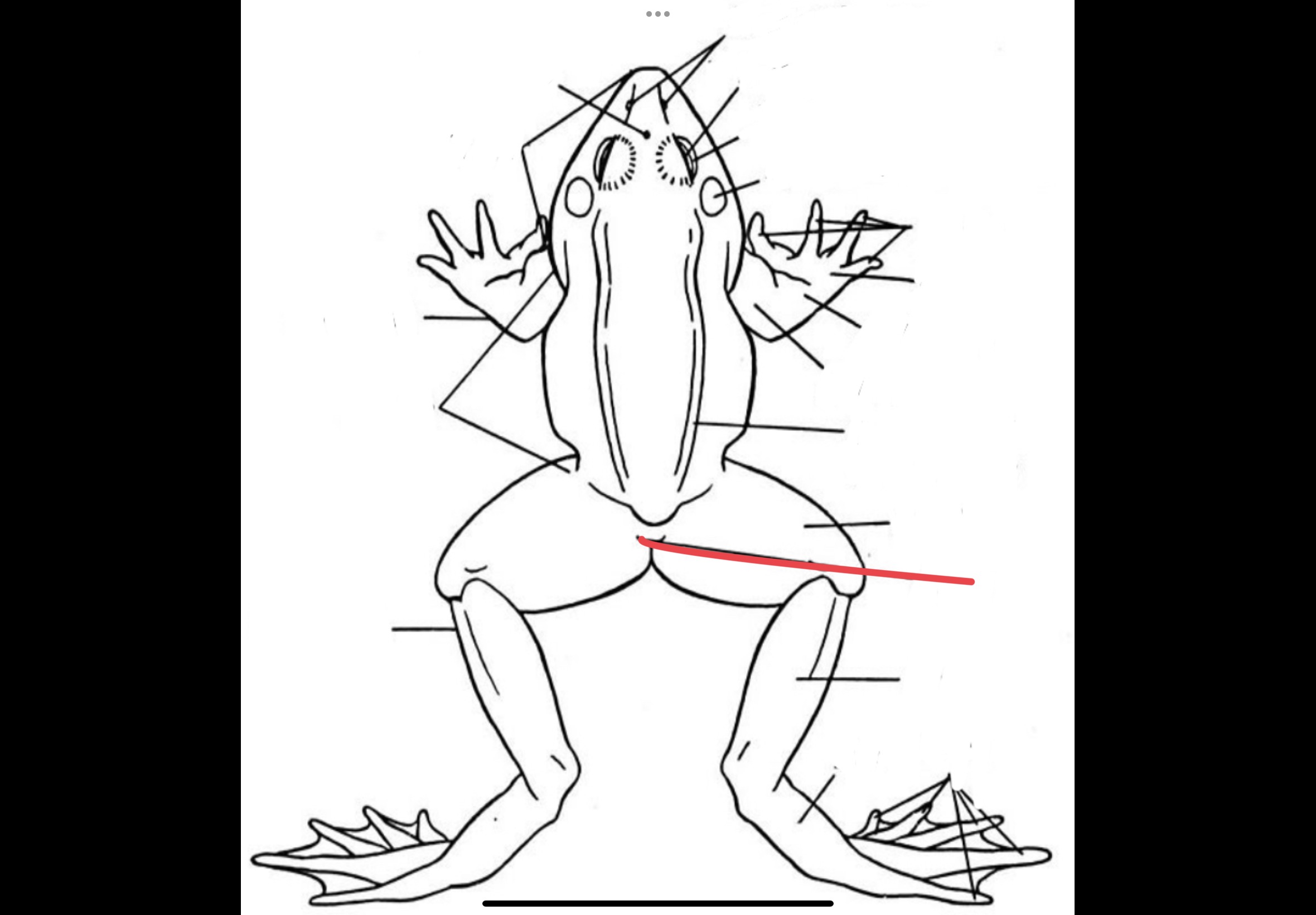
Snout
Most anterior border of the head of the frog
Browspot
Vestigial eye located between the eyeballs
Eyes
Bulging structures which consists of three eyelids
Nictitating membrane
Transparent structure that covers the eye to protect it and keep it moist
External Nares
Two openings which are anteriorly located that serves as an entry and exit way of air
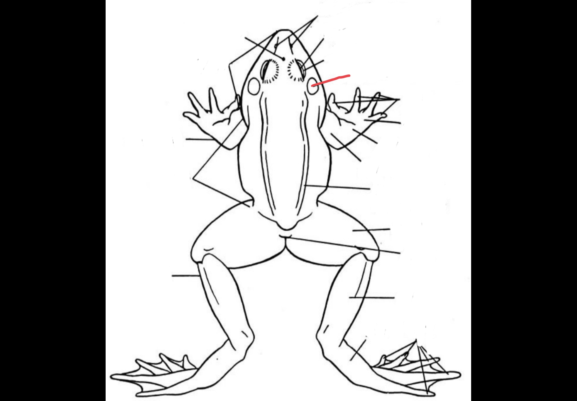
Tympanic Membrane
Two flat and rounded structures located laterally behind the eyeballs that receives sound waves
Trunk
main mass of the body
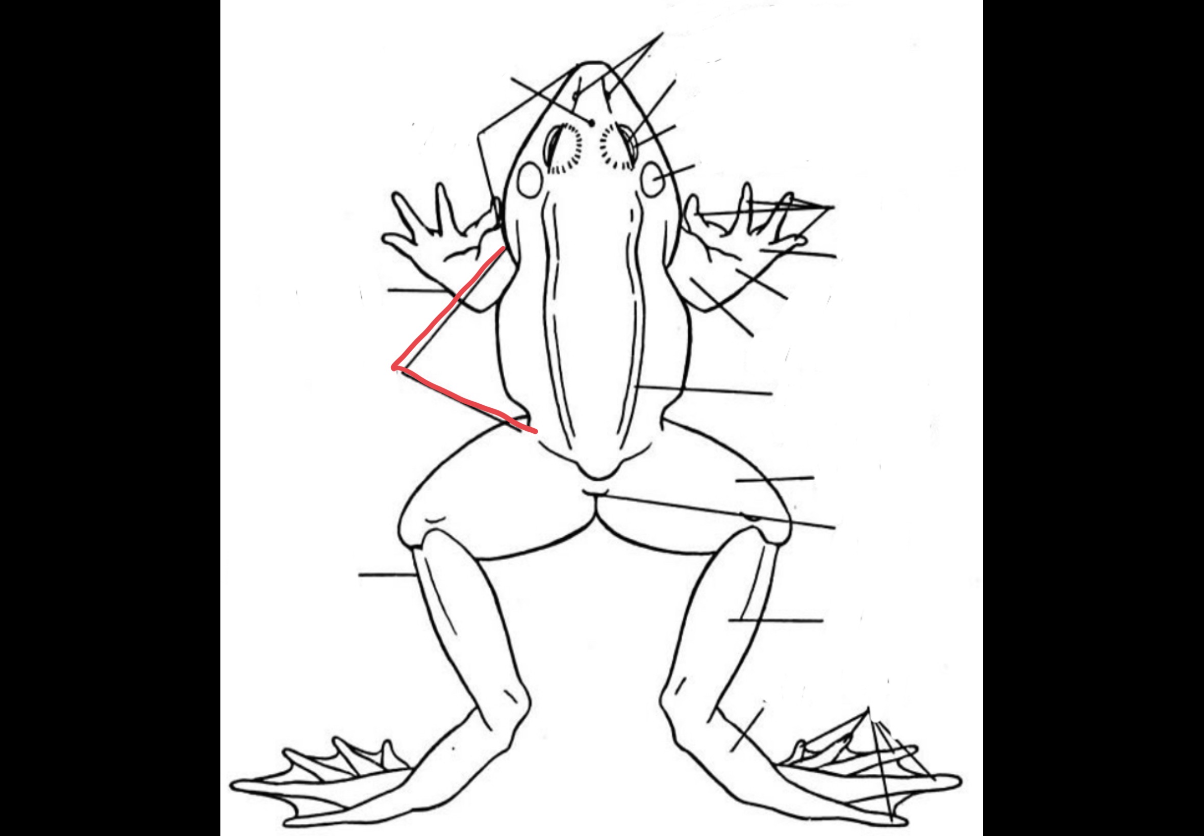
Forelimb
composed of the upper arm, lower arm, carpus, and manus
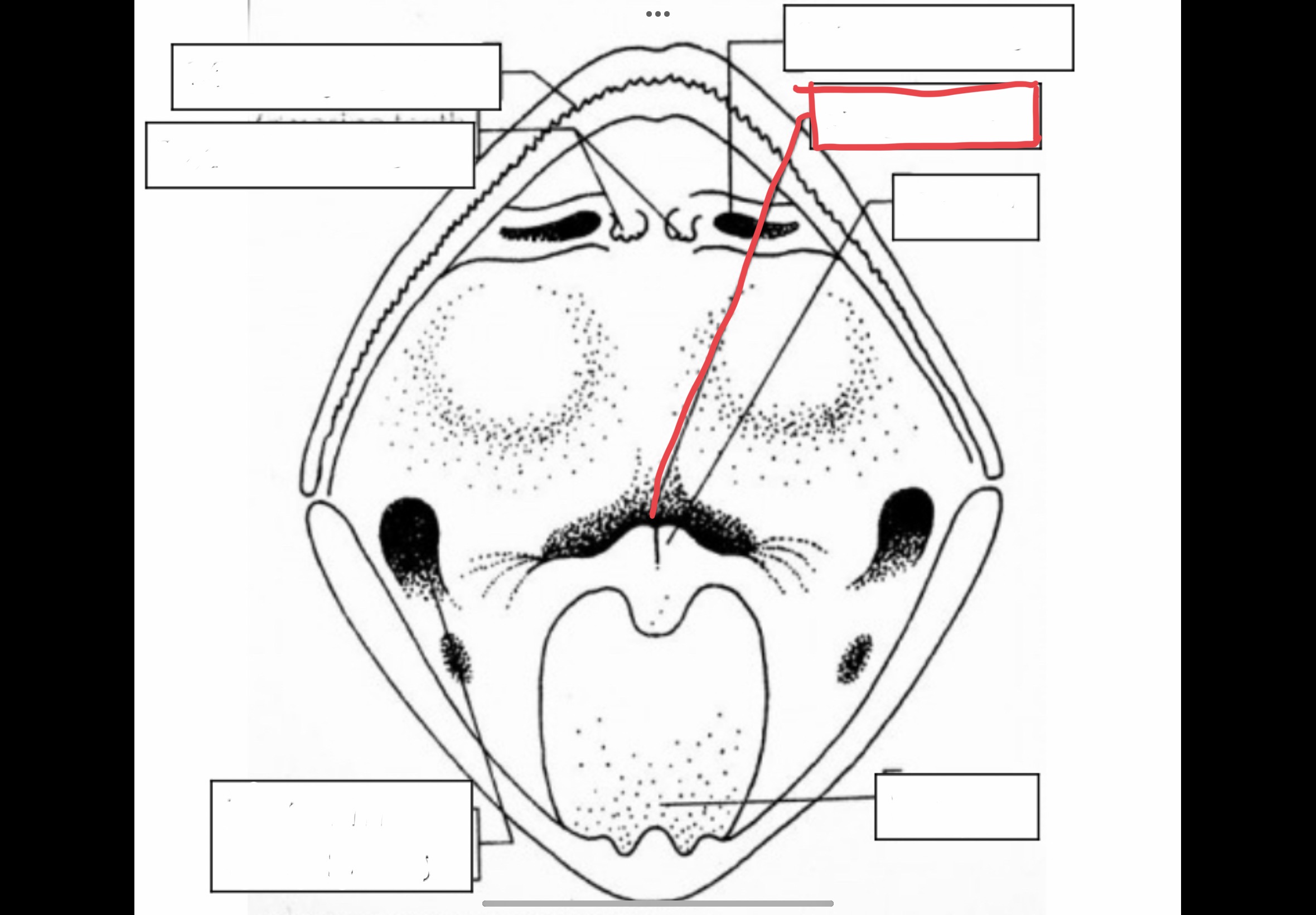
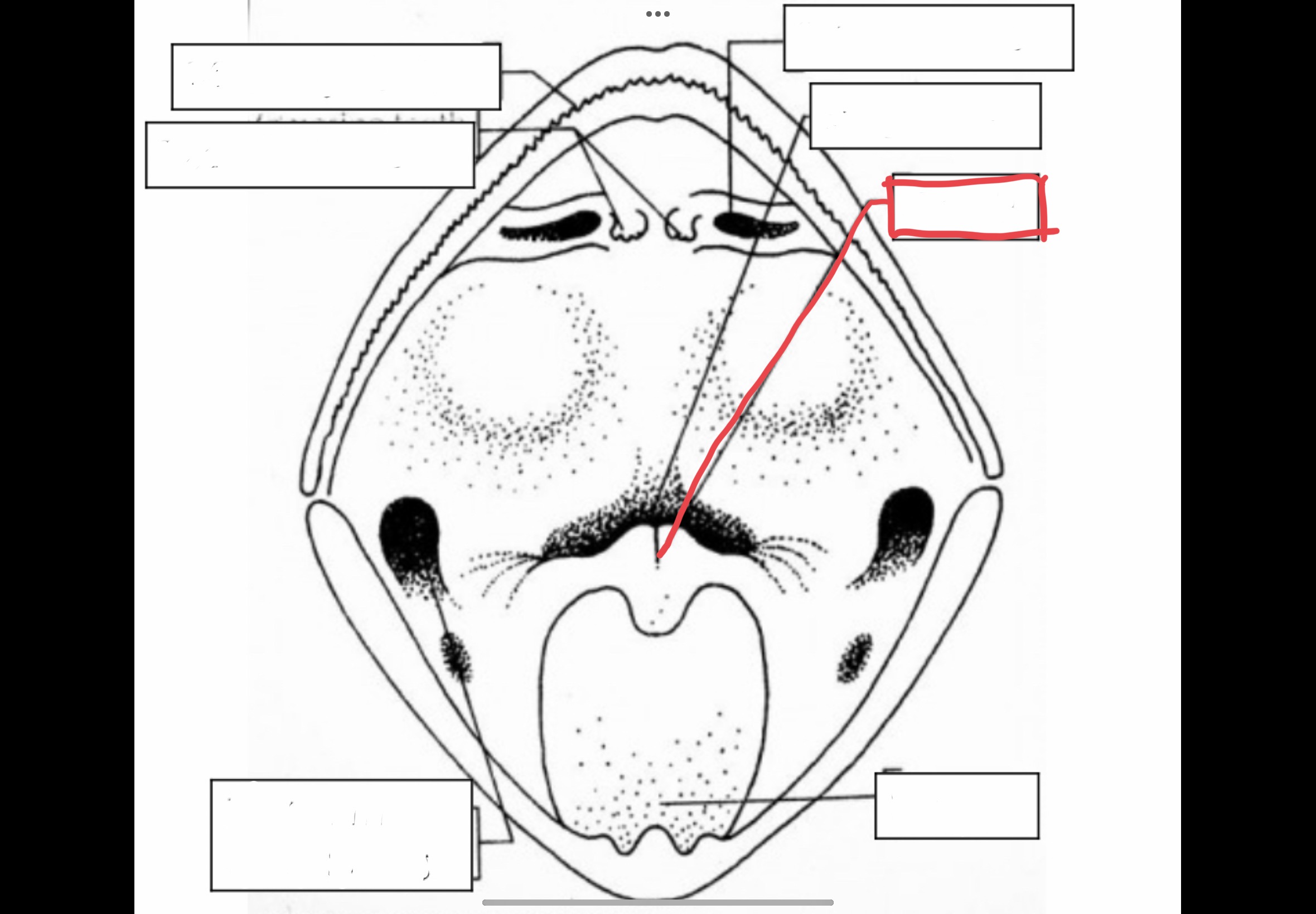
Mouth
Anterior opening of the frog's body
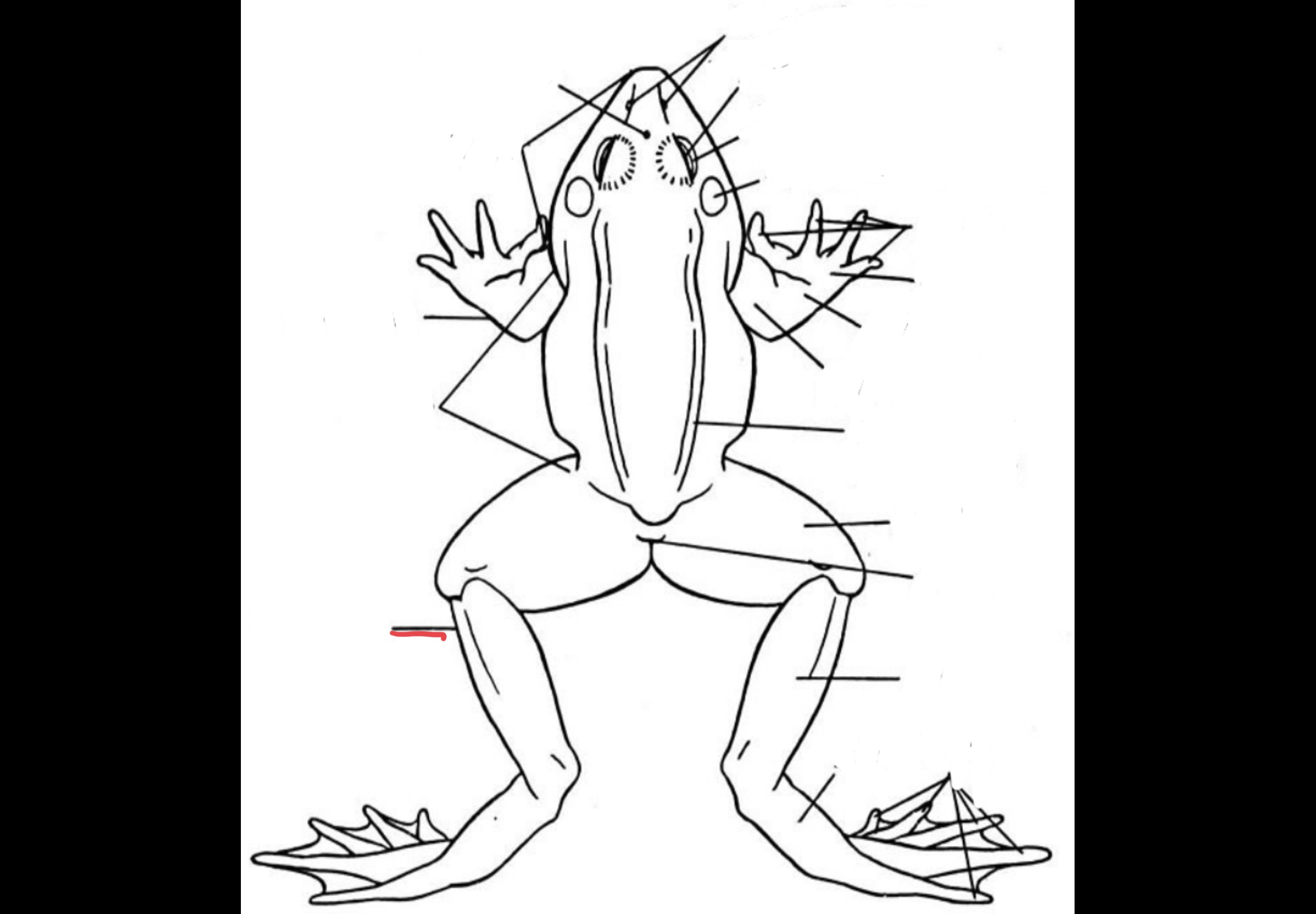
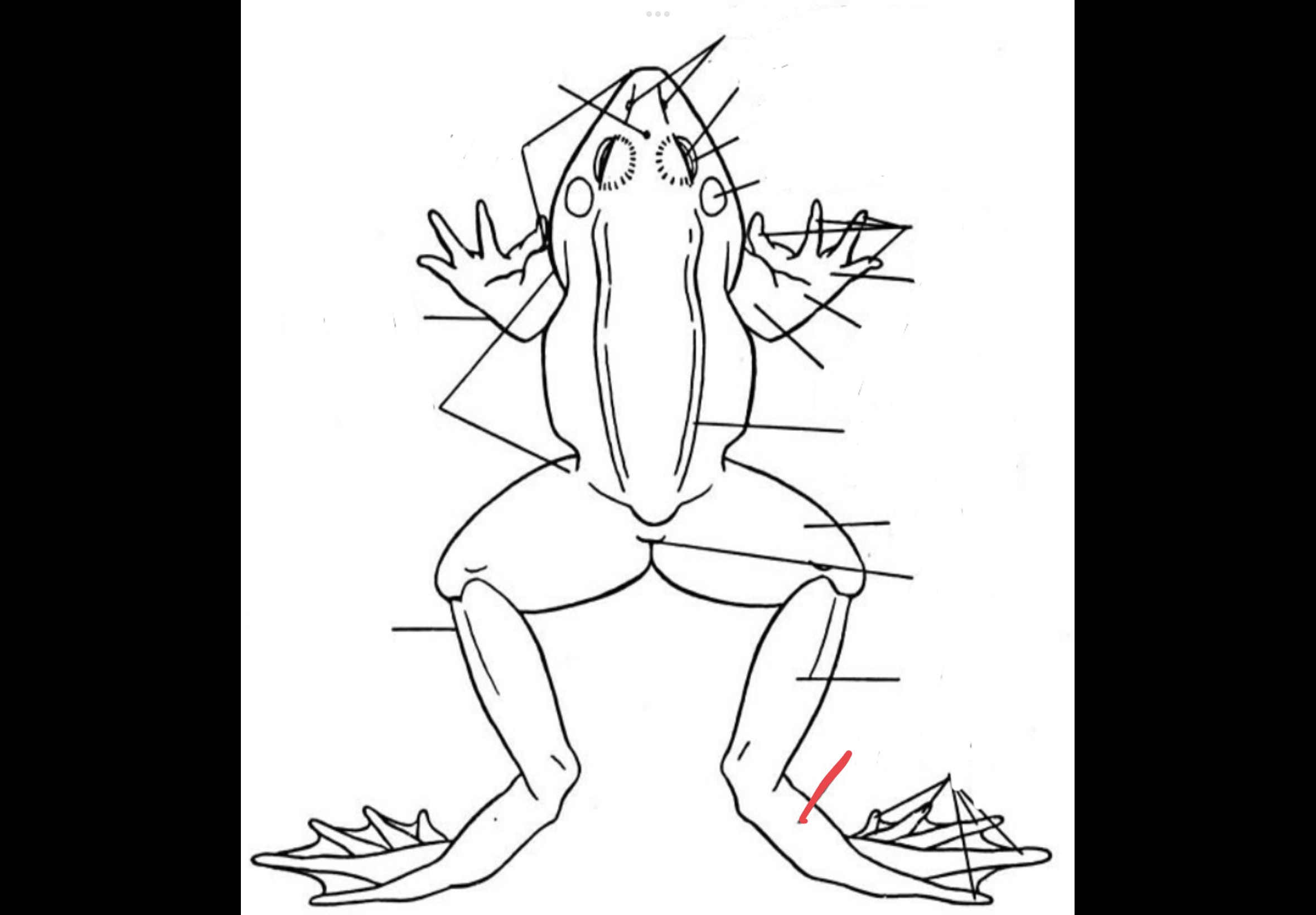
Hindlimb
Made up of the thigh, shanks, tarsus, and pes
Pes
Feet of the frog
Tarsus
Ankle of the frog
Carpus
Wrist of the frog
Manus
Hand of the frog
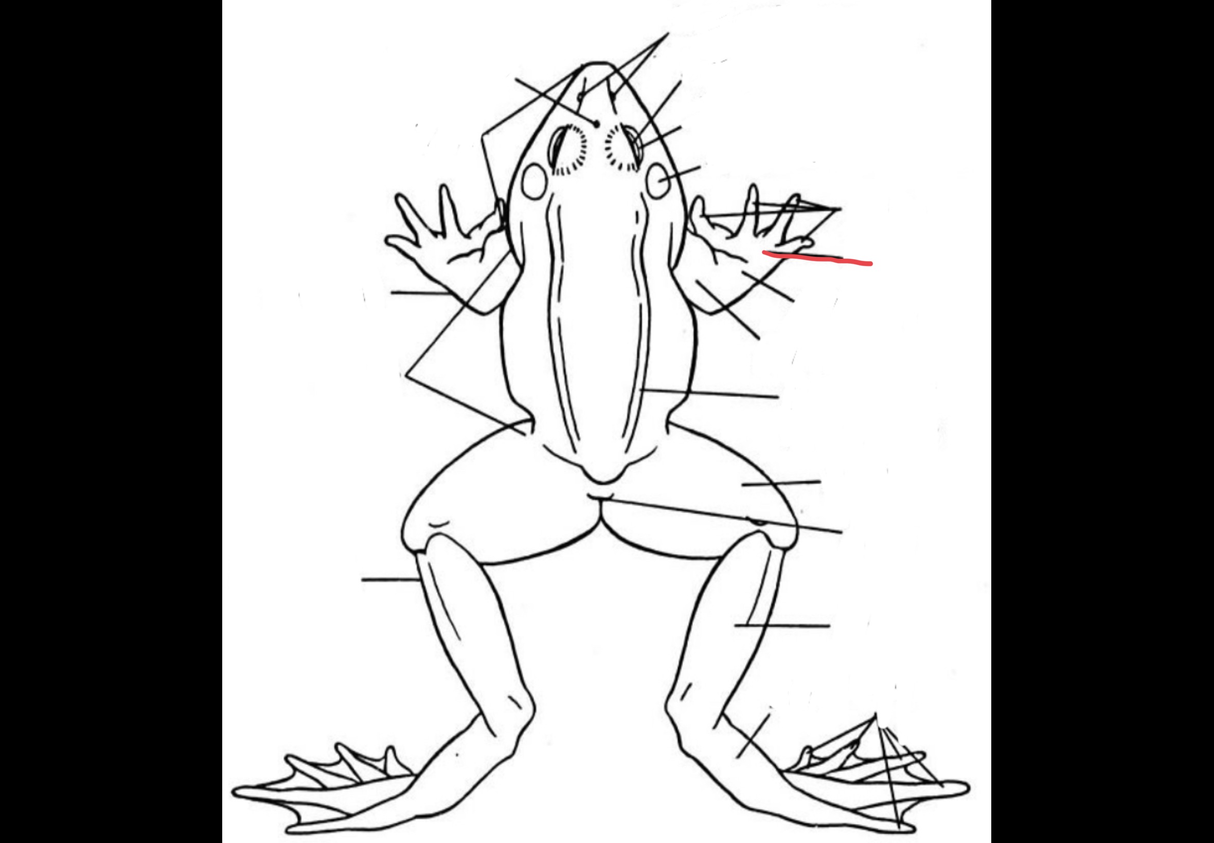
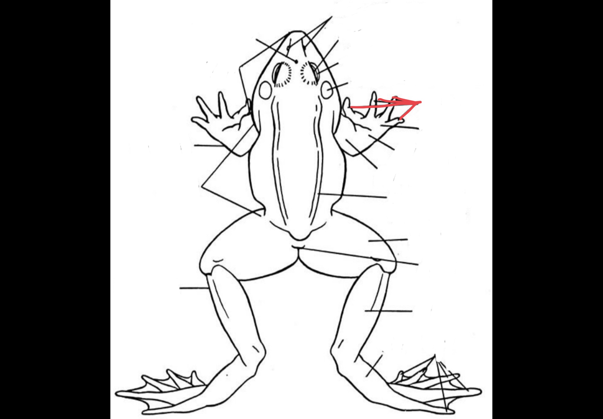
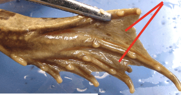
Digits
Fingers and toes of the frog
Web
Located in between the digits in the hindlimb which allows them to push themselves in the water for swimming
Esophagus
Tube that connects the mouth and the stomach
Glottis
opening from the mouth in the respiratory system, to the vocal cord or lungs
Tongue
Muscular structure attached tot he front of the mouth which can be extended
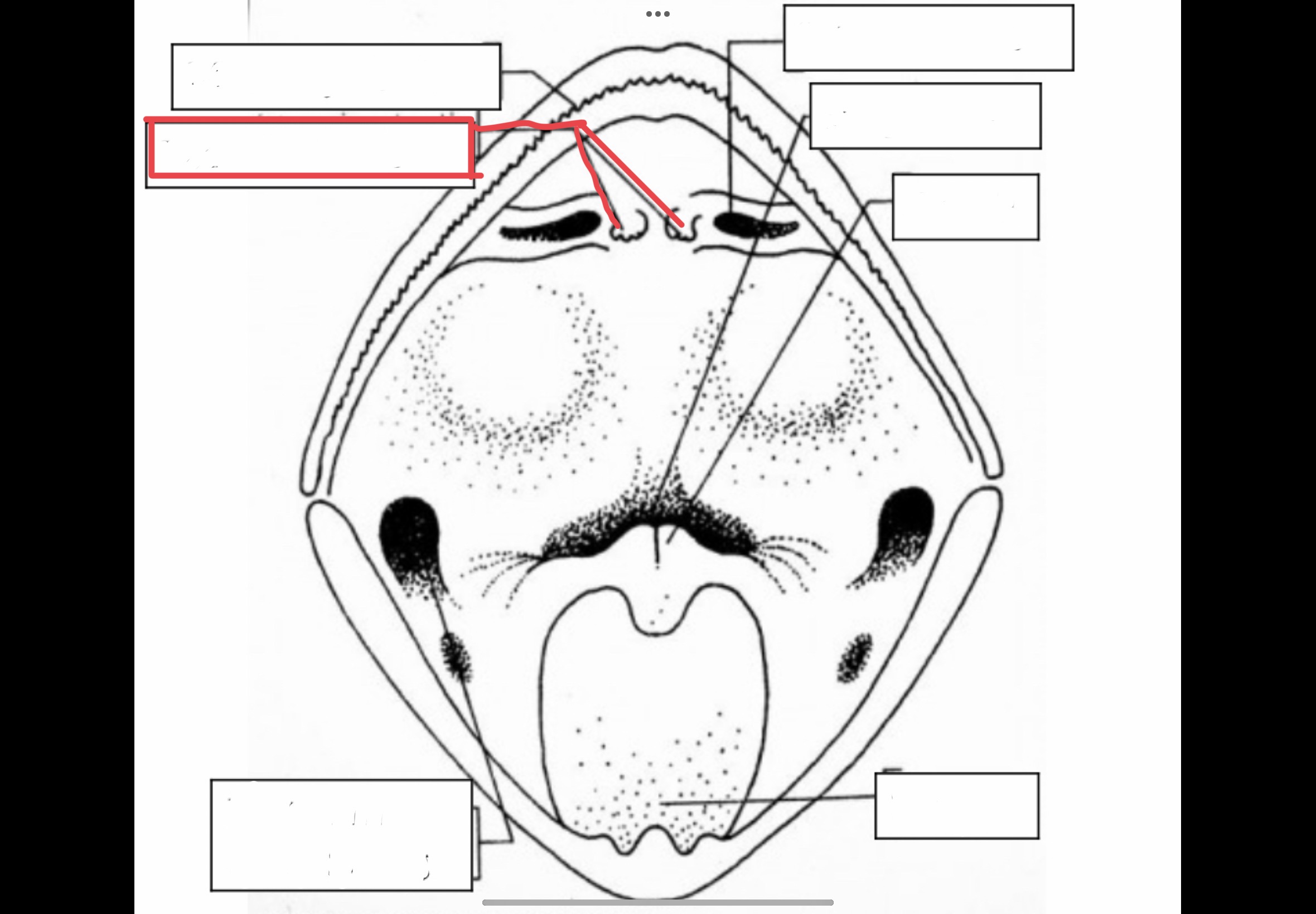
Maxillary Teeth
Sharp teeth in the maxillary that holds captured prey
Vomerine Teeth
Small projections in the top of a frog's mouth that holds captured prey
Eustachian Tube Opening
Mouth openings that lead to tubes that connect to the middle ear to equalize air pressure
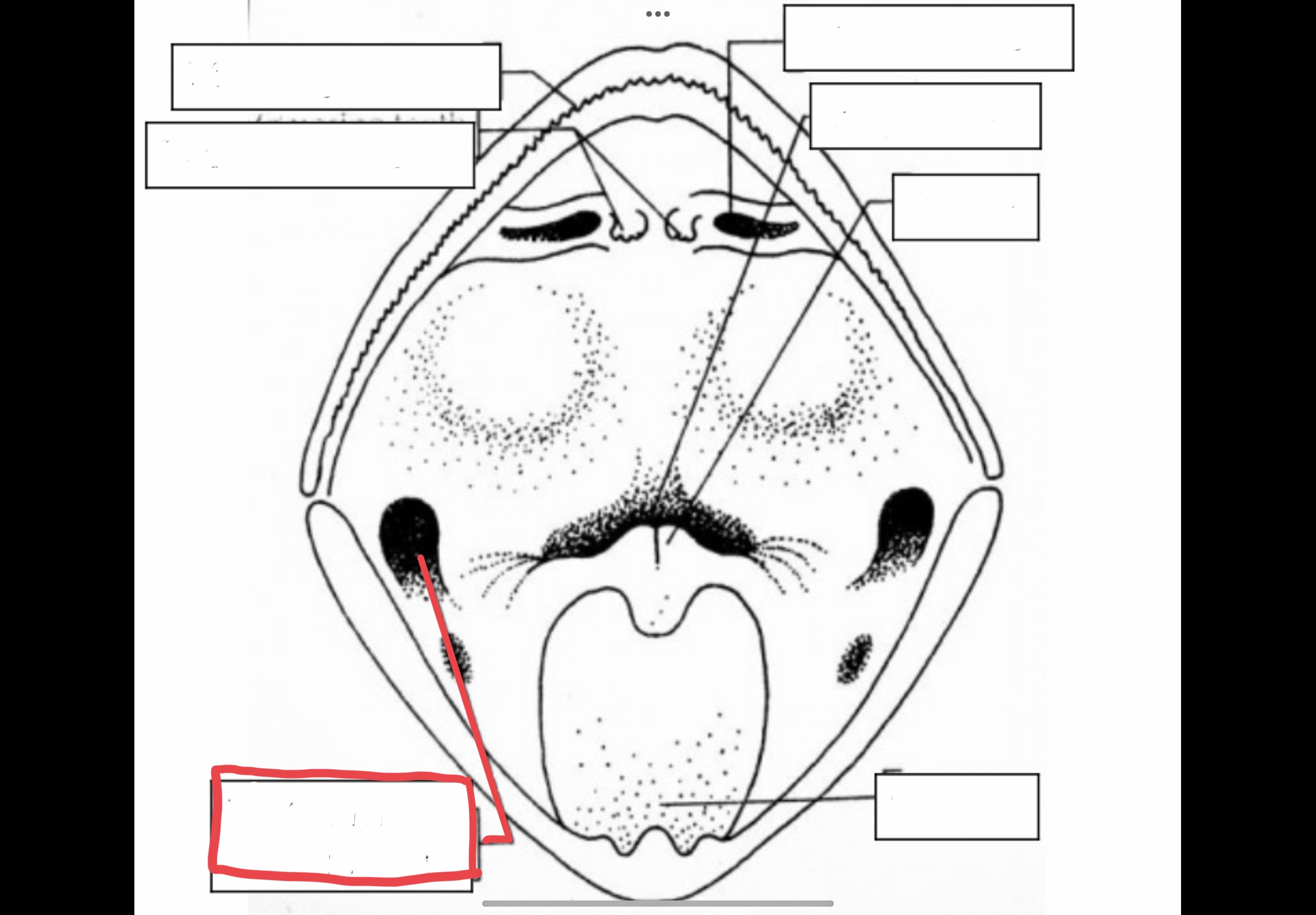
Cloacal Opening
Opening of cloaca through which indigested food, urine, and sperm are passed
Vocal Sacs
Flexible membrane of skin that is used for amplification of a male's mating call
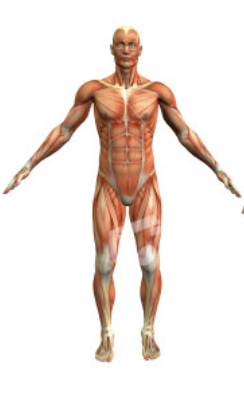
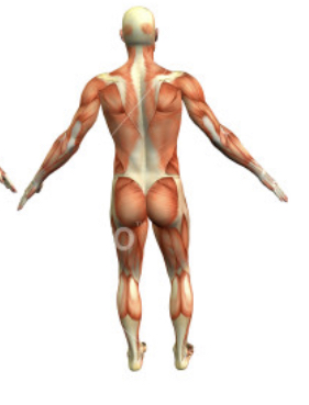
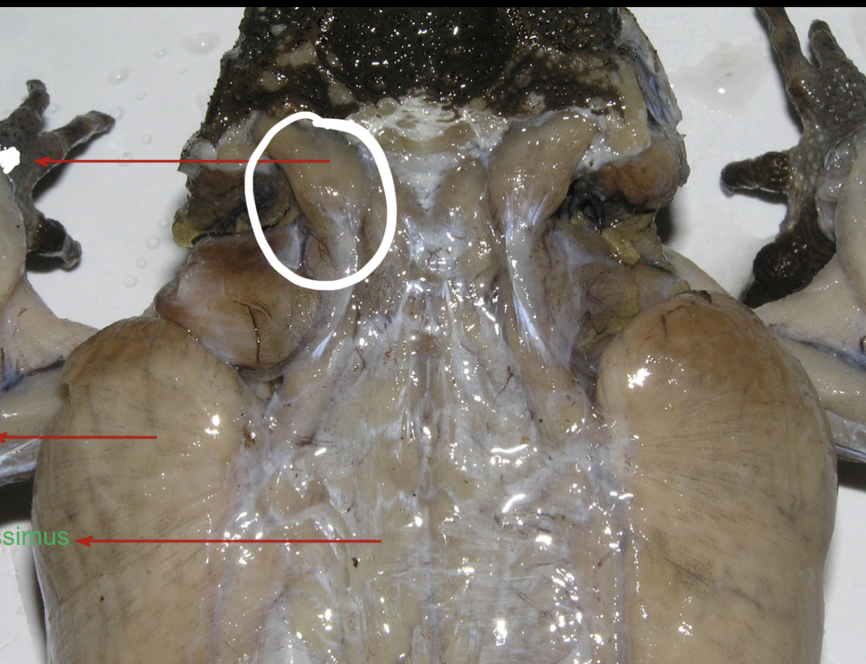
Muscular System
Organ system that is responsible for the movement of the body; made out of muscle fibers
Skeletal muscle tissue
A type of muscle tissue that has striations and is involved in voluntary movements
Cardiac Muscle Tissue
A type of muscle tissue that has striations and is involved in involuntary movements like propelling blood in circulation
Smooth Muscle Tissue
A type of muscle tissue that does not have striations and is involved in involuntary movements like giving birth
40%
Percentage of muscles to our body weight
Gluteus maximus
largest muscle in the body
Ear
Part of the body that contains the smallest muscle and bone
Masseter
Strongest muscle by weight, located in the jaw
Cardiac Muscle
the hardest-working muscle in the body
Myology
the study of the structure and functions of muscles
Fascia
connective tissue membrane lining the outer surface of the muscles, dense fibrous, connective tissue
Tendon
Formed by fascia
Aponeuroses
flat tendons
Protractor
pushes a part away from the base
Supinator
rotator that turns a part upward
Pronator
rotator the turns a part downward
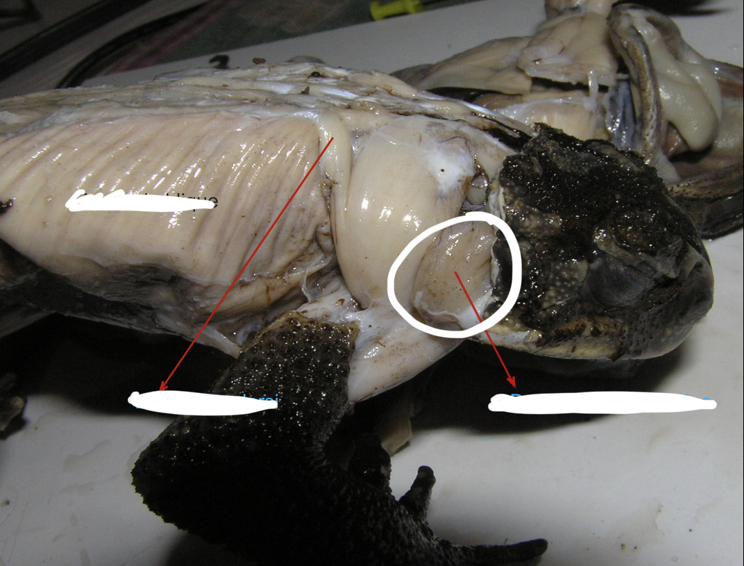
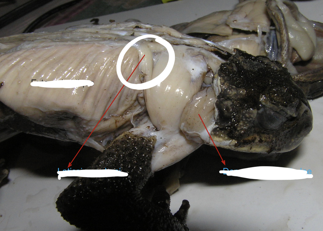
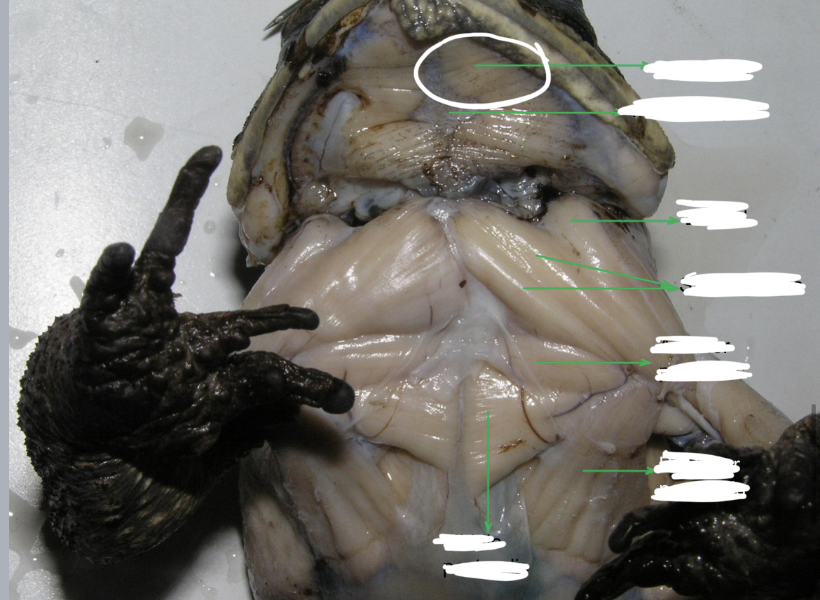
Temporalis
broad muscle posterior to the eye and on the same level as the tympanum
Depressor Mandibulae
flat fan shaped muscles posterior to the temporalis that originates from the tough fascia in the middorsal line. It inserts into the lower jaw and serves as a jaw depressor.
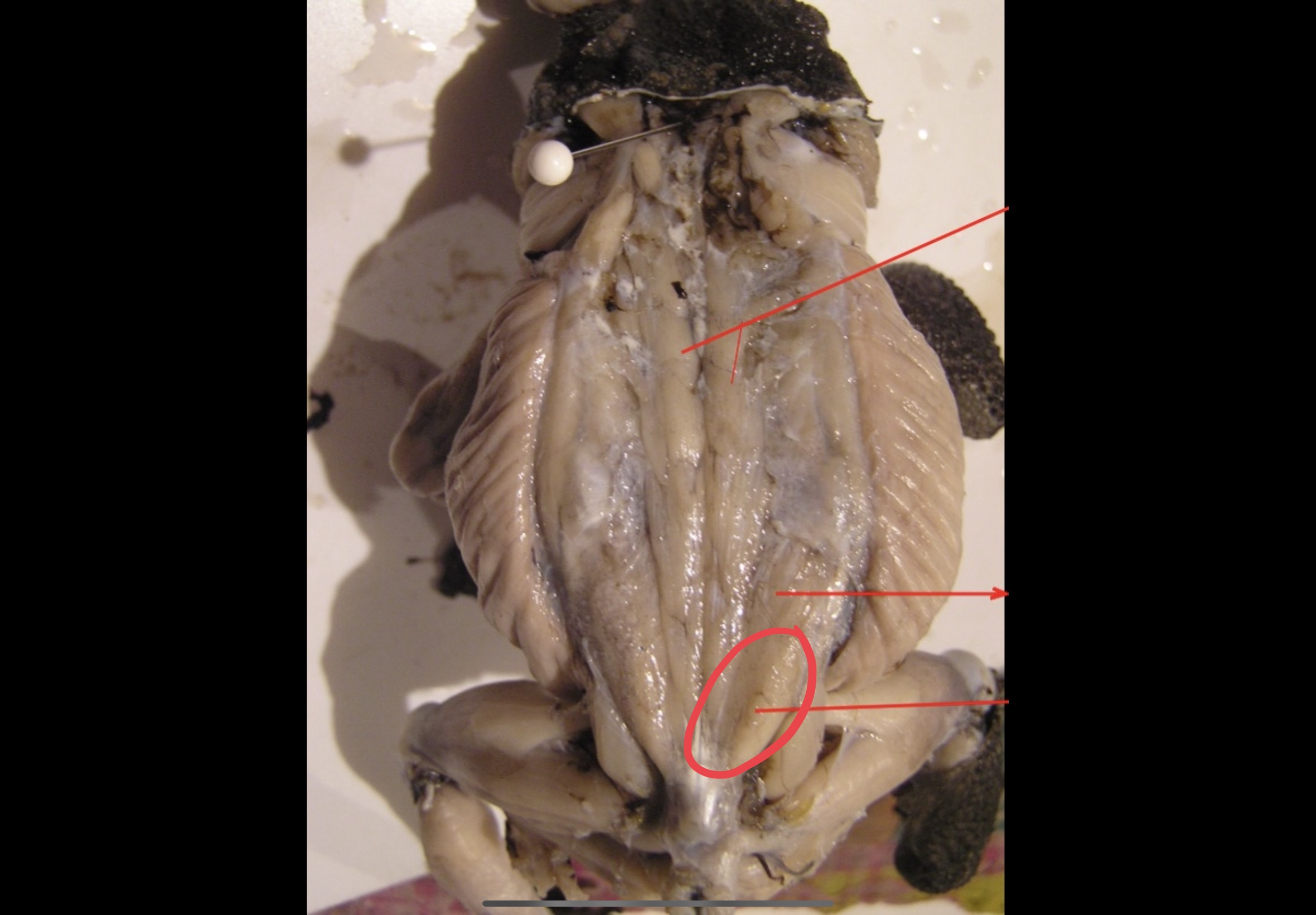
Latissimus dorsi
Broad muscles posterior to and partly covered by the depressor mandibulae
Longissimus Dorsi
Posterior to the latissimus dorsi these muscles are attached to the anterior third of the urostyle and skull. They are inserted along the vertebral column and they serve as extensor of the back and levator of the head.
Coccygeosacralis
A pair of narrow V – shaped muscles posterior to the longissimus dorsi
Coccygeoilliacus
A pair of broad V shaped muscles posterior to coccygeo sacralis.
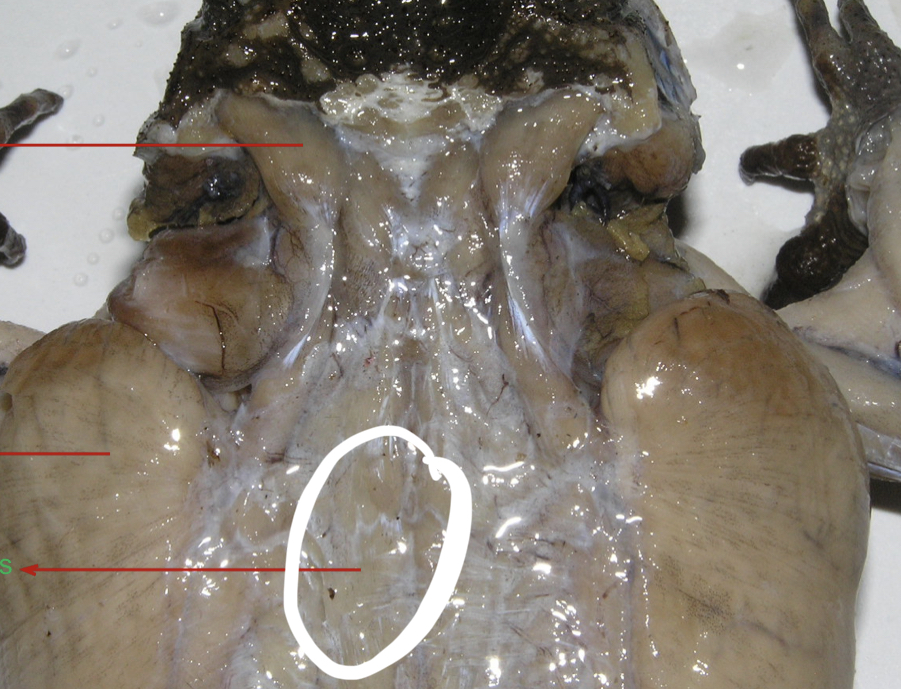
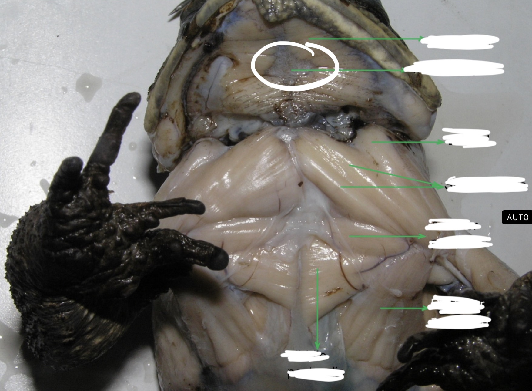
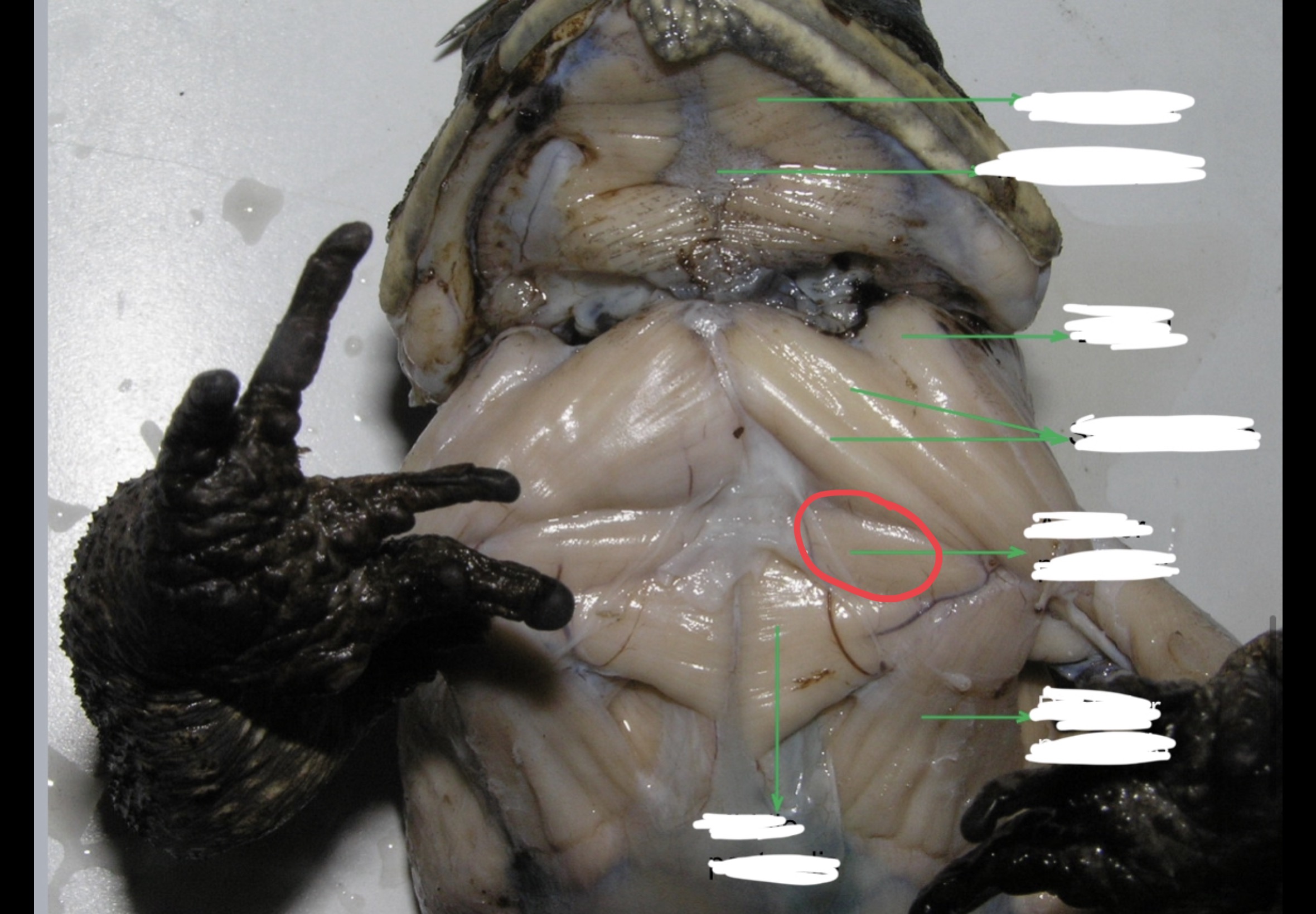
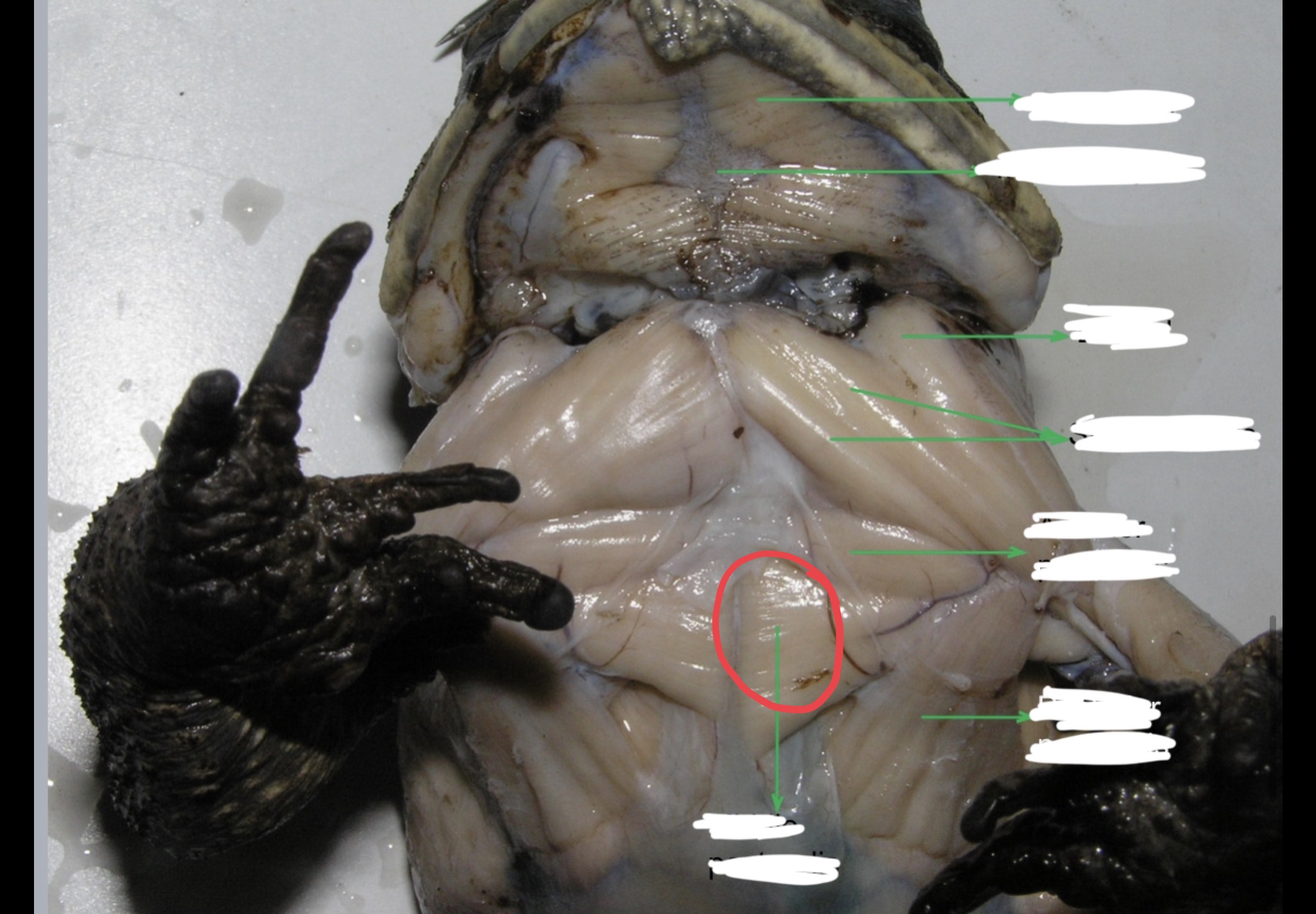
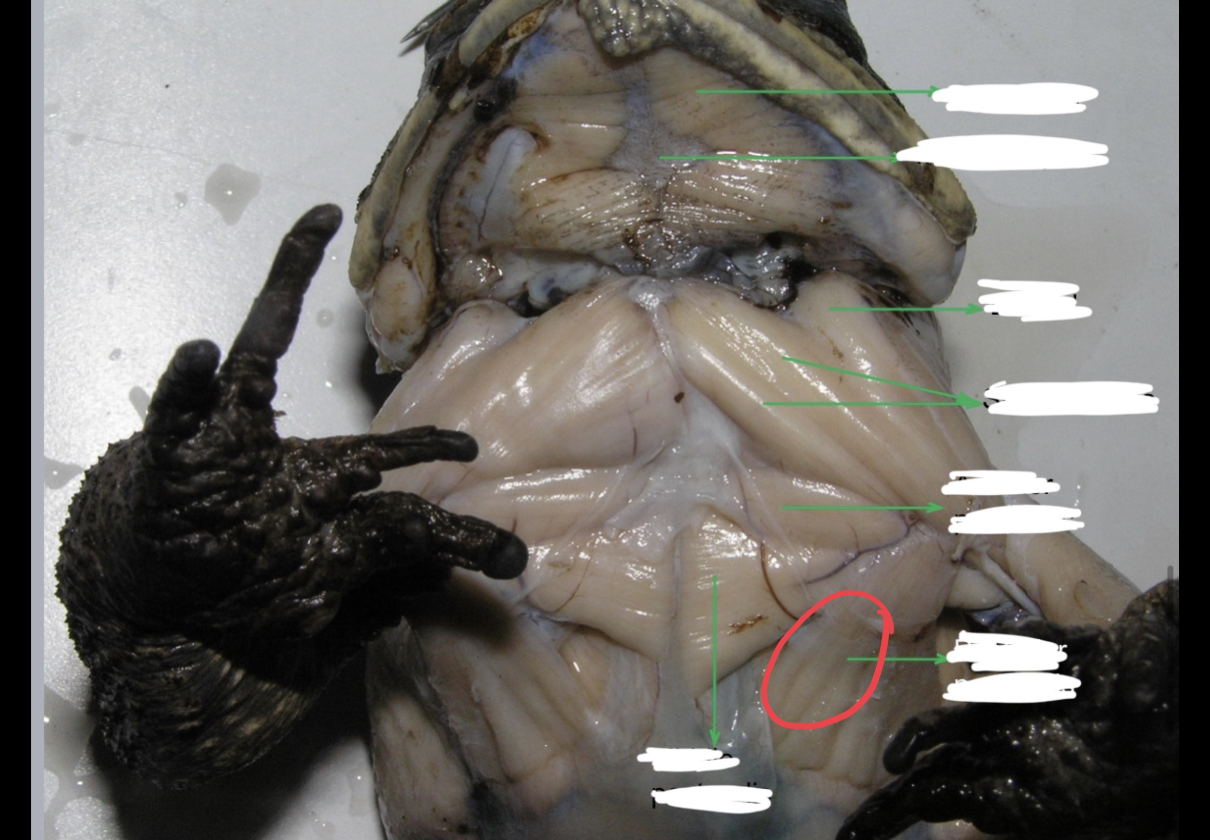
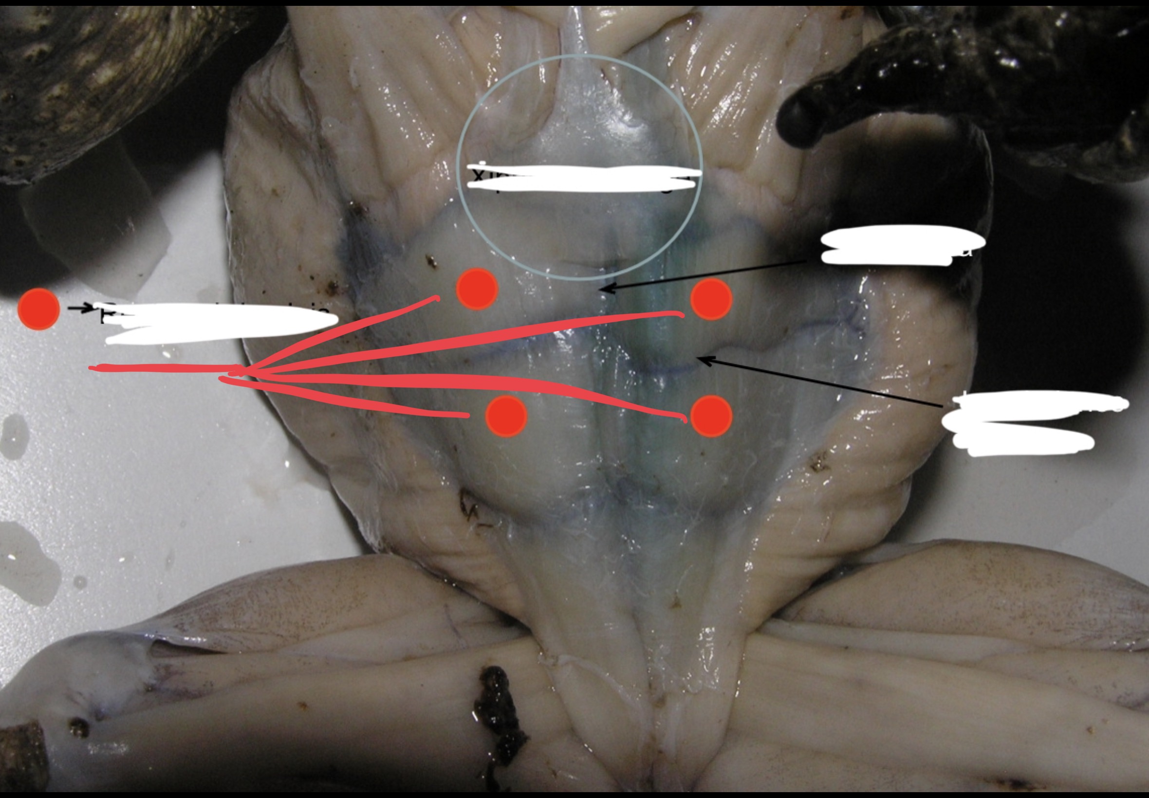
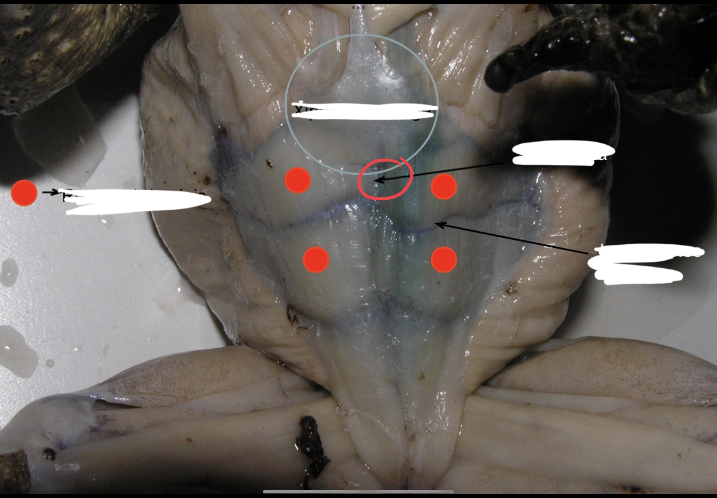
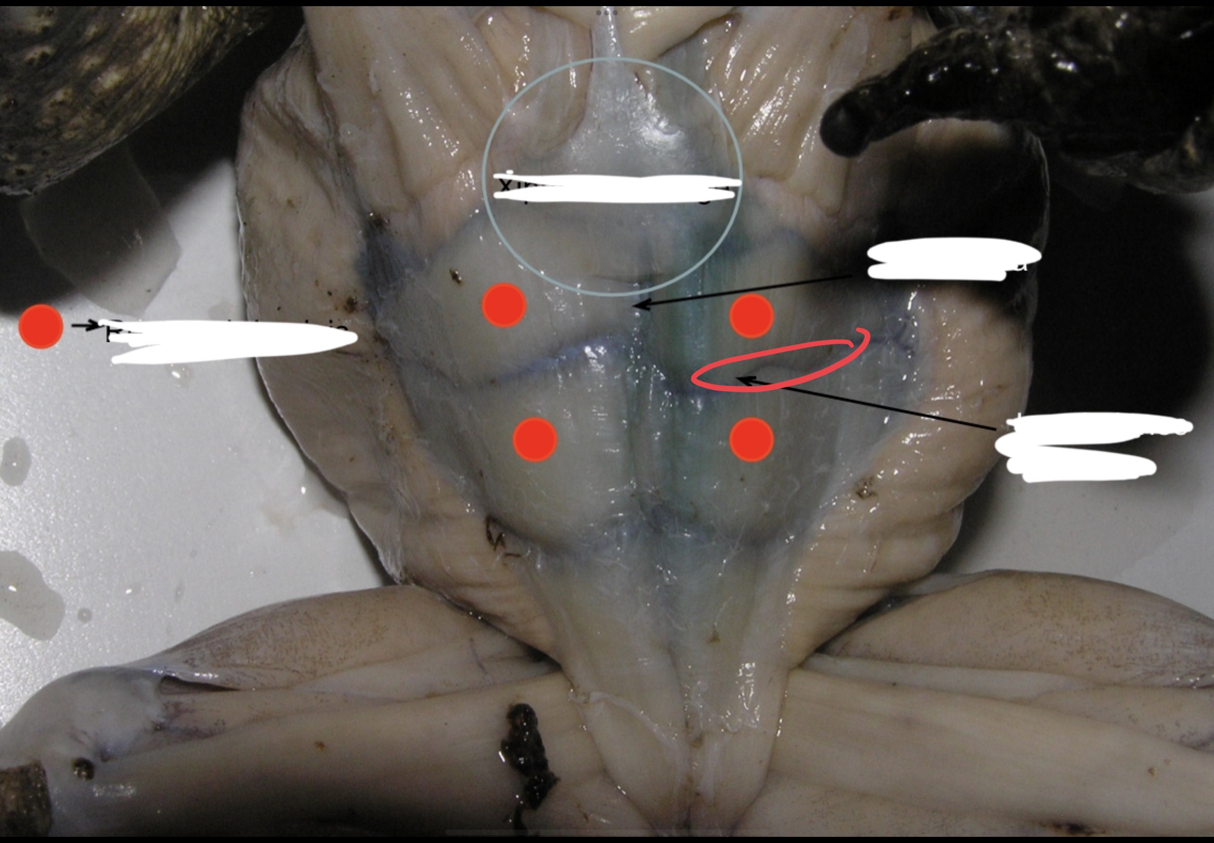
Mylohyoid
A large transverse muscle on the ventral surface of the mouth floor this muscle
Median Raphe
A midventral connective tissue partition that divides the mylohyoid into left and right portions
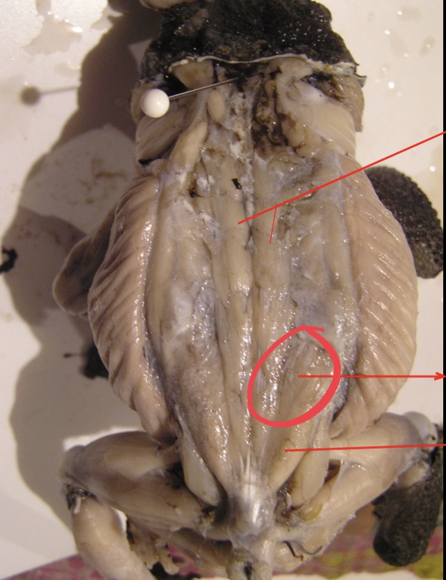
Anterior Pectoralis
This muscle lies immediately posterior to the strenoradialis and the most anterior of the chest muscle. It originates from the epicoracoidea and it is inserted into the deltoid ridge; it acts as adductor and rotator of the arm.
Middle Pectoralis
Posterior to the anterior pectoralis. this muscle takes origin from the mesosternum and xiphisternum. It is inserted into the ventral portion of the proximal end of the humerus and adducts and rotates the arm.
Posterior Pectoralis
Postero – lateral to the middle pectoralis. it extends to almost the entire portion of the median surface of the trunk. It is inserted into the deltoid ridge; it serves as adductor and rotator of the arm.
Rectus Adbominis
This longitudinal muscle extends to each side of the linea alba
Linea Alba
a strip of connective tissue that acts as a partition on the midventral line.
Inscriptiones tendinae
Divides the rectus adbominis into muscle segments.