Looks like no one added any tags here yet for you.
What are five components that many signal-transduction cascades have in common?
Release of a primary messenger as a response to a
physiological circumstance.
Reception of the primary messenger by a receptor, usually an
integral membrane protein (cell surface receptor).
Relay of the detection of the primary messenger to the cell
interior by the generation of an intracellular second
messenger.
Activation of effector molecules by the second messenger that
result in a physiological response.
Termination of the signal cascade.
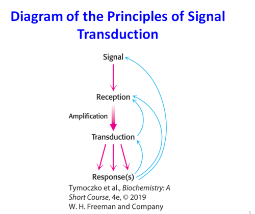
Describe this diagram.
Diagram of the Principles of Signal Transduction.
Signaling cells produce signaling molecules (primary messenger) →
Target cells detect the signal using a receptor protein →
Extracellular signal is converted to an intracellular signal
Cells are typically exposed to many different types of _________________________.
signaling molecules
Do cells respond to all signals?
No. The presence of a receptor determines whether a cell will respond.
What are two kinds of receptors that extracellular signal molecules can bind to?
surface receptors (integral protein)
intracellular receptors
Where are intracellular receptors located?
in the cytosol or in nucleus
What are the three major classes of cell surface receptors?
seven-transmembrane-helix receptors (G-Protein coupled receptors) associated with heterotrimeric G-proteins
dimeric membrane receptors that recruit protein kinases
dimeric protein receptors that are protein kinases
Dr. Kim has discovered a molecule she believes to be a signaling molecule associated with signal transduction. This molecule is hydrophillic; therefore, you expect it to react with a receptor where?
on the surface of the cell
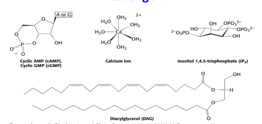
What are these structures examples of?
second messengers
What can a signal be amplified significantly by?
The generation of second messengers.
Second messengers can diffuse to other compartments of the cell
Seven-transmembrane-helix (7TM) receptors (G-Protein coupled receptors - GPCR)
mediate a host of biological functions by responding to a variety of signal molecules (ligands), including hormones, tastants, and even photons
What does the binding of a ligand outside the cell do to the receptor?
it induces a structural change that can be detected inside the cell
What fraction of drugs work through GPCRs?
About one-third because they are attractive for the development of drugs to treat many disorders
What is the largest class of receptors?
GPCRs
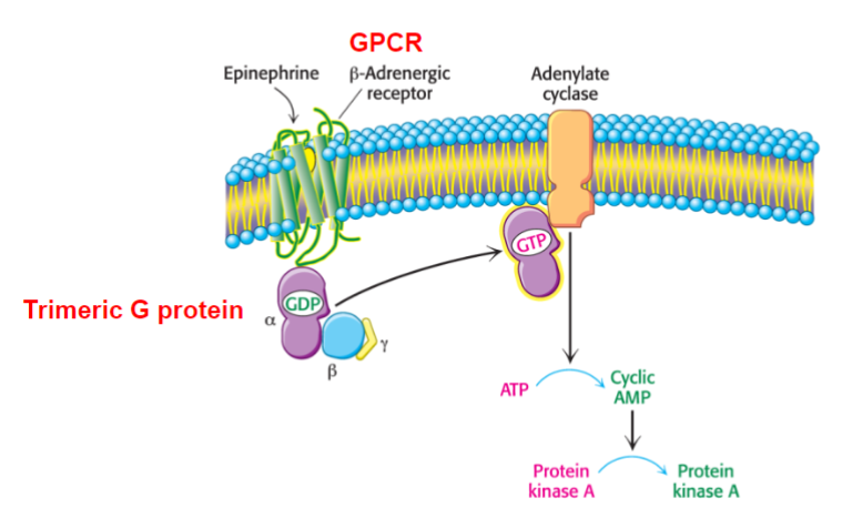
Describe this diagram.
This explains the activation of Protein Kinase A by a G-protein Pathway
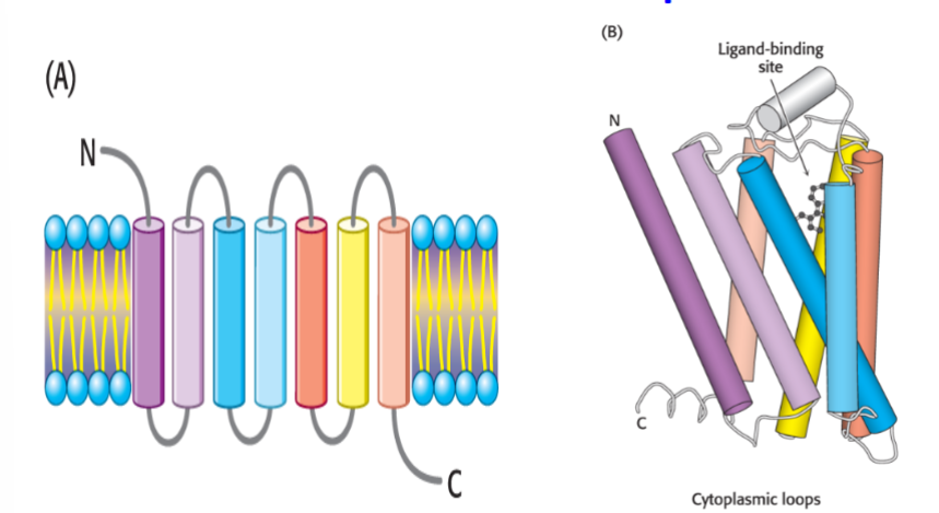
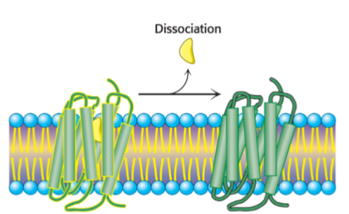
Describe this diagram.
This shows the structures of 7TM Receptors.
What occurs when the 7TM receptor activates?
the α subunit dissociates from the βγ dimer and exchanges GDP for GTP (GTPase turns signal off by converting GTP to GDP)
The GTP bound α-subunit transmits the signal to other cellular components.
Because 7TM receptors are always associated with G-proteins, they are often called G-protein coupled receptors (GPCRs)
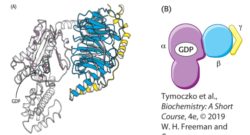
Describe these structures?
Heterotrimeric G-proteins
(A) shows a G-protein in active mode (alpha, beta, gamma subunits from left to right)
(B) shows a G-protein in inactive mode
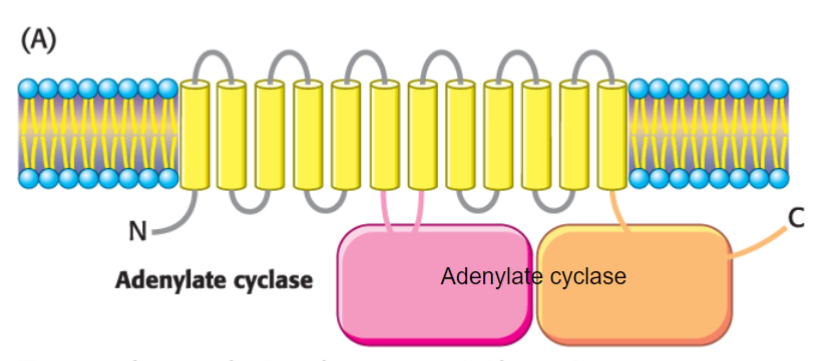
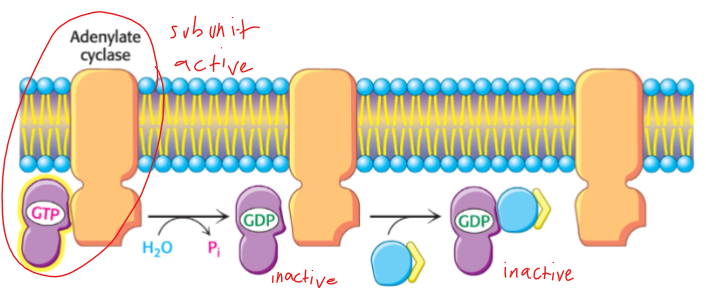
In the case of the β-adrenergic receptor signal transduction pathway, the activated G protein (Gαs) stimulates what?
the integral membrane enzyme, adenylate cyclase
Activation of the cyclase leads to the synthesis of what?
the second messenger, cyclic adenosine monophosphate (cAMP)
What does this diagram show?
adenylate cyclase activation
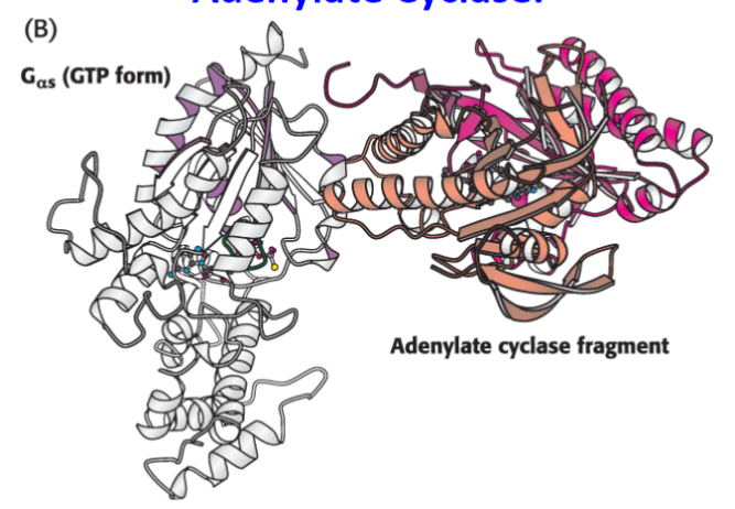
What does this diagram show?
the Complex Between Gα in its GTP Form Bound to a Catalytic Fragment of Adenylate Cyclase
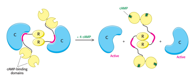
What does cyclic AMP activate?
protein kinase A
What two pairs of subunits does protein kinase A consists of?
2 catalytic (C) subunits
2 regulatory (R) subunits
What happens after binding of cAMP by the regulatory subunits?
these same subunits dissociate from the complex which results in the activation of the 2 C subunits
How do activated C subunits continue the epinephrine signal transduction pathway?
by phosphorylating protein targets that alter physiological functions of the cell
Describe this diagram.
This describes the regulation of protein kinase A
What is Cushing’s Syndrome?
results from excess cortisol secretion.
a constellation of diseases with a variety of symptoms including muscle weakness, thin, easily bruised skin and osteoporosis
sometimes caused by constitutively active protein kinase A. the catalytic subunit does not bind the regulatory subunit and is thus never inhibited
What are the three ways in which the epinephrine-imitated pathway is shut down?
1. Gα has inherent GTPase activity that cleaves the bound GTP to GDP. The Gα bound to GDP spontaneously reassociates with the βγ subunits, terminating the activity of the G protein.
2. Cyclic AMP phosphodiesterase converts cAMP to AMP, which does not activate protein kinase A.
3. Epinephrine-β-adrenergic receptor interaction is reversible. Once the concentration of epinephrine falls, the receptor will no longer be active.
G + P + H2O → GDP + phosphate
What does this diagram show?
resetting of Gα
What does this diagram show?
signal termination
What process does NOT terminate the adenylase cyclase cascade?
a) dissociaton of the ligand-receptor complex
b) deactivation of the G-protein by conversion of GTP into GDP
c) conversion of cAMP into AMP by the cAMP phosphodiesterase
d) dissociation of the Protein Kinase A to subunits
d) dissociation of the Protein Kinase A to subunits
How are cholera and whooping cough (pertussis) due to altered G-protein activity?
Choleragen, the bacterial toxin, modifies a Gαs protein such that it is trapped in the active GTP-bound form. The net result is a loss of NaCl and water into the intestine. ]
Pertussis toxin, the cause of whopping cough, also modifies a G protein. In the case of pertussis toxin, the G protein, Gαi, is trapped in the inactive form. Gαi, which normally inhibits a host of biochemical targets, is thus rendered inactive.
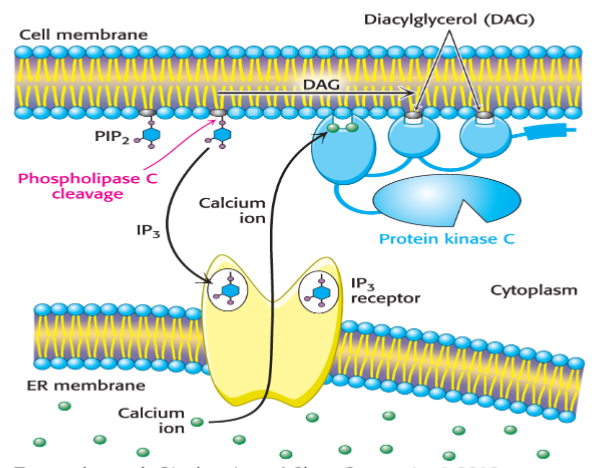
What does the phosphoinositide pathway involve?
(activated by some G-protein-coupled receptors)
it involves the Gαq protein as a component of the trimeric G–protein complex
What does Gαq activate?
phospholipase C, which cleaves the membrane lipid phosphatidylinositol bisphosphate (PIP2) into two second messengers
What two second messengers is phosphatidylinositol bisphosphate (PIP2) cleaved into?
inositol 1,4,5-trisphosphate (IP3) and diacylglycerol (DAG)
What does IP3 bind to and what results from the binding?
IP3-gated channel (IP3 receptor) in the endoplasmic reticulum, allowing an influx of Ca2+ ions (which regulate lots of cellular functions) into the cytoplasm
What does DAG do in conjunction with Ca2+?
it activates protein kinase C, a serine/threonine kinase
Describe this diagram.
This shows the phosphoinositide cascade.
What may result from receptor dimerization?
tyrosine kinase recruitment
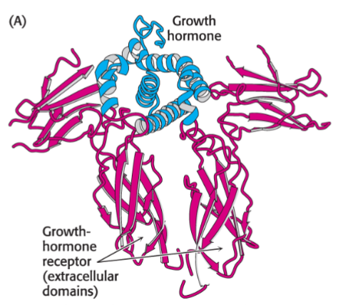
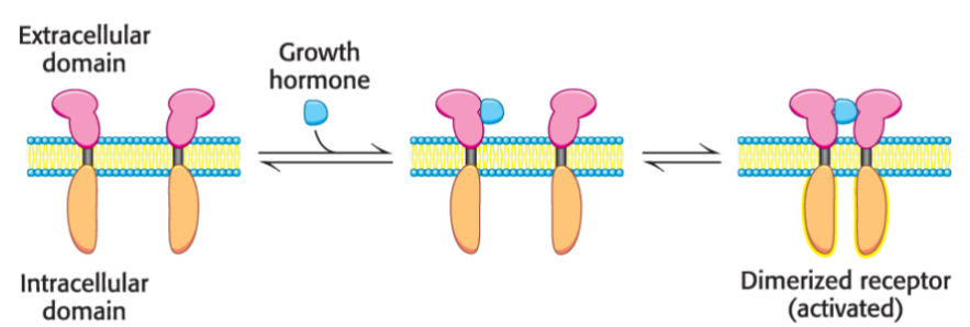
human growth receptor
monomeric integral membrane protein with an extracellular and intracellular domain joined by an intramembrane α helix
upon hormone binding, the receptor dimerizes
Describe this diagram.
This shows the binding of a growth hormone leading to receptor dimerization
Describe this diagram.
This shows receptor dimerization from the binding of a growth hormone
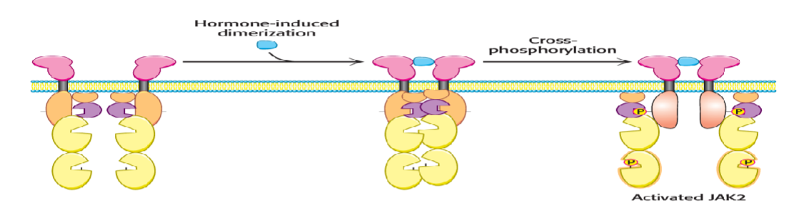
What does dimerization do to the extracellular domains of the receptor?
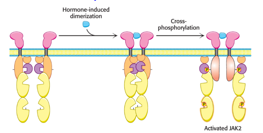
this brings the intracellular domains together, which are associated with Janus kinase 2 (JAK2)
When each JAK phosphorylates its partner on a ___________ residue, what happens?
tyrosine, this activates the two kinases
The activated kinases then ________________ other targets, including a regulator of gene expression called ___________________ and activator of ____________________. ______ further propagates the signal by altering gene expression
phosphorylate, signal transducer, transcription 5 (STAT5), STAT5
Describe this diagram.
This shows the cross-phosphorylation of two molecules of JAK2 induced by receptor dimerization
Explain how receptor tyrosine kinases (RTKs) work?
some growth factors and hormone receptors, such as the epidermal growth factor and insulin, bind to receptors that are tyrosine kinases
upon growth factor or hormone binding, these receptors form dimers
mutations in these receptors in humans cause a variety pathologies
How does cross-phosphorylation and activation of the two intracellular kinase domains arise?
from receptor dimerization
Phosphorylated kinases form docking platforms for other components of what?
the signal transduction pathway
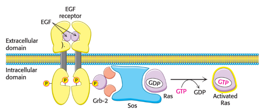
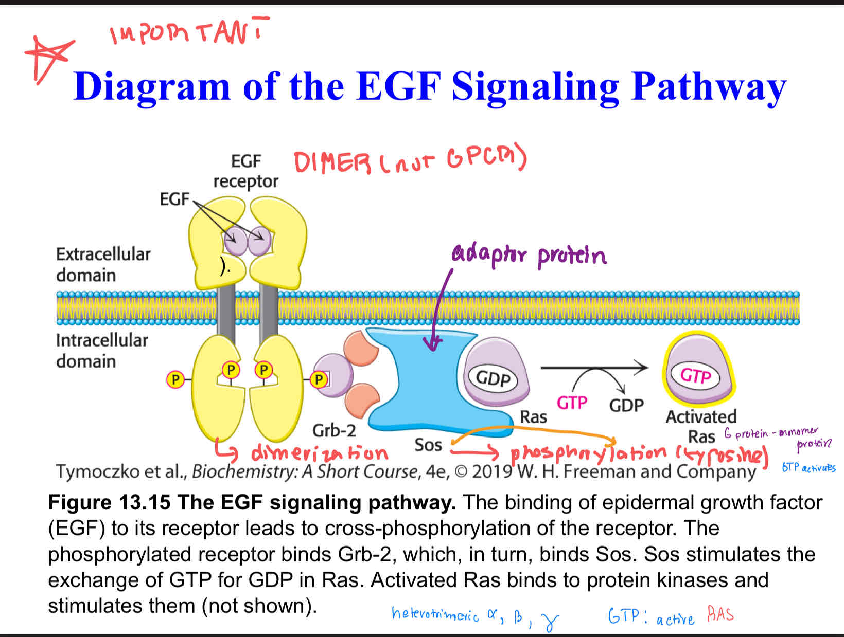
Describe this diagram (IMPORTANT)
EGF Signaling Pathway.
the binding of epidermal growth factor (EGF) to its receptor leads to cross-phosphorylation of the receptor. The phosphorylated receptor binds Grb-2, which, in turn, binds Sos. Sos stimulates the exchange of GTP for GDP in Ras. Activated Ras binds to protein kinases and stimulates them (not shown).
What protein is a key component of the EGF pathway, as well as other signal-transduction pathways?
Ras
What is Ras?
a member of the family of signal proteins called small G proteins or small GTPases. The small G proteins are monomeric
members of this family control a variety of cellular processes
like the Gα protein, Ras is active when bound to GTP and inactive when bound to GDP. Ras also has intrinsic GTPase activity, which controls signal duration
other proteins function to modulate the GTPase activity of Ras
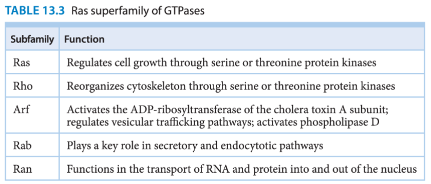
What are the five subfamilies of the Ras superfamily of GTPases and their functions?
When is the polypeptide hormone insulin secreted?
when the blood is rich in glucose.
Insulin is the biochemical signal for the fed state.
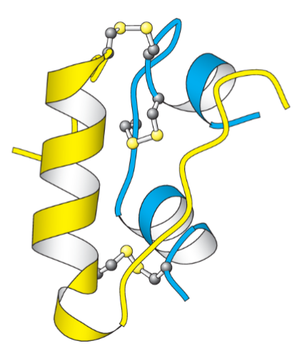
What are the two polypeptide chain in insulin linked by?
disulfide bonds
What kind of kinase in the insulin receptor?
it is a receptor tyrosine kinase
Unlike the other members of the receptor tyrosine kinase class, the insulin receptor exists as a ________ even in the absence of _________
dimer, insulin
Describe this diagram.
this shows the structure of insulin
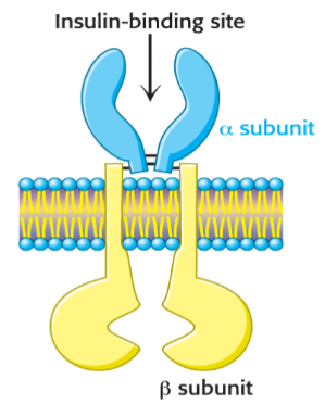
Describe this diagram.
this is an insulin receptor.
The receptor consists of two units, each of which consists of an α subunit and a β subunit linked by a disulfide bond. The α subunit lies outside the cell, and two α subunits come together to form a binding site for insulin. Each β subunit lies primarily inside the cell and includes a protein kinase domain.
What does insulin binding cause?
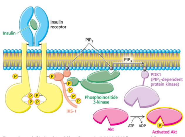
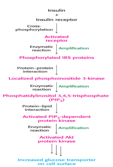
a change in the quaternary structure that results in cross-phosphorylation by the two kinase domains, activating the kinase. The activated kinase of the insulin receptor phosphorylates insulin-receptor substrates (IRSs)
What are phosphorylated IRSs?
they are adaptor proteins used to convey the insulin signal
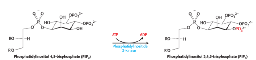
What happens when phosphoinositide-3 kinase binds IRS?
it then phosphorylates phosphatidylinositol 4,5-bisphophate (PIP2) to form phosphatidylinositol 3,4,5-trisphosphate (PIP3)
What happens when PIP3 activates PIP3-dependednt kinase?
it then phosphorylates and activates the kinase Akt
What do the enzymes phosphorylated by Akt control?
the trafficking of the glucose transporter (GLUT4), increasing glucose uptake by the cells, as well a enzymes that convert glucose into glycogen
Describe this diagram.
insulin signaling
Describe this diagram.
this describes the action of a lipid kinase in insulin signaling
Describe this diagram.
the insulin signaling pathway
How do protein phosphatases terminate the insulin signal?
by removing phosphates from the activated proteins in the insulin signal-transduction pathway
How do lipid phosphatases contribute to signal termination?
by converting PIP3 into PIP2
Ca2+ is an important _____________________ in eukaryotic signal transduction pathways.
second messenger
What is an example of a common Ca2+ sensor?
the protein calmodulin
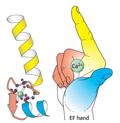
Calmodulin is activated upon binding Ca2+. What are its four Ca2+ binding sites called?
EF hands
What are two examples of biochemical targets that the Ca2+-calmodulin complex activates?
pumps (such as the plasma membrane Ca2+ ATPase)
calmodulin-dependent protein kinase (CaM kinase)
Recall that the Ca2+ also plays a role in the phosphoinositide cascade.
Describe this diagram.
structure of an EF Hand
What are genes that control cell growth called?
proto-oncogenes
What happens when a proto-oncogene is mutated?
leads to unrestrained cell growth (oncogene)
What are genes that normally restrain cancer called?
tumor-suppressor genes
Most signal molecules are too large and too polar to pass through the cell membrane. Thus, the information presented by signal molecules must be transmitted across the cell membrane WITHOUT the molecules themselves entering the cell. The binding site for these signals is located on the ________ surface of the responding cell and cell surface receptors span the plasma membrane. We call them______________. Such membrane receptors are ___________ proteins have both extracellular and intracellular domains.
outside, transmembrane receptors, integral membrane
Structural changes in receptors lead to changes in the concentration of small molecules, called _________________ that are used to relay information from the receptor- ligand complex.
second messengers
G-protein-coupled receptors (GPCRs) all have a similar structure with ____________ transmembrane domains. When a GPCR binds an extracellular signal, these receptors undergo a change in __________ in response to ligand(signal) binding.
seven, confirmation
Dimeric membrane receptors that recruit protein kinases…
Signal binding to receptor → receptor _________ → recruits tyrosine kinase→ _____________ and activation → propagate the signal.
What is an example of this process?
dimerization, cross-phosphorylation
example: growth hormone receptor
The binding of one hormone molecule to two receptors leads to the formation of a receptor dimer. ___________ is a key step in this signal transduction pathway. _____________ brings two molecules of Janus kinase 2 together so that each phosphorylates key residues on the other (cross- phosphorylation), Janus kinase 2 is a tyrosine-protein kinase and phosphorylate proteins on the hydroxyl group of tyrosine residues.
dimerization, dimerization, tyrosine
What are dimeric protein receptors that are protein kinases also known as?
receptor tyrosine kinases (RTKs)