
Human Reproduction
Human beings reproduce sexually and are viviparous.
In humans, the reproductive phase starts after puberty.
It involves:
○ Gametogenesis
○ Insemination
○ Fertilization
○ Implantation
○ Gestation
○ Parturition
The Male Reproductive System →
It is located in the pelvic region.
It consists of:
○ A pair of testes
○ Accessory glands and ducts
○ External genitalia
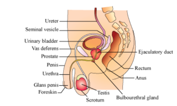
Testes:
Situated within the scrotum, which protects the testes and also helps in maintaining the temperature.
Each testis is 4 to 5 cm in length and 2 to 3 cm in width and has about 250 compartments called testicular lobules.
Testicular lobules have seminiferous tubules, which are the sites of sperm formation.
Two types of cells line seminiferous tubules:
○ Male germ cells − They undergo meiosis to form sperms.
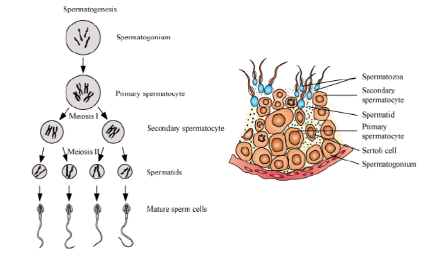
○ Sertoli cells − They provide nourishment to the germ cells.The region outside the seminiferous tubules is called the interstitial space, containing Leydig cells (interstitial cells). The Leydig cells produce androgens.
Accessory Glands and Ducts:
Accessory ducts include:
○ Rete testis
○ Vasa efferentia
○ Epididymis
○ Vas deferensThe seminiferous tubules open into the vasa differential through the rete testis.
The vasa efferentia open into the epididymis, which leads to the vas deferens. The vas deferens open into the urethra along with a duct from the seminal vesicle called the ejaculatory duct.
The ejaculatory duct stores the sperms and transports them to the outside.
The urethra starts from the urinary bladder, extends through the penis, and opens via the urethral meatus.
Accessory glands include:
○ A pair of seminal vesicles
○ Prostate gland
○ A pair of bulbourethral glandsThe secretions of these glands make up the seminal plasma and provide nutrition and a medium of motility to the sperms.
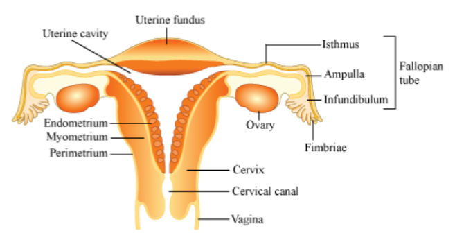
The Female Reproductive System →
It is located in the pelvic region:
It includes:
A pair of ovaries
A pair of oviducts
Uterus
Cervix
Vagina
External genitalia
Mammary glands (not part of the reproductive system but aid in child care)
Ovaries:
They are the primary female sex organs. They produce the ovum and other ovarian hormones.
They are located in the lower abdomen and are 2 to 4 cm in length.
They are connected by ligaments to the pelvic walls and to the uterus.
Each ovary is covered by epithelium and contains the ovarian stroma.
The ovarian stroma is made up of:
○ Peripheral cortex
○ Inner medulla
Oviducts:
They are also called fallopian tubes.
They are 10 to 12 cm long and extend from the ovary to the uterus.
The part of each oviduct lying towards the ovary is funnel-shaped and is called the infundibulum. It has finger-like projections called fimbriae.
The infundibulum leads to the ampulla and then to the isthmus, which has a narrow lumen opening into the uterus.
Uterus:
It is also called a womb and is pear-shaped.
It is connected to the pelvic walls by ligaments.
The uterine wall consists of:
○ External perimetrium
○ Middle myometrium
○ Internal endometrium, which lines the uterine cavityThe endometrium undergoes changes during the menstrual cycle.
Cervix and VaginaThe cervix connects the uterus to the vagina.
The cervix and the vagina constitute the birth canal.
External Genitalia:
Consists of:
○ Mons pubis − Fatty tissue covered by skin and pubic hair
○ Labia majora − Extends from mons pubis and surrounds the vaginal opening
○ Labia minora − Fold of skin beneath the labia majora
○ Hymen − Partially covers the vaginal opening
○ Clitoris − Lies at the junction of labia minora
Mammary Gland:
Present in all-female mammals
It is paired and glandular.
Each breast contains 15 to 20 mammary lobes with alveoli that secrete milk.
The alveoli open into the mammary tubules, which unite to form a mammary duct.
Many mammary ducts constitute the mammary ampulla, which is connected to the lactiferous duct.
Gametogenesis →
The testis and ovary produce the male and female gametes respectively by gametogenesis (spermatogenesis in males and oogenesis in females).
Spermatogenesis:
In males, sperms are produced by the spermatogonia (immature germ cells), which are present in the inner walls of the seminiferous tubules.
Spermatogonia increase in number by mitosis. These are diploid.
Some of the spermatogonia called primary spermatocytes periodically undergo meiosis.
After the first meiotic division, two haploid and equal secondary spermatocytes are formed.
These further undergo meiosis to give rise to four haploid spermatids.
These spermatids are converted into sperms by spermiogenesis.
The sperm head gets embedded in the Sertoli cells after spermiogenesis and is released from the seminiferous tubules by spermiation.
Spermatogenesis starts at puberty by the action of the gonadotropin-releasing hormone (GnRH), which in turn causes the release of two gonadotropins called Luteinizing Hormone (LH) and Follicle Stimulating Hormone (FSH).
LH acts on Leydig cells and causes them to release androgens, which stimulate the process of spermatogenesis while the FSH acts on the Sertoli cells, which help in spermiogenesis.
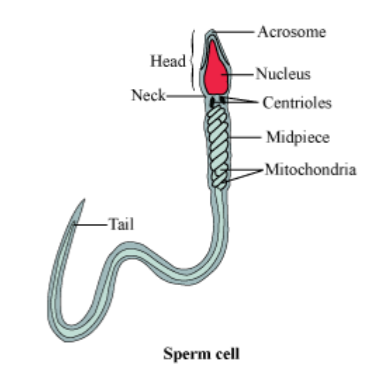
Structure of Sperm:
A mature sperm consists of:
○ Head
○ Neck
○ Middle piece
○ TailThe whole sperm is enclosed in a plasma membrane.
The head consists of a haploid nucleus and a cap-like acrosome, which contains enzymes that aid in fertilization.
The middle piece contains several mitochondria, which produce energy for the motility of the sperm.
Sperms released by the seminiferous tubules are transported by the accessory ducts.
Secretions of the epididymis, vas deferens, seminal vesicles, and prostate are essential for the maturation and motility of sperms.
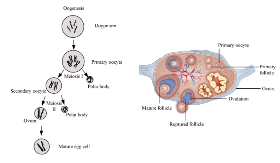
Oogenesis:
The ovum is formed by the process of oogenesis.
It starts during embryonic growth and millions of gamete mother cells (oogonia) are formed in the foetal ovary.These cells undergo meiosis but get temporarily arrested at the prophase and are called primary oocytes.
Before reaching puberty, a large number of primary oocytes degenerate and the remaining ones get surrounded by layers of granulosa cells and new theca and are called secondary follicles.
The secondary follicles are then converted into tertiary follicles that have a characteristic fluid-filled cavity called the antrum. At this stage, the primary oocyte present within the tertiary follicle completes meiosis, which results in the formation of a haploid secondary oocyte and a tiny polar body.
This tertiary follicle further changes into the Graafian follicle. The secondary oocyte is surrounded by the zone pellucida.
Then the Graafian follicle ruptures to release the ovum by ovulation.
Menstrual Cycle and Fertilization:
The menstrual cycle is the reproductive cycle in all primates and begins at puberty (menarche).
In human females, menstruation occurs once in 28 to 29 days. The cycle of events starting from one menstruation to the next one is called the menstrual cycle.
During the middle of the menstrual cycle, one ovum is released (ovulation).
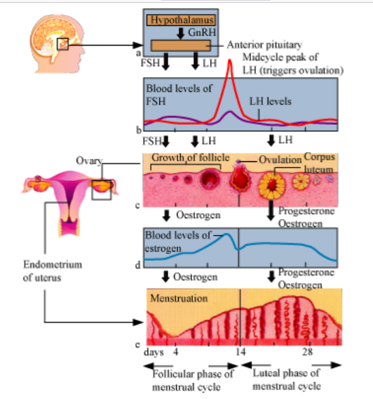
The cycle starts with the menstrual flow (3 to 5 days), caused due to the breakdown of the endometrium of the uterus. Blood vessels in the liquid state are discharged, but this occurs only when the ovum is not fertilized.
It is followed by the follicular phase. In this phase, the primary follicles mature into the Graffian follicles. This causes the regeneration of the endometrium.
These changes are brought about by ovarian and pituitary hormones. In this phase, the release of gonadotropins (LH and FSH) increases. This causes follicular growth and the growing follicles produce oestrogen.
The LH and FSH are at their peak in the middle of the cycle (14th day) and cause the rupture of the Graffian follicles to release the ovum. This phase is called the ovulatory phase.
The remains of the Graffian follicles get converted into the corpus luteum, which secretes progesterone for the maintenance of the endometrium.
In the absence of fertilization, the corpus luteum degenerates, thereby causing the disintegration of the
endometrium and the start of a new cycle.In humans, the menstrual cycle ceases to operate at the age of 50 years. This phase is known as menopause.
Fertilization and Implantation:
During coitus, the semen is released into the vagina, passes through the cervix of the uterus, and reaches the ampullary isthmic junction of the fallopian tube.
The ovum is also released into the junction for fertilization to occur.
The process of fusion of the sperm and the ovum is known as fertilization.
During fertilization, the sperm induces changes in the zona pellucida and blocks the entry of other sperms. This ensures that only one sperm fertilizes an ovum.
The enzymatic secretions of the acrosomes help the sperm enter the cytoplasm of the ovum.
This causes the completion of meiotic division of the secondary oocyte, resulting in the formation of a haploid ovum (ootid) and a secondary polar body.
Then, the haploid sperm nucleus fuses with the haploid nucleus of the ovum to form a diploid zygote.
Mitosis starts as the zygote moves through the isthmus of the oviduct (cleavage) and forms 2, 4, 8, and 16 daughter cells called blastomeres.
The 8−16 cell embryo is called a morula, which continues to divide to form the blastocyst. The morula moves further into the uterus.
The cells in the blastocyst are arranged into an outer trophoblast and an inner cell mass.
The trophoblast gets attached to the uterine endometrium, and the process is called implantation. This leads to pregnancy.
The inner cell mass gets differentiated to form the embryo.
Pregnancy:
After implantation, the trophoblast forms finger-like projections called chorionic villi, surrounded by the uterine tissue and maternal blood.
The chorionic villi and the uterine tissue get integrated to form the placenta, which helps in supplying the developing embryo with oxygen and nutrients, and is also involved in the removal of wastes.
The placenta is connected to the embryo by the umbilical cord. The placenta acts as an endocrine gland and produces the human chorionic gonadotropins, human placental lactogen, oestrogen, progesterone, and relaxin (later stages of pregnancy).
These hormones support foetal growth and help in the maintenance of pregnancy. Hormones like oestrogen, progestogen, cortisol, prolactin, etc., are increased several folds in the maternal blood.
Immediately after implantation, the inner cell mass (embryo) gets differentiated into the ectoderm, mesoderm, and endoderm, which give rise to the different tissues. This ability of the inner cell mass is due to the presence of multi potent cells called stem cells.
Most of the major organs are formed at the end of 12 weeks of pregnancy; during the 5th month, the limbs and body hair are formed; by the 24th week, the eyelids separate, and eyelashes are formed. At the end of nine months, the foetus is fully formed.
Parturition and Lactation:
Human pregnancy has a duration of 9 months. This duration is called the gestation period.
At the end of this period, vigorous uterine contractions lead to the delivery of the foetus. This process is called parturition.
Parturition is a neuroendocrine mechanism and is started by the signals from the developing foetus and the placenta, which produce the foetal ejection reflex.
This causes the release of oxytocin from the pituitary, which causes stronger uterine contractions.
This leads to the expulsion of the baby along with the placenta.
During pregnancy, the mammary glands undergo differentiation, and milk is produced at the end of
pregnancy.The milk produced during the first few days of lactation is known as colostrums. It contains several antibodies that aid the newborn to develop resistance.