


Chapter 21 - Reproduction in Humans
Male Reproductive System:
![]()
Testes (singular: testis) – The male reproductive organs (gonads). Produces sperms (male gametes) and male sex hormones e.g. testosterone. Male sex hormones are responsible for development and maintenance of secondary sexual characteristics. Leading from the end of each testis is a narrow tightly- coiled tube called the epididymis in which sperms are stored.
Scrotum – The two testes are held in a pouch-like sac outside the body called the scrotum. The lower temperature in the scrotum is essential for sperm production.
Sperm ducts – The sperm ducts (vas deferens) lead from the epididymis. During ejaculation, they transport sperm from the epididymis to the urethra.
Prostate gland – The prostate gland is a large gland which secretes directly into the urethra through several small ducts. The fluid contributes to semen. Semen is a composition of sperm and fluids from the sex glands containing nutrients and enzymes which nourish and activate the sperm, allowing them to swim actively.
Seminal vesicles – Ducts from the seminal vesicles join the vas deferens. The seminal vesicles are a pair of glands that secrete a fluid that makes up a proportion of semen.
Urethra – The urethra is a common passage for urine and semen to pass out of the body. The sphincter muscle at the base of the bladder prevents urine from passing out of the bladder during ejaculation of semen.
Penis – The penis consists of cylinders of spongy erectile tissue around the urethra. The tissue contains numerous spaces that allow it to fill up with blood. When that happens, the penis becomes erect and hard, allowing it to enter the vagina of a woman during sexual intercourse to deposit semen.
Sperm:
![]()
The male gamete, the sperm, consists of a head, middle piece and tail.
The head contains:
(a) An acrosome, an enzyme-containing sac. The acrosome contains digestive enzymes which break down the outer membrane of the ovum, allowing for fertilisation.
(b) A small amount of cytoplasm and a large haploid nucleus
The middle piece contains numerous mitochondria arranged spirally to provide energy for the sperm to swim to the egg.
The tail (flagellum) beats to propel the sperm towards the egg.
Female Reproductive System:
![]()
Ovaries – The female reproductive organs (gonads). Produces ova (singular: ovum) and female sex hormones e.g. estrogen and progesterone. Female sex hormones are responsible for development and maintenance of secondary sexual characteristics. Mature eggs are released from the ovaries into the oviducts.
Oviducts – The oviduct (fallopian tube) is a narrow muscular tube leading from the ovary to the uterus. The oviduct has a funnel-like opening to make it easier for ova to enter the oviduct. Cilia on the inner lining help move the ovum to the uterus. The ovum is usually fertilised in the oviduct.
Uterus – The uterus is a thick muscular organ that can stretch as the fetus increases in size during pregnancy. The smooth muscles in the uterine wall contract to expel the fetus during birth. The uterus is lined by a lining called the endometrium (uterine lining). The endometrium is richly supplied with blood vessels and is the site of implantation of the embryo post-fertilisation. It is broken down every month and flows out of the body in the process called menstruation.
Cervix – The cervix is a circular ring of muscle at the neck of the uterus. It opens into the vagina. It enlarges during birth to allow the passage of the fetus.
Vagina – The vagina is a thin-walled chamber where sperm is deposited during sexual intercourse. It forms the birth canal through which the baby is born.
Ovum:

The female gamete, the ovum, is a large cell containing abundant cytoplasm.
It has a large nucleus containing a haploid set of chromosomes.
It is surrounded by a plasma membrane and an outer membrane.
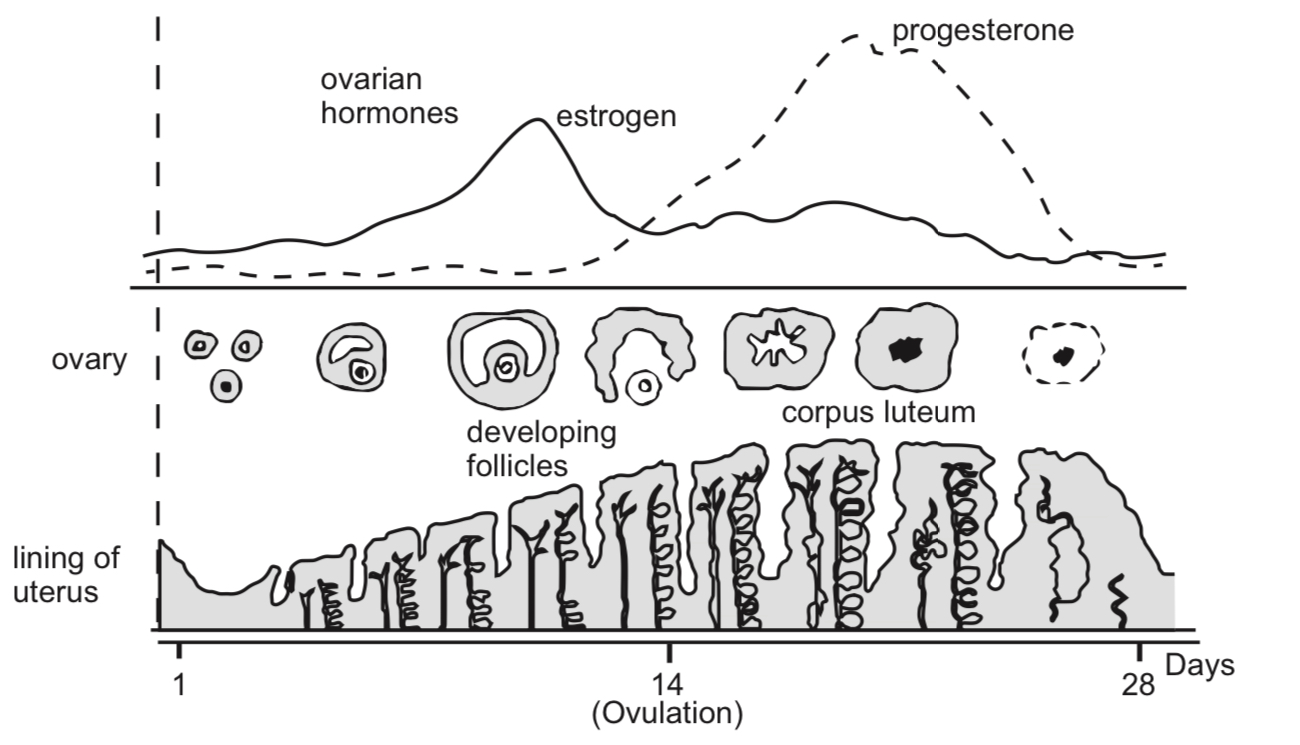
The menstrual cycle
The menstrual cycle normally spans over 28 days. There is natural variation in the length of the menstrual cycle, and it can range from 21 to 33 days.
Day 1 to 5: Menstruation lasts for 5 days. The first day of menstruation is day 1 of the menstrual cycle. The endometrium breaks down and flows out of the body.
Day 6 to 13: The ovaries secrete estrogen which causes the repair and growth of the endometrium. The endometrium becomes thicker.
Day 14: A mature ovum is released from the ovaries. Secretion of progesterone is stimulated. The ovum dies after about 1 to 2 days if it is not fertilized.
Day 15 to 28: Progesterone and estrogen are continually being secreted for continued development and maintenance of the endometrium. Progesterone maintains the endometrium by causing it to become thicker. The endometrium readies for implantation. Towards the end of the cycle, secretion of progesterone and estrogen decline sharply. The endometrium is no longer maintained and disintegrates. It flows out from the uterus together with some blood through the vagina. This marks the beginning of another cycle.
The fertile phase of the cycle is from day 11 to 17. This is because sperms can survive for 2 to 3 days in the female reproductive system. Sperms deposited in the vagina from day 11 onwards can fertilise the ovum which is released from the ovaries on day 14. The ovum can survive for 1 to 2 days after ovulation; hence fertilization is possible up till day 17.
The rest of the days make up the infertile phase of the menstrual cycle. Sexual intercourse during this period is unlikely to result in fertilisation since no ovum is present.
Fertilization:
During sexual intercourse, semen containing sperms is deposited into the vagina of a woman. The fluids from the male sex glands that make up semen provide nutrients and protection for the sperms, as well as a medium for them to swim in.
The sperms swim up the oviducts and encounter the ovum.
The acrosome of the sperms release enzymes to disperse the layer of cells surrounding the ovum and break down the outer membrane of the ovum.
Only 1 sperm will enter the ovum. The plasma membranes of the sperm and the ovum fuse and the sperm nucleus enters the ovum. The plasma membrane of the egg undergoes a change as soon as a single sperm has entered, preventing other sperms from entering.
The sperm nucleus fuses with the egg nucleus, forming a fertilised ovum known as a zygote.
The remaining sperms eventually die.
Development of the zygote
The cilia on the oviduct lining help move the zygote towards the uterus.
In the meantime, the zygote divides many times to form a hollow ball of cells called the embryo.
5 to 7 days after fertilisation, the embryo comes into contact with the endometrium and becomes embedded. This process is known as implantation.
Tissues growing out from the embryo invade the endometrium, forming the placenta. The placenta is an organ that contains both maternal and embryonic blood vessels. It allows for diffusion between the maternal blood circulation and embryonic blood circulation.
The placenta:
(a) Provides nutrients (glucose, amino acids and mineral salts) and oxygen for the embryo.
(b) Removes waste materials such as urea and carbon dioxide
(c) Allows protective antibodies to diffuse from maternal blood into embryonic blood
(d) Provides a barrier preventing maternal blood and embryonic blood from mixing. Reasons for this include:
(i) Maternal blood pressure is much higher than embryonic blood pressure and would damage vital tissues.
(ii) The embryo might have a different blood group, resulting in agglutination if mixing of blood occurs.
(e) Produces progesterone which maintains the endometrium during pregnancy.
The embryo eventually becomes connected to the placenta by the umbilical cord. Embryonic blood travels to the placenta via the arteries of the umbilical cord and returns with oxygen and dissolved food substances via the umbilical vein.
A membrane called the amniotic sac begins development at the same time as the placenta, and encloses the embryo in a fluid-filled space. The fluid is known as amniotic fluid.
The amniotic fluid functions to: (a) Absorb shock, support and protect the embryo from physical injury.
(b) Lubricate the vagina during birth to reduce friction.
(c) Allow the fetus to move freely during development.
About 9 weeks after fertilisation, the embryo has developed into a fetus.
Human Immunodeficiency Virus
Acquired Immune Deficiency Syndrome (AIDS) is a disease that can be spread through sexual intercourse.
It is caused by a virus called Human Immunodeficiency Virus (HIV).
HIV progressively reduces the effectiveness of the infected person’s immune system in protecting him from infection.
HIV infection progresses to AIDS, the last stage of the infection, in about 9 to 10 years after infection.
Symptoms of AIDS include:
(a) Persistent fever, sweat, swollen glands, chills, weakness and weight loss
(b) Pneumonia
(c) Tuberculosis
(d) Chronic diarrhoea
(e) Brain infection
(f) Tumours such as Kaposi’s sarcoma (cancer of the blood vessels) and cervical cancer in women
HIV is transmitted: (a) By sexual intercourse with an infected person (b) By sharing and reusing contaminated needles during intravenous drug use, tattoos and piercing (c) By receiving a blood transfusion from an infected donor (d) During pregnancy and childbirth. An infected mother could pass on the disease to her child
Spread of HIV can be prevented by:
(a) Having protected sexual intercourse. A condom reduces the risk of infection.
(b) Abstinence from sex or having sex with only one partner.(c) Not sharing objects that could be contaminated with blood or bodily fluids such as hypodermic syringes, razors and toothbrushes.
(d) Screening of blood in a blood bank for HIV infection to reduce chances of transmission during blood transfusions.(e) Infected mothers should undergo antiretroviral therapies and give birth by caesarean section to minimise risk of transmission to the foetus. Breastfeeding should be avoided after birth.
(f) Visiting reliable operators for tattoos, piercings or acupuncture where needles are sterilised or disposable.